Copyright ⓒ 2008, The Korean Academy of Oral Biology
117
Journal of Oral Biology
Mechanisms of tert-Buthyl Hydroperoxide-induced Membrane Depolarization in Rat Spinal Substantia Gelatinosa Neurons
Seong-Jun Lim and Sang-Woo Chun*
Dept. of Oral Physiology, College of Dentistry, Wonkwang University (Received September 19, 2008 ; Accepted September 29, 2008)
Reactive oxygen species (ROS) are toxic agents that may be involved in various neurodegenerative diseases. Recent studies indicate that ROS can act as modulators of neuronal activity, and are critically involved in persistent pain primarily through spinal mechanisms. In the present study, whole cell patch clamp recordings were carried out to investigate the effects of tert-buthyl hydroperoxide (t- BuOOH), an ROS, on neuronal excitability and the mechanisms underlying changes of membrane excitability.
In current clamp condition, application of t-BuOOH caused a reversible membrane depolarization and firing activity in substantia gelatinosa (SG) neurons. When slices were pretreated with phenyl-N-tert-buthylnitrone (PBN) and ascorbate, ROS scavengers, t-BuOOH failed to induce membrane depolarization. However, isoascorbate did not prevent t-BuOOH-induced depolarization, suggesting that the site of ROS action is intracellular. The t-BuOOH- induced depolarization was not blocked by pretreatment with dithiothreitol (DTT), a sulfhydryl-reducing agent. The membrane-impermeant thiol oxidant 5,5-dithiobis 2-nitro- benzoic acid (DTNB) failed to induce membrane de- polarization, suggesting that the changes of neuronal excitability by t-BuOOH are not caused by the modification of extrathiol group. The t-BuOOH-induced depolarization was suppressed by the phospholipase C (PLC) blocker U- 73122 and inositol triphosphate (IP
3) receptor antagonist 2- aminoethoxydiphenylbolate (APB), and after depletion of intracellular Ca
2+pool by thapsigargin. These data suggest that ROS generated by peripheral nerve injury can induce central sensitization in spinal cord, and t-BuOOH-induced
depolarization may be regulated by intracellular Ca
2+store mainly via PLC-IP
3pathway.
Key words: tert-buthyl hydroperoxide, substantia gelatinosa, depolarization, PLC-IP
3pathway, pain
서 론
조직손상으로부터 신체를 보호하기 위한 초기 경고신호 로 작용하여 유해한 환경에서 생명체가 생존하는데 중요 한 역할을 담당하는 통증은 일차적으로 일차 구심신경말 단에 존재하는 동통수용기의 활성화로 발생되지만, 임상 에서 발견되는 염증성 통증(inflammatory pain), 신경병 증성 통증(neuropathic pain) 등 지속성 통증의 유발원인 과 발생기전은 매우 다양하다(Hunt와 Mantyh, 2001). 지 속성 통증의 경우에는 말초감작(peripheral sensitization) 과 중추감작(central sensitization)의 발생과 관련된다. 말 초감작은 유해수용기에 의해 나타나는데 역치가 넘은 자 극에 대해서 정상적인 경우보다 더 강하게 반응하며 역 치하 자극에 대해서도 반응하는 경향을 보인다. 중추감작 은 중추신경계 특히 척수후각의 통증전달 신경세포에서 일어나는 유사한 현상이다(Cook 등, 1987; Woolf와 Thompson, 1991; Willis와 Coggeshall, 2004).
Hydrogen peroxide, superoxide, hydroxyl radical, 산 화질소를 포함하는 활성산소종(reactive oxygen species;
ROS)은 조직손상이나 증가된 유해반응과 관련 된다 (Levy 와 Zochodne, 1998; Khalil과 Khodr, 2001; Liu 등, 2004; Wang 등, 2004). 최근의 연구에서는 ROS가 염증 성 통증이나 신경병증성 통증과 같은 상태에서 통증발생 에 관여되어 있다고 보고되고 있다. 척수신경 결찰 모델 (spinal nerve ligation; SNL)에 의한 신경병증성 통증에
*Corresponding author: Sang-Woo Chun, Department of Oral Physiology, College of Dentistry, Wonkwang University, 344-2, Shinyong-Dong, Iksan 570-749, Korea.. Tel.: +82-63-850-6932;
E-mail: physio1@wonkwang.ac.kr
서 척수내의 ROS 생성 증가가 관찰되었으며(Park 등, 2006), 대표적 항산화제인 phenyl N-tert-buthylnitrone (PBN)과 5, 5-dimethyl-pyrroline-N-oxide (DMPO)의 투 여로 ROS의 제거에 의해 수 시간동안 지속되는 진통효 과가 나타남이 보고되었다(Kim 등, 2004). 또한 비타민 E가 인산화된 NMDA 수용체의 1 소단위(pNR1)를 감소 시키고 척수후각 뉴론의 반응성을 감소시켜 척수에서의 중추감작을 감소시킴에 의해 진통효과를 나타낸다고 하 였다(Kim 등, 2006).
전기생리학적인 방법을 이용하여 세포에서 ROS의 작 용을 규명하기 위한 연구의 결과는 아직 확실하지 않다.
예를 들면, 시상신경세포에서는 과산화수소가 흥분성 및 억제성 시냅스의 균형을 조절하여 과흥분을 유발하였고 (Frantseva 등, 1998), 흰쥐 해마에서도 NMDA 수용체 활성에 의해 과흥분성이 관찰되었다(Avshalumov와 Rice, 2002). Bao 등(2005)은 과산화수소가 TRP 통로를 활성 화하여 탈분극과 흥분성의 증가를 유발한다고 보고하였 으며, 중뇌 도파민 분비세포에서는 KATP 통로의 열림에 의해서 과분극을 유발한다고 보고하였다(Avshalumov 등, 2005). 척수의 아교질 세포에서는 과산화수소가 미세억제 성 시냅스후 전류의 빈도를 증가시킨다고(Takahashi 등, 2007) 보고되었고, ROS의 일종인 tert-buthyl hydro- peroxide (t-BuOOH) 투여에 의해 흥분성 시냅스후 전류 의 빈도가 증가하고 양전류 자극에 의해 유발되는 활동 전압의 빈도가 증가함이 보고되었다(Son과 Chun, 2007).
따라서 이 연구에서는 ROS의 일종인 t-BuOOH가 실 제로 척수에 작용하여 중추감작을 일으켜 통증을 유발할 수 있는지 확인하고자 patch clamp 방법을 이용하여 척 수 아교질 뉴론(substantia gelatinosa, SG; lamina II)의 흥분성에 대한 t-BuOOH의 효과를 알아보았고 또한 t- BuOOH에 의한 뉴론의 흥분성 증가는 어떤 기전에 의하 여 일어나는지를 조사하였다.
실험재료 및 방법
척수절편 제작
생후 13일-20일 된 Sprague-Dawley 흰쥐를 암수 구별 없이 사용하였다. 실험과정은 원광대학교의 동물실험 윤 리위원회의 승인을 받았고 동물실험 규정을 준수하였다.
흰쥐를 ether로 마취한 후 20% urethane (2 ml/Kg)을 복강 내 투여하였다. 흉추에서부터 천추까지 척추제거술(lami- nectomy)을 하여 척수를 노출한 후 요천수 팽대부 (lumbosacral enlargement)에서 1 cm 정도 길이의 척수 를 절단하였다. 조직절편기(vibratome 752M, Campden, 영국)의 고정대에 agar block을 먼저 고정한 후 순간접 착제를 이용하여 척수절편을 고정하였다. 95% O2-5%
CO2를 공급하면서 두께 350 µm의 척수절편을 얻었는데,
절단 중 계속 온도 조절기(model 765, Campden, 영국) 를 이용하여 용액의 온도를 1-2oC 정도로 낮게 유지 시 켰다. 척수절편은 32oC의 인공 뇌척수액 용액에 1시간 정도 보관하여 정상상태로 회복시켰고, 이후에 실온에서 실험을 시행하였다. 기록은 척수절편을 현미경(BX50WI, Olympus, 일본) 위의 기록용기(1 ml)에 옮긴 후 치실로 만든 그물로 움직이지 않도록 고정한 후 시행하였고, 실 험기간 동안 계속해서 95% O2-5% CO2가 포함된 용액 을 관류펌프(Minipuls 3, Gilson, 프랑스)를 이용하여 관 류시켰다(2-3 ml/min).
실험용액
척수절편 제작에 사용했던 절단용액의 조성(mM)은 252 Sucrose, 2.5 KCl, 0.1 CaCl2, 2 MgCl2, 10 glucose, 26 NaHCO3, 1.25 NaH2PO4등으로 구성되었으며, 막전압을 기록하기 위한 세포외 용액의 조성은 117 NaCl, 3.6 KCl, 2.5 CaCl2, 1.2 MgCl2, 1.2 NaH2PO4, 25 NaHCO3, 11 Glucose 이었고 95% O2-5% CO2를 공급하여 pH를 7.4 로 유지하였다. 세포내 용액은 150 K-Glu, 10 HEPES, 5 KCl, 0.1 EGTA, 5 MgATP, 0.3 Na GTP를 사용하였 고, pH는 KOH를 첨가하여 7.3으로 조정하였다. 실험에 사용한 t-BuOOH, PBN, ascorbate, isoascorbate, dithio- threitol (DTT), 5,5-dithiobis (2-nitro-benzoic acid) (DTNB), thapsigargin, 1,2-bis (2-aminophenoxy)ethane- N,N,N',N'-tetraacetic acid (BAPTA) 등은 Sigma사 (미국 )에서 구입하였고, U-73122, 2-aminoethoxydiphenylbolate (APB) 등은 Tocris사(영국)에서 구입하여 사용하였다. t- BuOOH, PBN, ascorbate, isoascorbate, DTT, DTNB 등은 세포외 용액에 녹여 사용하였으며, thapsigargin, APB 등은 DMSO (dimethyl sulfoxide; Sigma)에 먼저 녹인 후 최종농도로 실험직전에 세포외 용액에 희석하여 사용 하였고, BAPTA는 세포내 용액에 녹여 사용하였다. 세포 에 대한 실험용액의 적용은 중력을 이용한 관류장치(BPS- 4SG, Ala Scientific Instruments, 미국)를 이용하여 기록 용기 내 용액을 교환하였다.
전기생리학적 기록방법
막전압기록은 whole cell patch clamp 방법을 사용하 였다. 미세 유리전극 제조기(PP-830, Narishige, 일본)와 microforge (MF-830, Narishige, 일본)를 이용하여 외경 1.5 mm의 연질 유리미세관(TW150-3, WPI, 미국)을 저 항이 5-8 MΩ이 되도록 기록전극을 제작하였다. 10배의 대물렌즈로 다른 부위에 비해 밝은 띠를 형성하고 있는 척수 아교질 부위를 확인한 후 전극에 양압을 가하면서 미세 전극조절기(ROE-200, Sutter, 미국)를 이용하여 30o 경사를 유지하면서 세포에 접근하였다. Seal test를 시행 하면서 세포에 접근하여 피펫의 저항이 순간적으로 증가 하는 것으로 세포에 근접함을 확인한 후 양압을 풀고 음
압을 가하여 세포와의 gigaohm seal을 이루었다. 전압과 전류측정에는 Axopatch 200B 증폭기(Axon, 미국)를 사 용하였고, 이 증폭기는 Digidata 1200B (Axon, 미국) AD 변환기를 통하여 컴퓨터에 연결하였으며, pCLAMP software (version 9.0, Axon, 미국)를 사용하여 실험수행의 명령 과 얻어진 전기신호의 저장 및 분석에 이용하였다. 발생 된 전류는 low pass 8-pole Bessel filter로 2kHz로 여 과하였다. 모든 실험은 실온에서 시행하였다.
실험자료의 분석
막전압의 분석은 Clampfit (Version 8.0, Axon, 미국) 을 이용하였다. 대조군과 약물처리군 사이에 통계적으로 유의한 차이가 존재하는지의 여부는 paired t-test 혹은 non-paired t-test를 이용하였고, p<0.05에서 통계적으로 유의하다고 판정하였다. 통계자료의 값은 평균값 ± 표준 오차(mean ± S.E.M.)로 표시하였다.
실험 결과
아교질 세포의 흥분성에 대한 t-BuOOH의 효과 척수 아교질 세포에 대한 t-BuOOH의 효과를 조사하기 위하여 whole cell patch clamp 방법을 이용하여 막전압 을 기록하였다. 전류기록법으로 지속적으로 막전압을 기 록하면서 t-BuOOH를 2 mM 처리 하였을 때 55개의 세 포 중 45개의 세포에서(82%) 탈분극(3.1 ± 0.5 mV)이 발 생되었고 나머지 10개의 세포는 과분극되거나(n = 6) 반 응이 없었다(n = 4). 탈분극된 세포의 경우 약물 주입 후 1-5분 후부터 1-15 mV의 탈분극이 발생되었고 그 중 일 부의 세포에서는 활동전압이 발생되었으며 이는 약물이 포함되지 않은 용액으로 처리하였을 때 탈분극이 5분-30 분 동안 지속된 후 서서히 원상태로 회복되었다(Fig. 1A).
대표적인 광범위 항산화제인 PBN을 1 mM 전 처리한 후 t-BuOOH를 투여 하였을 때는 탈분극이 발생되지 않 았으며 (0.2 ± 0.8 mV, p<0.01)(n = 20), 과분극을 보이는 세포의 비율(8/20)이 증가하였다(Fig. 1B). 또한 ascorbate 500µM를 전 처리한 군 (Fig. 1C)에서도 t-BuOOH에 의 해 유발되는 탈분극은 발생되지 않았으나 (−1.0 ± 0.8 mV, p<0.01)(n = 21), 세포막을 통과하지 못하는 isoascorbate 500µM을 전 처리한 군에서는 t-BuOOH에 의해 유발되 는 탈분극의 정도가 감소되기는 하였으나 여전히 발생하 여(1.4 ± 1.0 mV, n = 19)(Fig 1D, E) ascorbate의 항산 화 작용은 세포내에서 주로 이루어짐을 알 수 있었다.
t-BuOOH에 의해 발생하는 탈분극의 기전
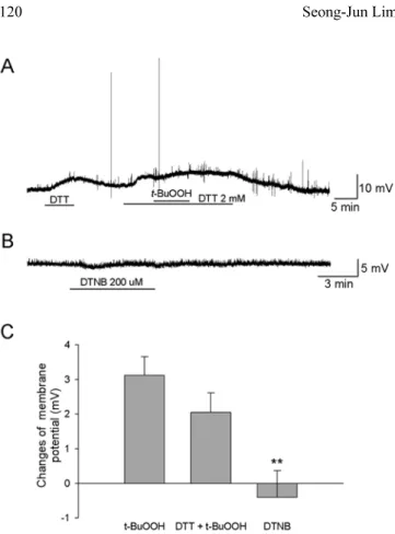
활성산소는 세포막의 단백질에 직접 작용하여 산화작용 을 일으켜 효과를 나타낸다(Hong 등, 2006; Andrea 등, 2008). 척수 아교질 세포에서 t-BuOOH에 의한 흥분성의
증가가 직접적인 산화작용에 의한 것인지를 알아보기 위 하여 세포막 단백질에 작용하는 환원제인 DTT를 전처리 한 후 t-BuOOH를 투여하였을 때 1.9 ± 0.6 mV의 탈분 극이 발생하여 t-BuOOH만 투여하였을 때와 유의한 변 화를 보이지 않았다(n = 20)(Fig. 2A, C). 또한 세포막을 통과하지 못하는 산화제인 DTNB를 투여하였을 때 탈분
Fig. 1. Effects of antioxidants on the t-BuOOH-induced membrane depolarization in substantia gelatinosa (SG) neurons. (A) t-BuOOH (2 mM) applied for 7 min caused a reversible membrane depolar- ization and firing activity. When slices were pretreated with 1 mM of PBN (B) and 500 µM of ascorbate (C), ROS scavengers, t- BuOOH failed to induce membrane depolarization, but isoascor- bate (500 µM) did not prevent t-BuOOH-induced membrane depo- larization (D). (E) t-BuOOH-induced membrane potential changes under control condition and pretreatment of antioxidants.
**: Values are significantly different from the control by indepen- dent t-test (p<0.01).
† :
Values are significantly different from the ascorbate by inde-
pendent t-test (p<0.05). Means ±SEM.
극이 유발되지 않아서(−0.8 ± 0.8 mV, n = 10), t-BuOOH 에 의한 흥분성의 증가는 세포내 신호전달 과정에 의해 발생하였을 것으로 추측할 수 있다(Fig. 2B, C).
t-BuOOH에 의한 탈분극이 세포내 칼슘과 관련된 세포 내 신호전달 경로를 통하는지를 확인하고자 세포내 칼슘 chelator BAPTA, 세포내 칼슘저장소의 칼슘을 고갈시키는 작용이 있는 thapsigargin, phopholipase C (PLC) 차단제 U-73122, 칼슘저장소의 IP3 수용체 차단제 APB 등을 전 처리 한 후 t-BuOOH의 투여효과를 확인하였다(Fig. 3).
이 연구에서는 세포내액의 칼슘을 제거시키기 위하여 세포내 용액에 칼슘 chelator인 BAPTA 10 mM을 사용 하였다. BAPTA가 포함된 전극으로 기록시 t-BuOOH에 의해 −2.0 ± 0.6 mV (n = 13)(p<0.01)로 과분극이 발생 되었다. Thapsigargin은 세포내 칼슘저장소로 Ca2+을 reuptake 하는 과정을 차단함으로써 칼슘저장소내의 칼슘 을 고갈시킨다. Thapsigargin 1 µM을 45분 이상 전처리 하고 t-BuOOH를 투여하였을 때 0.4 ± 0.5 mV (n = 11)
의 전압변화가 발생하여 대조군에 비해 유의하게 감소하 였다(p<0.05). U-73122 10 µM을 t-BuOOH 적용 5-10 분전에 전처리 한 후 t-BuOOH를 투여 하였을 때 0.9 ± 0.9 mV (n = 14)의 탈분극이 발생하여 대조군에 비해 유 의하게 감소하여(p<0.05) PLC 경로를 통한 세포내 칼슘 의 증가가 탈분극의 발생에 중요하게 작용한 것으로 추 측된다. PLC의 활성은 IP3를 생성하여 세포내 칼슘저장 소로부터 세포질로 칼슘방출을 매개한다. IP3수용체 차단
Fig. 2. The changes of neuronal excitability by t-BuOOH are not
caused by the modification of extrathiol group. (A) The t-BuOOH- induced depolarization was not blocked by pretreatment with dithiothreitol (DTT; 2 mM), a sulfhydryl-reducing agent. (B) The membrane-impermeant thiol oxidant DTNB (200 µM) did not induce membrane depolarization. (C) Membrane potential changes by application of t-BuOOH alone (t-BuOOH), t-BuOOH in the presence of DTT (DTT + t-BuOOH) and DTNB alone (DTNB).
**: Values are significantly different from the control by indepen- dent t-test (p<0.01). Means ±SEM.
Fig. 3. t-BuOOH-induced membrane depolarization requires PLC activation and Ca
2+release from internal stores. (A) t-BuOOH- induced membrane depolarization was blocked with intracellular calcium chelator BAPTA (10 mM) (a), Ca
2+ATPase inhibitor thapsigargin (1 µM) (b), the PLC blocker U-73122 (10 µM) (c) and IP
3receptor antagonist APB (50 µM) (d). (B) Mean t- BuOOH-induced membrane depolarization in control and in neu- rons pretreated with BAPTA, thapsigargin, U-73122 and APB.
**: Values are significantly different from the control by indepen-
dent t-test (p<0.01), *: (p<0.05). Means ±SEM.
제인 APB 50 µM을 전처리 후 t-BuOOH를 투여하였을 때 −1.5 ± 0.6 mV (n = 18)의 전압변화를 기록하여 대조 군에 비해 유의하게 감소하였다 (p<0.01).
고 찰
척수후각 세포(dorsal horn neuron, DHN) 중 표층의 척수 아교질 신경세포는 주로 말초로부터 일차 감각신경 섬유를 통하여 직접 감각정보를 전달받고 이를 통합한다.
특히 신체에 유해한 통각정보는 얇은 수초를 가진 Aδ- 섬유와 무수섬유인 C-섬유와 같은 일차 감각신경섬유를 통해 척수 표층의 DHN으로 전달되며 이웃하는 lamina I과 IV등의 투사신경세포(projection neuron)를 통하여 시상 (thalamus) 등의 상위중추로 통각정보를 전달한다(Kumazawa 와 Perl, 1978; Yoshimura와 Jessell, 1989). 만성통증은 이러한 정보전달 체계 중에서 지속적 신경자극에 의해 시냅스 가소성의 변화가 유발되며, 이러한 변화에 의해 오랜 시간 지속되는 세포 반응성의 증가가 만성통증의 세포기전이라고 알려져 있다. 중추에서는 척수후각 뉴론 이 감작되어지고 말초자극에 의하여 더 민감하게 반응하며 더 넓은 수용야로부터의 입력신호를 받는다(Cook 등, 1987;
Woolf와 Thomson, 1991). NMDA 수용체는 중추감작을 매개하고 많은 이차전달자들이 일련의 변화에 관여되어 있다(Woolf와 Thompson, 1991; Willis, 1994; Woolf와 Costigan, 1999).
ROS의 한 종류인 t-BuOOH는 세포막을 쉽게 통과할 수 있는 유기 과산화수소(hydrogen peroxide)로 뇌와 척 수에 많이 분포되어 있다(Sohn 등, 2005). 이것은 t-BuOO·
혹은 t-BuO·로 대사되어 지질과산화반응(lipid peroxi- dation)을 유발하여 세포에 손상을 준다(Abe와 Saito, 1998). 그러나 ROS는 세포독성과 관련된 역할 이외에 여러 가지 생리적 기능을 조절하는 세포내 신호전달물질 로도 작용하고 있다(Droge, 2002). 예를 들면, TGF-β1 (transforming growth factor)이나 EGF (epidermal growth factor)와 같은 성장인자에 의한 수용체 자극은 일시적으 로 과산화수소 생성을 유발하여 세포증식, 혈관확장과 같 은 정상적인 세포의 기능을 조절하는데 중요한 역할을 한다 (Sobey 등, 1997; Varela 등, 2004). 또한 최근에 는 ROS가 통증발생에 관여되어 있다고 보고되고 있는데, tirilazad (Khalil 등, 1999), superoxide dismutase (Wang 등, 2004), PBN (Kim 등, 2004), vitamin E (Kim 등, 2006)과 같은 다양한 항산화제를 전신투여 혹은 척수내 로 투여함에 의해 진통효과를 가짐이 보고되었다. 이러한 결과는 척수의 활성산소가 통증의 발생과 전달에 관여하고 있음을 의미하는 것으로 이 연구에서는 실제로 척수절편 에서 외부에서 투여한 t-BuOOH가 아교질 세포의 막전 압을 탈분극시켜 흥분성을 증가시킴을 확인하였다(Fig. 1).
t-BuOOH에 의한 반응은 약물제거 후 원상태로 회복되 는 가역적인 반응을 보였다. 이는 통증에 관여하는 ROS 의 작용은 영구적인 세포손상을 일으키는 것이 아닌 정 상적인 세포내 신호조절물질로써의 작용 혹은 경미한 산 화자극에 의한 일시적인 기능변화를 일으킨 상태라 할 수 있다. 최근의 연구에서 ROS는 protein phospatase, protein kinase, transcription factor 등을 이용하여 세포내의 단 백질활성을 조절함으로서 세포활성을 조절할 수 있음이 보고되었다(Maher와 Schubert, 2000; Guedes 등, 2006).
ROS에 의해 유발되는 안정막 전압의 변화에 관한 일 부 연구에서는 약물투여 초기에 과분극이 발생됨이 보고 되고 있다. 해마의 CA1 뉴론이나 중뇌의 도파민 분비 뉴론에서 과산화수소 투여에 의해 과분극을 유발하였는데 이는 칼슘 의존성 K+ 통로의 활성화나 ATP 민감성 K+ 통로의 활성화에 의해 발생하여 산화자극에 대하여 세포를 보호하는 역할을 한다고 하였다(Park 등, 2003; Avshalumov 등, 2005). 그러나 이 연구에서는 세포내 용액의 ATP 농 도를 5 mM로 사용하여 기록한 세포 중 10% 이하의 세 포에서만 t-BuOOH의 투여에 의한 과분극이 관찰되었다.
이 연구에서 세포내 칼슘 chelator인 BAPTA를 세포내 용액에 첨가하고 기록하였을 때는 t-BuOOH에 의한 탈 분극이 나타나지 않는 것으로 보아 t-BuOOH에 의한 탈 분극은 세포내 칼슘의 양과 밀접한 관계를 보였다(Fig. 3).
ROS는 다양한 세포에서 세포내 칼슘농도의 증가를 일으 키는데, 이는 L형의 칼슘통로를 활성화 시키거나(Akaishi 등, 2004), TRPM2 (Wehage 등, 2002), ryanodine 수용체 (Favero 등, 1995), IP3 의존성 통로(Hu 등, 2000)와 같 은 칼슘방출통로를 조절하거나, SERCA와 같은 칼슘펌 프의 조절(Redondo 등, 2004)에 의한다. 세포내의 증가 된 칼슘은 이차전달물질로 작용할 수 있고 Na+−Ca2+ 교 환기전의 활성화(Hirono 등, 1998), 비특이성 양이온 전 도도의 증가(Chuang 등, 2000)를 유발하여 막전압의 탈 분극을 일으킬 수 있다.
세포내에서 칼슘이 이동하는 주요한 경로로는 일반적으 로 PLC-IP3 경로이다. Ligand가 세포막에 존재하는 G 단백과 연결된 수용체와 결합하면 Gq/11 단백질이 활성 화되고 활성화된 G 단백은 PLCβ를 활성화시키고 활성 화된 PLCβ는 PIP2를 가수분해한다. PIP2는 가수분해 되 어 IP3와 diacylglycerol (DAG)를 생성하는데 IP3는 세 포내 칼슘저장고인 내형질세망(endoplasmic reticulum)의 막에 존재하는 IP3 수용체에 결합하여 세포내로 칼슘을 분비시킨다. 이 연구에서도 세포내 칼슘저장소의 칼슘을 고갈시키는 작용이 있는 thapsigargin, phopholipase C (PLC) 차단제 U-73122, 칼슘저장소의 IP3 수용체 차단 제 APB 등을 전처리한 후 t-BuOOH를 투여한 결과 탈 분극이 발생되지 않아(Fig. 3), t-BuOOH에 의한 탈분극 은 PLC-IP3 경로를 통하여 발생하였으리라 사료된다.
종합하면 이 연구에서 t-BuOOH의 투여는 SG 뉴론에
서 막전압을 탈분극 시키고 활동전압을 발생시켰는데 이 러한 결과는 주로 t-BuOOH가 직접적으로 시냅스후 뉴 론에 작용하여 세포내 PLC-IP3경로를 통하여 세포내 칼 슘농도를 증가시키고 그에 따른 일련의 반응에 의해 유 발되었다고 할 수 있다. 또한 이러한 점은 ROS가 이전 의 역치하 입력신호에 대하여 더 쉽게 시냅스후 뉴론의 활동전압을 발생시킬 수 있어 척수의 정보전달과정에 중 요하게 관여할 수 있음을 시사한다.
감사의 글
이 논문은 2006년도 원광대학교의 교비지원에 의해서 수행되었음.
참고문헌