Naringin Protects Ovalbumin-induced Asthma through the Down-regulation of MMP-9 Activity and GATA-3 Gene
Chang-Min Lee, Jeong Hyun Chang
1, In Duk Jung, Young-Il Jeong, Noh Kyung Tae, Hee-ju Park
2, Jong-Suk Kim
3, Yong Kyoo Shin
4, Sung Nam Park and Yeong-Min Park*
Department of Microbiology and Immunology & National Research Laboratory of Dendritic Cell Differentiation & Regulation, and Medical Research Institute, Pusan National University School of Medicine, Busan 602-739, South Korea
1
Department of clinical laboratory Science, College of Health & Therapy, Daegu Haany University, Gyeong San 712-715, South Korea
2
Department of Pediatrics, Pusan National University School of Medicine, Busan 602-739, South Korea
3
Department of Biochemistry, Chonbuk National University Medical School, Jeonju 561-180, South Korea
4
Department of Pharmacology, Chungang University College of Medicine, Seoul 156-756, South Korea.
Received April 2, 2009 /Accepted June 22, 2009
The common word flavonoids is often used to classify a family of natural compounds, highly abun- dant in all higher plants, that have received significant therapeutic interest in recent years. Naringin is associated with a reduced risk of heart disease, neurodegenerative disease, cancer and other chronic diseases; however the molecular basis of this effect remains to be elucidated. Thus we attempted to elucidate the anti-allergic effect of Naringin in ovalbumin (OVA)-induced asthma model mice. The OVA-induced mice showed allergic reactions in the airways. These included an increase in the num- ber of eosinophils in bronchoalveolar lavage (BAL) fluid, an increase in inflammatory cell infiltration into the lung around blood vessels and airways, airway luminal narrowing, and the development of airway hyper-responsiveness (AHR). The administration of Naringin before the last airway OVA chal- lenge resulted in a significant inhibition of all asthmatic reactions. Accordingly, this study may pro- vide evidence that Naringin plays a critical role in the amelioration of the pathogenetic process of asthma in mice. These findings provide new insight into the immunopharmacological role of Naringin in terms of its effects on asthma in mice.
Key words : Naringin, asthma, GATA-3, AHR, IgE
*Corresponding author
*Tel:+82-51-240-7557, Fax:+82-51-243-2259
*E-mail : [email protected]
Introduction
Asthma is an inflammatory disease characterized by bron- chial hyper-responsiveness that can proceed to life-threat- ening airway obstruction. The T helper 2 (Th2)-type cytokines interleukins-4 (IL-4), IL-5, and IL-13, produced by activated CD4
+T cells play a central role in the pathogenesis of asthma by controlling the key process of immunoglobulin E (IgE) production, growth of mast cells and the differentiation and activation of mast cells and eosinophils [7,15,32-34]. In con- trast, Th1 cytokines such as interferon-γ (IFN-γ) and IL-12, which down-regulate Th2 responses, inhibit the development of allergic lung inflammation [13,39]. Thus, interventions that inhibit Th2 cytokines by enhancing Th1 cytokine production, may be useful in the treatment of allergic asthma [36].
OVA-induced asthma is characterized by AHR and in- flammation of the airways [20]. This inflammation is asso-
ciated with the infiltration of eosinophils, neutrophils, and lymphocytes into the bronchial lumen and lung tissues [4,21].
These cellular infiltrates release various chemical mediators
that can cause AHR [5,16]. Recruitment of these inflammatory
cells from the blood to the site of inflammation is regarded
as a critical event in the development and prolongation of
airway inflammation. Inflammatory cells have to cross the
basement membrane and move through connective tissue
until they finally reach inflammatory sites, and require the
involvement of adhesion molecules, cytokines, chemokine
and enzymes including matrix metalloproteinases (MMPs)
in this journey. MMPs are a family of zinc- and cal-
cium-dependent endopeptidases capable of proteolytically
degrading many of the components of the extracellular ma-
trix [27]. MMPs are produced by not only structural cells,
[26,40] but also inflammatory cells [18,23]. They are secreted
as latent forms followed by proteolytic processing to active
forms [8]. Of the MMP family, MMP-2 (gelatinase A, 72-kDa
gelatinase) and MMP-9 (gelatinase B, 92-kDa gelatinase) are
MMPs that share similar domain structures and in vitro ma-
trix substrate specificities [35], and appear to induce the mi- gration of eosinophils, lymphocytes, neutrophils, and den- dritic cells across basement membranes during tissue injury and repair [22,29]. Recruitment of leukocytes from the circu- lating blood into tissues requires a series of cell adhesion molecules, such as ICAM-1 and VCAM-1, which are shown to play key roles in the induction of airway inflammation.
We reported in a previous work that a variety of phy- tochemicals exhibit profound immunoregulatory activities both in vitro and in vivo, particularly in DCs [17,41].
The flavonoids comprise a family of common phenolic plant pigments that have been identified as dietary anti- carcinogens and antioxidants [6]. We reported in a previous study that a variety of phytochemicals exhibit pro-found im- munoregulatory activity, particularly in the DC. Naringin, one of the most common flavonoids, is found in a variety of fruits and vegetables, including onions, parsley, and or- anges as well as chamomile tea, wheat sprouts, and certain seasonings [9]. Naringin has demonstrated anti-in- flammatory, anticarcinogenic, and free radical-scavenging activities in a variety of in vitro systems [2]. In a recent study, investigators identified Naringin as a potent inhibitor of the nuclear transcription factor nuclear factor-κB (NF-κB), which may perform a pivotal function in the regulation of cell growth, apoptosis, and the regulation of the cell cycle [12]. Studies using human leukemia cells as well as carcino- ma cells in the breast, colon, and elsewhere have revealed that Naringin inhibits cell growth via the induction of cell cycle arrest and apoptosis [14]. It also attenuates proin- flammatory cytokine production in LPS-stimulated periph- eral blood mononuclear cells via the selective elimination of monocytes and macrophages, inhibits TNF-induced inter- cellular adhesion molecule-1 up-regulation in vivo, and in- hibits IL-1-induced prostaglandin synthesis and TNF- -induced IL-6 and IL-8 production [11]. Moreover, it actively inhibits IkB kinase activity, IkB degradation, NF-kB DNA protein-binding activity, NF-B luciferase activity, and mi- togen-activated protein kinase (MAPK) activity.
It has been shown using animal models that allergic air- way inflammation is increased by Th2 cytokine production and decreased Th1 cytokine production. In a recent study, it was suggested that T-bet might protect against asthma through increased expression of GATA-3 mRNA in asth- matic airways [3,24,28,31,37].
In this study, we have attempted to characterize the ef- fects of a noncytotoxic concentration of Naringin in a
murine model of asthma. Our findings demonstrated, for the first time, that Naringin treatment inhibited asthmatic syndrome, and suppressed the OVA-induced gelatinolytic activity of MMP-9, and the translocation of GATA-3 in the cytosol.
Materials and Methods Animals and experimental protocol
Female BALB/c mice, 6-8weeks of age and free of mur- ine-specific pathogens, were obtained from the Charles River Laboratories (Yokohama, Japan). All experimental animals used in this study were maintained under a protocol ap- proved by the Institutional Animal Care and Use Committee of the Pusan National University Medical School. Mice were immunized intraperitoneally (i.p.) with 20 μg of OVA (Sigma-Aldrich, St. Louis, MO) emulsified in 1 mg of alumi- num hydroxide (Pierce Chemical Co., Rockford, IL) on day 1 and 15. Mice were challenged for 30 min via the airway with OVA (5% OVA) each day from days 21-23 on consec- utive days. BAL fluid was obtained at 24 hr after the last challenge. At the time of lavage, the mice (6 mice in each group) were killed with an overdose of ether. The chest cav- ity was exposed to allow for expansion, after which the tra- chea was carefully incubated and the catheter secured with ligatures. Prewarmed saline solution was slowly infused into the lungs and withdrawn. The aliquots were pooled and then kept at 4
oC. A part of each pool was then centrifuged, and the supernatants were kept at -70
oC until use.
Administration of Naringin
Mice were injected i.p with 3 or 6 mg/kg/day in 200 μl of Naringin (Sigma, St Louis, Mo) each day from days 18-20 on consecutive days.
Total cell counting
The total cell numbers were counted with a hemocyto- meter. Smears of BAL cells prepared with Cytospin II (Shandon, Runcorn, UK) were stained with Diff-Quik sol- ution (Dade Diagnostics of P.R. Inc, Aguada, PR) for differ- ential cell counting. Two independent, blinded investigators counted the cells, using a microscope. Approximately 200 cells were counted in each of four different random locations.
Histopathology
At 48h after the last challenge, lungs were removed from
the mice after they had been sacrified. Prior to the removal of the lungs, the lungs and trachea were filled intratracheally with a fixative (4% paraformaldehyde) using a ligature around the trachea. Lung tissues were fixed with 10% (v/v) paraformaldehyde. The specimens were dehydrated and em- bedded in paraffin. For histological examination, 4 μm sec- tions of fixed embedded tissues were cut on a Leica model 2165 rotary microtome (Leica, Nussloch, Germany), placed on glass slides, deparaffinized, and sequentially stained with hematoxylin 2 and eosin-Y (Richard-Allan Scientific, Kalamazoo, MI).
RNA preparation and real-time PCR
The total RNA from lung tissues was isolated with the use of a rapid extraction method (TRI-Reagent) (Invitrogen Life Technologies, CA, U.S.A), as previously described. [14]
Real-time PCR was performed on cDNA samples using the SYBR Green system (Bio Rad, Richmond, CA). Primers used were GATA-3 sense 5’-GAG GTG GAC GTA CTT TTT AAC ATC G-3’, GATA-3 antisense 5’-GGC ATA CCT GGC TCC CGT-3’. Cycling conditions were 1 cycle at 50
oC for 2 min, 1 cycle at 95
oC for 10 min, and 40 cycles each corresponding to 15s at 95
oC and 1 min at 60
oC. Analysis used the sequence detection software supplied with the instrument. The rela- tive quantitation value is expressed as 2±DcT, where DCT is the difference between the mean CT value of duplicates of the sample and of the GAPDH control.
Measurement of Th1/Th2 cytokines and IgE levels
Levels of IL-4 and IL-5 were quantified in the super- natants of BAL fluids by enzyme immunoassays performed according to the manufacturer’s protocol of the manu- facturer (IL-4, IL-5; R&D Systems, Inc., Minneapolis, MN).
Levels of IgE were quantified in the supernatants of whole blood by enzyme immunoassays according to the manu- facturer’s protocol (R&D Systems; Minneapolis, MN).
Determination of airway responsiveness to methacholine
Airway responsiveness was measured in mice 24 hr after the last challenge in an unrestrained conscious state, as de- scribed previously. [24] Mice were placed in a barometric plethysmographic chamber (All Medicus Co., Seoul, Korea) and baseline readings were taken and averaged for 3 min.
Aerosolized methacholine in increasing concentrations (2.5 to 50 mg/ml) was nebulized through an inlet of the main
chamber for 3 min. Readings were taken and averaged for 3 min after each nebulization. Enhanced pause (Penh), calcu- lated as (expiratory time/relaxation time-1) × (peak ex- piratory flow/peak inspiratory flow), according to protocol of the manufacturers, is a dimensionless value that repre- sents a function of the proportion of maximal expiratory to maximal inspiratory box pressure signals and a function of the timing of expiration. Penh was used as a measure of airway responsiveness to methacholine. Results were ex- pressed as the percent increase of Penh following challenge with each concentration of methacholine, where the baseline Penh (after saline challenge) was expressed as 100%. Penh values were averaged for 3 min after each nebulization and evaluated.
Western blot analysis
The lung tissues were homogenized, washed with PBS, and incubated in lysis buffer plus a protease inhibitor cock- tail (Sigma, St Louis, Mo) to obtain extracts of lung proteins.
A western blot analysis was performed as described pre- viously [40]. The samples were loaded to 10% SDS-PAGE gels and were separated at 120 V for 90 minutes. The blots were incubated with an anti MMP-9 antibody, anti GATA-3 diluted at a ratio of 1:800, (Santa Cruz Biotechnology, Santa Cruz, CA) overnight at 4
oC. The membranes were stripped and reblotted with anti-actin antibody (Sigma) to verify the equal loading of protein in each lane.
Densitometric analysis and statistics
Experiments were repeated at least three times with con- sistent results. Unless otherwise stated, data are expressed as the mean±S.E.M. ANOVA was used to compare ex- perimental groups to control values while comparisons be- tween multiple groups were performed using Tukey’s Multiple Comparison test. Statistical significance was in- dicated by a P value less than 0.05.P
***<0.001, P
**<0.05, P
*<0.01.
Results
Naringin reduces inflammatory cells in BAL fluids
Numbers of total cells, eosinophils, lymphocytes, and
macrophages in BAL fluids were increased significantly at
24 hr after OVA inhalation compared with the numbers after
saline inhalation (Fig. 1). The increased numbers of eosino-
phils were significantly reduced by the administration of
Naringin.
Fig. 1. Effect of Naringin on total and differential cellular com- ponents of BAL fluids of OVA-sensitized and OVA-chal- lenged mice. Mice were treated with the PBS (CON), OVA plus Naringin 3 mg/kg/day, 6 mg/kg (OVA + Nrg 3 mg, OVA + Nrg 6 mg) and OVA (OVA), respectively, as described in Materials and Methods. The BAL cells were collected 1 day after the OVA challenge. The differ- ent cell types were enumerated. The results were from one representative experiment out of 5 performed. This experiment used 5 mice (n=5). ***P<0.001 vs. OVA. NEU, neutrophil; EOS, eosinophil; LYM, lymphocyte; MAC, macrophages.
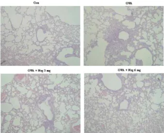
Naringin ameliorates pathological changes of OVA-induced asthma
Histological analyses revealed typical pathologic features of asthma in the OVA-exposed mice. Numerous in- flammatory cells, including eosinophils infiltrated around the bronchioles as compared with the control (Fig. 2). Mice treated with Naringin showed marked reductions in the in- filtration of inflammatory cells in the lung tissues. Total lung inflammations were increased significantly at 24 hr after OVA inhalation compared with the scores after saline inhalation. The total lung inflammations were significantly reduced by the administration of Naringin. The scores of peribronchial, perivascular, and total lung inflammation were increased significantly at 72 hr after OVA inhalation compared with scores after saline inhalation (Fig. 2). The in- creased peribronchial, perivascular, and total lung in- flammation after OVA inhalation were significantly de- creased by the administration of Naringin. These results sug- gest that Naringin inhibits OVA-induced inflammation in the lungs, including the influx of eosinophils.
Naringin decreases MMP-9 and GATA-3 mRNA levels in lung tissues of OVA-sensitized and -challenged mice
Real-time PCR analysis revealed that expression of
Fig. 2. Naringin inhibits lung inflammation and inflammatory cells infiltration. Mice were sensitized and challenged as described in Materials and Methods. Sections were ob- tained from the lungs of mice receiving the control (CON), OVA plus Naringin 3 mg/kg/day (OVA + Nrg 3 mg), OVA plus Naringin 6 mg/kg/day (OVA + Nrg 6 mg) and OVA (OVA). Lungs were removed 2 days after the last airway challenge. Sections were stained by haematoxylin and eosin staining (x200).
MMP-9, T-bet, and GATA3 mRNA in lung tissues was sig- nificantly increased at 24 hr after OVA inhalation compared with the levels after saline inhalation (Fig. 3). The increased mRNA expression of MMP-9, GATA-3 was decreased by the administration of Naringin.
Naringin reduces levels of Th2 cytokine (IL-4, IL-5) in lung tissues of OVA-sensitized and -challenged mice
BAL fluids were obtained 4 hr after the last airway challenge. The levels of IL-4 and IL-5 in the BAL fluids were significantly increased by airway challenge with OVA when compared with that with that of the control. The admin- istration of Naringin reduced the concentration of IL-4, IL-5 secretion (Fig. 4). The levels of Th2 cytokines, IL-4 and IL-5, were found to be increased in OVA-sensitized and -challenged mice, but that of the Th1 cytokine, IFN-γ (Data not shown) was not changed as compared to saline-sensitized and -challenged mice. These results indicate that Naringin treatment inhibits Th2 cytokine levels in the BAL fluids.
Naringin decreases airway hyper-responsiveness
Airway responsiveness was assessed as the percent increase
of Penh in response to increasing doses of methacholine.
Fig. 3. Effect of Naringin on MMP-9, GATA3 mRNA expression in lung tissues of OVA-sensitized and -challenged mice. Sampling was performed at 24 hr after the last challenge in saline-inhaled mice administered saline (CON), OVA-inhaled mice ad- ministered saline (OVA), OVA-inhaled mice administered Naringin 3 mg/kg/day (OVA+Nrg 3) and OVA inhaled mice administered Naringin 6 mg/kg/day (OVA+Nrg 6). Data represent means±S.E.M. from 5 independent experiments.
***
P
<0.001.Fig. 4. The effect of Naringin treatment on Th2 cytokine. OVA- sensitized mice were treated as described Material and Method.
BAL fluid was performed 4 hrs after the last airway challenge as described by the manufacturer. IL-4, IL-5 cytokine levels in the BAL fluids were measured by ELISA Kit. Data represent mean±SEM from 6 independent experiments ***
P
<0.001 vs. OVA.In OVA-sensitized and -challenged mice, the dose-response curve of percent Penh was shifted to the left compared with that of control mice (Fig. 5). In addition, the percent Penh produced by methacholine administration (at doses from 2.5 mg/ml to 50 mg/ml) increased significantly in the OVA-sen- sitized and -challenged mice compared with the controls.
OVA-sensitized and -challenged mice treated with Naringin showed a dose-response curve of percent Penh that shifted to the right compared with that of untreated mice. The shift was dose-dependent. These results indicate that Naringin treatment reduces OVA-induced airway hyper- responsiveness.
Naringin inhibits MMP-9, GATA-3 production in the lung tissue
OVA-challenge induced a marked induction of matrix metalloproteinase-9, GATA-3 activity in BAL fluids in
comparison to control mice (Fig. 6). When the admin- istration Naringin 3 mg/kg/day, 6 mg/kg/day this in- creased MMP-9, GATA-3 production was significantly in- hibited (Fig. 6).
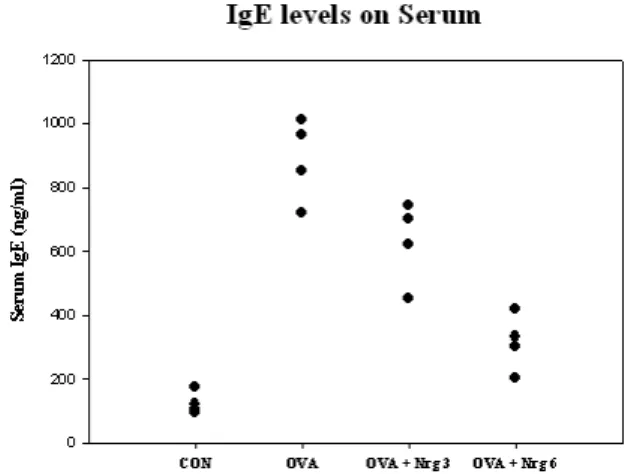
Naringin decreases IgE levels in serum
Because Th2 cytokines promote airway inflammation in asthma through increased IgE levels, we investigated the ex- pression of IgE in serum. IgE favors to Th2 in airway in- flammation, we measured how far Naringin modulated the levels of serum IgE in OVA mice. As shown in Fig. 9, the levels of serum IgE were found to be significantly increased in OVA mice compared with those of PBS mice (CON).
However, administration of Naringin significantly decreased
the levels of serum IgE. These data indicate that Naringin
modulates Th1/Th2 levels in an OVA-induced asthma mod-
el (Fig. 7).
Fig. 5. The effect of Naringin on airway responsiveness in OVA- sensitized and OVA-challenged mice. Airway responsive- ness was measured at 24 hr after the last challenge in saline-inhaled mice administered PBS (Con), OVA-sensi- tized mice administered saline (OVA) and OVA-sensi- tized mice administered Naringin (OVA+Nrg 3 mg/kg/day, 6 mg/kg/day). Airway responsiveness to aerosolized methacholine was measured in unrestrained, conscious mice. Mice were placed into the main chamber and were nebulized first with PBS, then with increasing doses (2.5 to 50 mg/ml) of methacholine for 3 min for each nebulization. Readings of breathing parameters were taken for 3 min after each nebulization during which Penh values were determined. Data represent means±S.E.M.
from 5 independent experiments. ***
P
<0.001, **P
<0.01.Fig. 6. The effect of Naringin on MMP-9 production in lung tis- sues of OVA-sensitized and OVA-challenged. Sampling was performed at 48 hrs after challenge with the PBS (Con), OVA (OVA), and OVA plus Naringin 3 or 6 mg/kg/day (OVA+Nrg 3 or 6) and analyzed by western blotting. All of the groups of the experiment showed MMP-9 production, but the OVA plus Naringin group only showed the active form of MMP-9.
Discussion
This study is the first to provide experimental evidence demonstrating that Naringin inhibits OVA-induced airway inflammation in a murine model of asthma. Naringin pro- foundly inhibited asthmatic reactions such as leukocytic re- cruitment into the airway and lung inflammation. We also
Fig. 7. The effect of Naringin on IgE levels in serum of OVA-sen- sitized and OVA-challenged. Blood was collected by car- diac puncture to measure serum IgE. All experiments were analyzed using ELISA (n=4).
demonstrated that Naringin regulates the Th1/Th2 balance, which can be mediated by the level of GATA3 levels.
Based on animal studies, the immunological processes in- volved in airway inflammation of asthma are characterized by the proliferation and activation of T cells of the subtype Th2 CD4+. Ultimately, mediators lead to degranulation of effector/proinflammatory cells with the release of mediators and oxidants, which lead to the injury and inflammation not- ed in asthma. OVA-induced asthma has been recognized as a disease that results from chronic airway inflammation characteristically associated with the infiltration of lympho- cytes, eosinophils, and neutrophils into the bronchial lumen [7,32]. In our experiment, we demonstrate that OVA-induced asthma increased levels of eosinophil infilteration, eosino- phils peroxidase activity, eotaxin, and the thicknesses of bronchial wall and area of smooth muscle. But they were significantly decreased by administration of Naringin.
It was recently demonstrated that the administration of an MMP inhibitor reduces the migration of inflammatory cells through the endothelial and epithelial basement mem- branes [22]. Additionally, an MMP inhibitor regulates in- flammatory cell migration by reducing ICAM-1 and VCAM-1 expression in a murine model of toluene diisocya- nate-induced asthma [29]. In our murine model of asthma, intraperitoneally injected mice with 3 mg/kg/day, 6 mg/kg/day Naringin to evaluate the effect of Naringin on the expression of MMP-9 and GATA3 mRNA. In this study, Naringin reduced levels of MMP-9, GATA3 in lung tissues of Naringin treated mice.
T-bet, a member of the T-box family of transcription fac-
tors, is a master determinant of Th1 lineage [1,38]. Indeed, T-bet deficient mice exhibit a profound lack of Th1 immune responses [25] and ectopic expression of T-bet in murine Th2 cells directs activation of IFN-γ, as well as the upregulation of IL-12Rβ [10,25]. Th1 cytokines are known to inhibit aller- gic responses [25]. Six members (GATA-1 to GATA-6) of this family have been identified in avians, with homologues in mammals and avians. Based on their expression profile and structure, the GATA proteins may be classified as haemato- poietic (GATA-1 to GATA-3) [19,30] or nonhaematopoietic (GATA-4 to GATA-6). Naive CD4+ T cells express low levels of GATA-3 mRNA. The expression of GATA-3 is, however, markedly upregulated in cells differentiating along the Th2 lineage, and is downregulated in cells differentiating along the Th1 pathway [37].
Our data demonstrate that Naringin reduces the increased levels of GATA3 mRNA in OVA-sensitized and -challenged mice (Fig. 5). Also, it suggests that Naringin treatment is a novel, selective way to simultaneously suppress GATA-3 in asthmatic reactions in vivo. Also, we examined Th1/Th2 cytokine production in BAL fluid cells. Naringin reduces the increased levels of IL-4, Th2 cytokine production in OVA-sensitized and -challenged mice. Taken together these suggest that GATA-3 might be a candidate gene for asthma, and a regulator of Th1/Th2 balance.
In conclusion, our results strongly indicate that Naringin reduces allergic airway inflammation due to the alteration of Th1/Th2 polarization via the suppression of GATA-3.
Therefore our data suggests that Naringin might offer a new therapeutic approach to allergic airway diseases.
Acknowledgement
This work was supported for two years by Pusan National University Research Grant.
References
1. Afkarian, M., J. R. Sedy, J. N. Yang, G. Jacobson, N. Cereb, S. Y. Yang, T. L. Murphy, and K. M. Murphy. 2002. T-bet is a STAT1-induced regulator of IL-12R expression in naive CD4+ T cells.
Nat. Immunol.
3, 549-557.2. Ali, M., M. F. Agha, G. N. El-Sammad, and K. Hassan. 2009.
Modulation of anticancer drug-induced P-glycoprotein ex- pression by naringin.
Z. Naturforsch C
. 64, 109-116.3. Bian, T., K. S. Yin, S. X. Jin, X. L. Zhang, J. Y. Zhou, X.
Q. Ma, J. J. Hu, and W. De. 2006. Treatment of allergic air- way inflammation and hyperresponsiveness by imiquimod
modulating transcription factors T-bet and GATA-3.
Chin.
Med. J. (Engl.)
119, 640-648.4. Bousquet, J., P. J. Chanez, Y. Lacoste, G.. Barneon, N.
Ghavanian, I. Enander, P. Venge, S. Ahlstedt, J.
Simony-Lafontaine, and P. Godard. 1990. Eosinophilic in- flammation in asthma.
N. Engl. J. Med.
323, 1033-1039.5. Busse, W. W., W. F. Calhoun, and J. D. Sedgwick. 1993.
Mechanism of airway inflammation in asthma.
Am. Rev.
Respir. Dis.
147, S20-24.6. Chen, C. C., M. P. Chow, W. C. Huang, Y. C. Lin, and Y.
J. Chang. 2004. Flavonoids inhibit tumor necrosis factor-al- pha-induced up-regulation of intercellular adhesion mole- cule-1 (ICAM-1) in respiratory epithelial cells through acti- vator protein-1 and nuclear factor-kappaB: structure-activ- ity relationships.
Mol. Pharmacol
. 66, 683-693.7. Corrigan, C. J and A. B. Kay. 1992. T cells and eosinophils in the pathogenesis of asthma.
Immunol. Today.
13, 501-507.8. Delclaux, C., C. Delacourt, M. P. D'Ortho, V. Boyer, C.
Lafuma, and A. Harf. 1996. Role of gelatinase B and elastase in human polymorphonuclear neutrophil migration across basement membrane.
Am. J. Respir. Cell Mol. Biol.
14, 288-295.9. Duthie, G. and A. Crozier. 2000. Plant-derived phenolic antioxidants.
Curr. Opin. Clin. Nutr. Metab. Care.
3, 447-451.10. Gavett, S. H., D. J. O'Hearn, X. Li, S. K. Huang, F. D.
Finkelman, and M. Wills-Karp. 1995. Interleukin 12 inhibits antigen-induced airway hyperresponsiveness, in- flammation, and Th2 cytokine expression in mice.
J. Exp.
Med.
182, 1527-1536.11. Hougee, S., A. Sanders, J. Faber, Y. M. Graus, van den W.
B. Berg, J., Garssen, H. F. Smit, and M. A. Hoijer. 2005.
Decreased pro-inflammatory cytokine production by LPS-stimulated PBMC upon
in vitro
incubation with the fla- vonoids Naringin, luteolin or chrysin, due to selective elimi- nation of monocytes/macrophages.Biochem. Pharmacol.
69, 241-248.12. Hsiao, Y.,C., W. H. Kuo, P. N. Chen, H. R. Chang, T. H.
Lin, W. E. Yang, Y. S. Hsieh, and S. C. Chu. 2007. Flavanone and 2'-OH flavanone inhibit metastasis of lung cancer cells via down-regulation of proteinases activities and MAPK pathway.
Chem. Biol. Interact
. 167, 193-206.13. Iwamoto, I., H. Nakajima, H. Endo, and S. Yoshida. 1993.
Interferon gamma regulates antigen-induced eosinophil re- cruitment into the mouse airways by inhibiting the infiltra- tion of CD4+ T cells.
J. Exp. Med
. 177, 573-576.14. Kanno, S., A. Shouji, A. Tomizawa, T. Hiura, Y. Osanai, M. Ujibe, Y. Obara, N. Nakahata, and M. Ishikawa. 2006.
Inhibitory effect of naringin on lipopolysaccharide (LPS)-induced endotoxin shock in mice and nitric oxide production in RAW 264.7 macrophages.
Life Sci
. 73, 671-681.15. Karp, M. and C. Oker-Blom. 1999. A streptavidin-luciferase fusion protein: comparisons and applications.
Biomol. Eng.
16, 101-104.
16. Kay, A. B. 1991. Asthma and inflammation.
J. Allergy Clin.
Immunol.
87, 893-910.17. Kim, G. Y., H. Cho, S. C. Ahn, Y. H. Oh, C. M. Lee, and Y. M. Park. 2004. Resveratrol inhibits phenotypic and func-
tional maturation of murine bone marrow-derived dendritic cells.
Int. Immunopharmacol.
4, 245-253.18. Leppert, D., E. Waubant, R. Galardy, N. W. Bunnett, and S. L. Hauser. 1995,. T cell gelatinases mediate basement membrane transmigration
in vitro
.J. Immunol.
154, 4379-4389.19. Li, X. M., R. K. Chopra, T. Y. Chou, B. H. Schofield, M.
Wills-Karp, and S. K. Huang. 1996. Mucosal IFN-gamma gene transfer inhibits pulmonary allergic responses in mice.
J. Immunol.
157, 3216-3219.20. Mapp, C. E., P. Boschetto, E. Zocca, G. F. Milani, F.
Pivirotto, V. Tegazzin, and L. M. Fabbri. 1987. Pathogenesis of late asthmatic reactions induced by exposure to isocyanates.
Bull. Eur. Physiopathol. Respir.
23, 583-586.21. Mapp, C. E., P. Boschetto, L. Dal Vecchio, P. Maestrelli, and L. M. Fabbri. 1988. Occupational asthma due to isocyanates.
Eur. Respir. J
. 1, 273-279.22. Matrisian, L. M. 1990. Metalloproteinases and their in- hibitors in matrix remodeling.
Trends Genet.
6, 121-125.23. Mautino, G., N. Oliver, P. Chanez, J. Bousquet, and F.
Capony. 1997. Increased release of matrix metal- loproteinase-9 in bronchoalveolar lavage fluid and by alveo- lar macrophages of asthmatics.
Am. J. Respir. Cell Mol. Biol.
17, 583-591.
24. Montefort, S. and S. T. Holgate. 1991. Adhesion molecules and their role in inflammation.
Respir. Med.
85, 91-99.25. Mullen, A. C., F. A. High, A. S., Hutchins, H. W. Lee, A.
V. Villarino, D. M. Livingston, A. L. Kung, N. Cereb, T.
P. Yao, S. Y. Yang, and S. L. Reiner. 2001. Role of T-bet in commitment of TH1 cells before IL-12-dependent selection.
Science
292, 1907-1910.26. Murphy, G. and A. J. Docherty. 1992. The matrix metal- loproteinases and their inhibitors,
Am. J. Respir. Cell Mol.
Biol.
7, 120-125.27. Nagase, H. 1997. Activation mechanisms of matrix metalloproteinases.
Biol. Chem.
378, 151-160.28. Nakamura, Y., O. Ghaffar, R. Olivenstein, R. A. Taha, A.
Soussi-Gounni, D. H. Zhang, A. Ray, and Q. Hamid. 1999.
Gene expression of the GATA-3 transcription factor is in- creased in atopic asthma,
J. Allergy Clin. Immunol.
103, 215-222.29. Okada, S., H. Kita, T. J. George, G. J. Gleich, and K. M.
Leiferman. 1997. Migration of eosinophils through base- ment membrane components
in vitro
: role of matrix metal- loproteinase-9.Am. J. Respir. Cell Mol. Biol.
17, 519-528.30. Ouyang, W., S. H. Ranganath, K. Weindel, D. Bhattacharya, T. L. Murphy, W. C. Sha, and K. M. Murphy. 1998.
Inhibition of Th1 development mediated by GATA-3 through an IL-4-independent mechanism.
Immunity
9,745-755.
31. Parronchi, P., M. De Carli, R. Manetti, C. Simonelli, S.
Sampognaro, M. P. Piccinni, D. Macchia, E. Maggi, G. Del Prete, and S. Romagnani. 1992. IL-4 and IFN (alpha and gamma) exert opposite regulatory effects on the develop- ment of cytolytic potential by Th1 or Th2 human T cell clones.
J. Immunol.
149, 2977-2983.32. Punnonen, J., G.. Aversa, B. G. Cocks, and de J. E. Vries.
1994. Role of interleukin-4 and interleukin-13 in synthesis of IgE and expression of CD23 by human B cells.
Allergy
49, 576-586.33. Renz, H., K. Bradley, J. Saloga, J. Loader, G. L. Larsen, and E. W. Gelfand. 1993. T cells expressing specific V beta ele- ments regulate immunoglobulin E production and airways responsiveness
in vivo
.J. Exp. Med.
177, 1175-1180.34. Saito, H., K. Hatake, A. M. Dvorak, K. M. Leiferman, A.
D. Donnenberg, N. Arai, K. Ishizaka, and T. Ishizaka. 1988.
Selective differentiation and proliferation of hematopoietic cells induced by recombinant human interleukins,
Proc.
Natl. Acad. Sci. USA
85, 2288-2292.35. Stahle-Backdahl, M., M. Inoue, G. J. Guidice, and W. C.
Parks. 1994. 92-kD gelatinase is produced by eosinophils at the site of blister formation in bullous pemphigoid and cleaves the extracellular domain of recombinant 180-kD bul- lous pemphigoid autoantigen.
J. Clin. Invest.
93, 2022-2030.36. Sur, S., J. Lam, P. Bouchard, A. Sigounas, D. Holbert, and W. J. Metzger. 1996. Immunomodulatory effects of IL-12 on allergic lung inflammation depend on timing of doses.
J.
Immunol.
157, 4173-4180.37. Szabo, S. J., S. T. Kim, G. L. Costa, X. Zhang, C. G. Fathman, and L. H. Glimcher. 2000. A novel transcription factor, T-bet, directs Th1 lineage commitment.
Cell
100, 655-669.38. Szabo, S. J., B. M. Sullivan, C. Stemmann, A. R. Satoskar, B. P. Sleckman, and L. H. Glimcher. 2002. Distinct effects of T-bet in TH1 lineage commitment and IFN-gamma pro- duction in CD4 and CD8 T cells.
Science
295, 338-342.39. Tanaka, H., M. Komai, K. Nagao, M. Ishizaki, D. Kajiwara, K. Takatsu, G.. Delespesse, and H. Nagai. 2004. Role of in- terleukin-5 and eosinophils in allergen-induced airway re- modeling in mice.
Am. J. Respir. Cell Mol. Biol.
31, 62-68.40. Yao, P. M., B. Maitre, C. Delacourt, J. M. Buhler, A. Harf, and C. Lafuma. 1997. Divergent regulation of 92-kDa gelati- nase and TIMP-1 by HBECs in response to IL-1beta and TNF-alpha.
Am. J. Physiol
. 273, L866-874.41. Yoon, M. S., J. S. Lee, B. M. Choi, Y. I. Jeong, C. M. Lee, J. H. Park, Y. S. Moon, C. Sung, S. K. Lee, Y. H. Chang, H. Y. Chung, and Y. M. Park. 2006. Naringin inhibits im- munostimulatory function of dendritic cells: Implication of immunotherapeutic adjuvant.