R E S E A R C H A R T I C L E Open Access
Immediate effects of first-line
thrombectomy devices for intracranial atherosclerosis-related occlusion: stent retriever versus contact aspiration
Joonsang Yoo 1,2 , Seong-Joon Lee 3 , Jeong-Ho Hong 1 , Yong-Won Kim 4 , Ji Man Hong 3 , Chang-Hyun Kim 5 , Dong-Hun Kang 6 , Jin Wook Choi 7 , Yong-Sun Kim 8 , Sung-Il Sohn 1 , Yang-Ha Hwang 4* and Jin Soo Lee 3*
Abstract
Background: Although stent retriever (SR) is recommended as a frontline device of endovascular treatment (EVT) for embolic large artery occlusion causing acute ischemic stroke, contact aspiration (CA) device showed similar efficacy in the recent trials. However, the efficacy of the both devices as first-line therapy for intracranial
atherosclerotic stenosis (ICAS)-related large vessel occlusion has not yet been established. Therefore, we compared the immediate effects and final outcomes of SR and CA as first-line devices for treating ICAS-related occlusions.
Methods: We retrospectively analyzed the data of patients who underwent EVT for acute ischemic stroke from the registry of three Korean hospitals. Patients with ICAS-related occlusion who were treated within 24 h of onset of the symptoms were included. We investigated immediate reperfusion performance, immediate safety outcomes, and 3- month clinical outcomes for the two first-line devices.
Results: Of the 720 registered patients, 111 were eligible for this study. Forty-nine patients (44.1%) used SR and 62 (55.9%) used CA as the first-line device. Achieving successful reperfusion immediately after first-line thrombectomy was more frequent in the SR group than that in the CA group (77.6% vs. 43.5%, p = 0.001), with fewer additional rescue treatments (12.2% vs. 59.7%, p < 0.001). The incidence of iatrogenic dissection or rupture was lower in the SR group than that in the CA group (8.2% vs. 29.0%, p = 0.012). After additional rescue treatments, however, the final successful reperfusion rate did not differ between the two groups (SR 87.8% vs. CA 77.4%, p = 0.247), and there was no significant difference in the 3-month good outcomes (modified Rankin Scale, p = 0.524).
(Continued on next page)
© The Author(s). 2020 Open Access This article is licensed under a Creative Commons Attribution 4.0 International License, which permits use, sharing, adaptation, distribution and reproduction in any medium or format, as long as you give appropriate credit to the original author(s) and the source, provide a link to the Creative Commons licence, and indicate if changes were made. The images or other third party material in this article are included in the article's Creative Commons licence, unless indicated otherwise in a credit line to the material. If material is not included in the article's Creative Commons licence and your intended use is not permitted by statutory regulation or exceeds the permitted use, you will need to obtain permission directly from the copyright holder. To view a copy of this licence, visit http://creativecommons.org/licenses/by/4.0/.
The Creative Commons Public Domain Dedication waiver (http://creativecommons.org/publicdomain/zero/1.0/) applies to the data made available in this article, unless otherwise stated in a credit line to the data.
* Correspondence: [email protected]; [email protected]
4
Department of Neurology, Kyungpook National University Hospital, School of Medicine, Kyungpook National University, 130 Dongdeok-ro, Jung-gu, Daegu 41944, South Korea
3
Department of Neurology, Ajou University Medical Center, Ajou University School of Medicine, 164 World cup-ro, Yeongtong-gu, Suwon 16499, South Korea
Full list of author information is available at the end of the article
(Continued from previous page)
Conclusions: First-line SR thrombectomy showed higher immediate reperfusion and less vessel injury for ICAS- related occlusions than CA. However, there was no significant difference in the final reperfusion status or 3-month outcomes from additional rescue treatments.
Keywords: Ischemic stroke, Reperfusion therapy, Stent retriever, Contact aspiration, Intracranial atherosclerosis, Endovascular treatment
Background
Endovascular treatment (EVT) has shown favorable results in major trials mainly using stent retriever (SR) [1]. There- fore, SR is recommended as a first-line device for treatment of large vessel occlusion in the AHA/ASA guidelines [2].
However, contact aspiration (CA) showed outcomes com- parable to those with SR in recent studies [3, 4].
Intracranial atherosclerotic stenosis (ICAS)-related large vessel occlusion (ICAS-LVO) is a common cause of stroke, especially among the Asian population [5–9].
In ICAS-LVO, immediate reocclusion occurs frequently, requiring further rescue treatment, and the final reperfu- sion rate tends to be lower than that of embolic occlu- sions [10, 11]. Because of these features, a different approach may be needed for ICAS-LVO than that for general embolic occlusions [12].
However, which device is suitable for first-line therapy in ICAS-LVO is unclear, because most of the previous randomized trials were based on Western populations.
Knowledge about which device is more effective in ICAS-LVO would be useful in selecting a more suitable device for patients with ICAS-LVO before EVT [13–15].
Recently, a study comparing outcome of SR and CA in ICAS-LVO patients at two hospitals was published [16].
However, studies on immediate performances and side effects are still insufficient. Therefore, we compared the immediate effects and final outcomes of using SR or CA as first-line devices in the treatment of ICAS-LVO.
Methods
Study population and inclusion criteria
All clinical and image data were de-identified and allo- cated study identification numbers. The protocol for data collection was approved by the Institutional Review Board of each hospital. Our study was implemented in accordance with the ethical standards of the 1964 Dec- laration of Helsinki and its later amendments. The need for written informed consent was waived because of the retrospective nature of this study. The data of this study are available from the corresponding author upon rea- sonable request.
This was a retrospective analysis of the ASIAN KR (Acute Stroke due to Intracranial Atherosclerotic occlu- sion and Neurointervention Korean Retrospective) regis- try. The details of the registry were previously published
[11]. In brief, the registry consists of data on consecutive patients who underwent emergency EVT for cervicocer- ebral artery occlusions causing acute ischemic stroke at three stroke centers in Korea. The patients were enrolled between January 2011 and February 2016. For the current study, the inclusion criteria were: 1) patients with intracranial large artery occlusions; 2) underlying etiology classified as ICAS; and 3) time from symptom onset to EVT start ≤24 h.
Classifying ICAS-LVO
Underlying ICAS, which should be differentiated from embolism or other etiologies, was defined based on the remaining fixed focal stenosis during EVT. The step-by- step evaluations for differentiation were described previ- ously [11]. Briefly, after confirmation of arterial occlu- sion, patients with uncommon stroke etiologies, such as dissection, Moyamoya disease, and vasculitis were ex- cluded. Embolic occlusion was classified based on complete vessel recanalization after thrombectomy, and ICAS-related occlusion was classified when a remnant stenosis of > 70%, or a lesser degree of stenosis with a tendency toward reocclusion and/or flow impairment after thrombectomy was observed [11, 17]. This classifi- cation was further confirmed by repeat angiography dur- ing admission.
EVT procedure
Devices were selected at the discretion of neuro- interventionists based on the consensus within each stroke team. In the current study, we divided patients into two groups based on whether SR or CA was used as the first-line device. Solitaire AB/FR (Medtronic, Irvine, CA, USA) or Trevo (Stryker, Kalamazoo, MI, USA) belonged to the SR group, and 1st generation or 2nd generation Penumbra MAX systems (Penumbra Inc., Alameda, CA, USA) belonged to the CA group. Balloon guide catheters, adjuvant local lytic infusion, intracranial angioplasty and/or stenting were implemented as needed.
Image and clinical assessment
Premorbid functional status, conventional vascular risk
factors, and laboratory findings assessed during admis-
sion were collected. Stroke severity was assessed using
the initial National Institutes of Health Stroke Scale (NIHSS) score. Clinical outcomes were measured using the modified Rankin Scale (mRS) score at 3-months.
mRS scores 0 to 2 or no change between premorbid and 3-month mRS were considered as good outcomes.
The location of initial occlusion site was determined using baseline computed tomography angiography or magnetic resonance angiography. Reperfusion per- formance was evaluated using modified treatment in cerebral ischemia (mTICI) grade [18]. Successful re- perfusion was defined as mTICI grade 2b or higher.
When the first-line device did not achieve the desired reperfusion status, rescue treatments were allowed, in- cluding other thrombectomy devices, fibrinolytics, angioplasty, and intracranial stenting. The number of EVT methods used until achievement of final reperfu- sion was also determined. Intracerebral hemorrhages were classified in accordance with the European Co- operative Acute Stroke Study criteria [19]. Subarach- noid hemorrhage (SAH) was classified using the modified Fisher scale [20].
Primary outcomes
Immediate reperfusion performance as a primary effi- cacy outcome was assessed immediately after the first at- tempt and full use of the first-line thrombectomy device, and final reperfusion status was assessed at the last angi- ography. We also assessed the immediate side effects, such as 1) vasospasm, 2) iatrogenic dissection or rupture by thrombectomy, and 3) new embolism in other vessels as primary safety outcomes. Angiographic lesions with surface irregularity or intimal flap were considered as iatrogenic vessel injuries.
Statistical analysis
Variables are expressed as mean ± standard deviations, medians (interquartile ranges), or numbers (percentages), as statistically appropriate. We compared baseline charac- teristics, as well as clinical and imaging results between the SR and CA groups using chi-squared tests and inde- pendent Student’s t-tests or Wilcoxon rank-sum tests, re- spectively. To determine the factors associated with good clinical outcome and iatrogenic dissection, we performed multivariate analyses after adjusting for age, sex, and vari- ables with p < 0.1 in the univariate analysis. p values were two-tailed, and variables were considered significant at p < 0.05. All statistical analyses were performed with R version 3.5.1 (http://www.R-project.org).
Results
Baseline characteristics
During the study period, 111 of the 720 registered pa- tients were included in the current study (Fig. 1). Mean age of the included patients was 65.2 ± 13.3 years, and 70 patients (63.1%) were men. Among them, 49 patients (44.1%) belonged to the SR group, and 62 (55.9%) to the CA group. Demographics and baseline characteristics of the patients between two groups did not differ (Table 1).
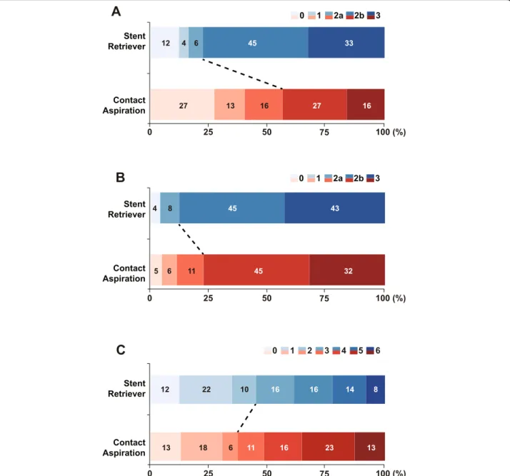
Immediate effects following first-line thrombectomy Table 2 summarizes the comparative results regarding treatment. Successful reperfusion after first attempt of thrombectomy was not significantly different between the two groups (SR 28.6% vs. CA 17.7%, p = 0.260). Im- mediate successful reperfusion after first-line thrombec- tomy was achieved more frequently in the SR group (SR 77.6% vs. CA 43.5%, p = 0.001) (Fig. 2a). Transient vaso- spasm occurred in four patients in the SR group (SR
Fig. 1 Flowchart of the current study. EVT: Endovascular treatment; ICAS: Intracranial atherosclerotic stenosis; ICAS-LVO: ICAS-related large
vessel occlusion
8.2% vs. CA 0%, p = 0.035). In contrast, iatrogenic dissec- tion or rupture occurred more frequently in the CA group (SR 8.2% vs. CA 29.0%, p = 0.012). Odds ratio of iatrogenic dissection or rupture in the CA group was 4.488 in a logistic regression analysis after adjusting age, sex, presence of atrial fibrillation, initial NIHSS score, procedural time, and total number of EVT methods
(95% confidence interval [CI] 1.394–17.676, p = 0.018) (Supplementary Table 1). This rate of iatrogenic dissec- tion or rupture was not different between the first- generation Penumbra and Penumbra MAX or between the Solitaire and Trevo stents; however, these results must be interpreted with caution owing to the small sample size. The frequency of new embolisms at other Table 1 Baseline characteristics of patients with intracranial atherosclerotic stenosis (ICAS)-related occlusions and endovascular treatments
Contact aspiration
( n = 62) Stent retriever
( n = 49) P
Demographics
Age, years 66.3 ± 11.7 63.9 ± 15.2 0.348
Sex, men 39 (62.9) 31 (63.3) > 0.999
Risk factors
Hypertension 38 (61.3) 33 (67.3) 0.645
Diabetes mellitus 20 (32.3) 14 (28.6) 0.833
Dyslipidemia 23 (37.1) 12 (24.5) 0.225
Atrial fibrillation 14 (22.6) 10 (20.4) 0.965
Smoker 26 (41.9) 13 (25.5) 0.103
Medications prior admission
Antiplatelets 11 (17.7) 7 (14.3) 0.817
Anticoagulants 2 (3.2) 4 (8.2) 0.403
Initial occlusion site 0.078
Internal carotid artery 12 (19.4) 2 (4.1)
Middle cerebral artery, M1 36 (58.1) 36 (73.5)
Middle cerebral artery, M2 2 (3.2) 1 (2.0)
Vertebral artery 2 (3.2) 4 (8.2)
Basilar artery 10 (16.1) 6 (12.2)
Initial NIHSS score 16.5 (11 –22) 15 (12 –19) 0.340
ASPECTS score
a7.5 (5 –9) 7 (6 –8) 0.625
Laboratory findings
Hemoglobin, g/dL 14.0 ± 1.8 13.9 ± 2.0 0.764
White blood cells, ×10
9/L 9.6 ± 3.6 11.5 ± 5.6 0.047
Platelets, ×10
9/L 240 ± 77 239 ± 59 0.928
Glucose, mmol/L 8.2 ± 3.3 8.4 ± 3.3 0.813
Intravenous tPA 27 (43.5) 23 (46.9) 0.869
Use of balloon guide catheter 36 (58.1) 26 (53.1) 0.738
Onset to door time, min 208 (111 –387) 172 (105 –560) 0.801
Door to puncture time, min 97.5 (77 –127) 111 (92 –138) 0.106
First-line thrombectomy device
Solitaire 41 (83.7)
Trevo 8 (16.3)
Penumbra, 1st generation 45 (72.6)
Penumbra, 2nd generation 17 (27.4)
ICAS Intracranial atherosclerotic stenosis; NIHSS National Institutes of Health Stroke Scale; ASPECTS Alberta Stroke Program Early CT Score; tPA tissue plasminogen activator
a
Baseline ASPECTS on quality imaging was evaluated in 81 patients (91.0% patients with anterior circulation infarction)
vessels was relatively rare and did not differ between the groups (SR 6.1% vs. CA 3.2%, p = 0.653).
Rescue treatments after first-line thrombectomy
The rate of switching to the other thrombectomy device (from SR to CA / from CA to SR) for rescue treatment was significantly lower in the SR group (SR 12.2% vs. CA 59.7%, p < 0.001). As other rescue treatments, tirofiban infusion (SR 46.9% vs. CA 46.8%, p > 0.999), balloon angioplasty (SR 10.2% vs. CA 11.3%, p > 0.999), and per- manent intracranial stenting (SR 12.2% vs. CA 9.7%, p = 0.901) were performed in both groups with no signifi- cant differences in their rates.
Final endovascular treatment results
Final successful reperfusion rate was similar between the two groups (SR 87.8% vs. CA 77.4%, p = 0.247) (Fig. 2b).
However, the endovascular techniques were fewer in the SR group (SR 2 [1, 2] vs. CA 2 [2, 3], p = 0.003). Puncture to final angiography time was not significantly different (SR 76 [48–116] min vs. CA 63 [45–92] min, p = 0.151).
Imaging and clinical outcomes after endovascular treatment
Intracerebral hemorrhagic transformation of any type (p = 0.659) and parenchymal hematoma type 2 (p = 0.403) occurred at similar rates in the two groups. Thick subarachnoid hemorrhages occurred in two patients in the only SR group. There was no significant difference in good clinical outcome at 3 months (SR 44.9% vs. CA 37.1%, p = 0.524) (Fig. 2c). In multivariate analysis, 3- month clinical outcome was not associated with the first-line thrombectomy device (Table 3). Instead, age, initial stroke severity, initial occlusion site and final suc- cessful reperfusion were independently associated with good clinical outcome at 3-months.
Discussion
In the current study, we investigated if there were differ- ences primarily in immediate reperfusion performance and side effects, and secondarily in post-procedural hemorrhagic complications and clinical outcomes, de- pending on the choice of the first-line devices between SR and CA in patients with ICAS-LVO. Based on the re- sults of our retrospective analysis of a multicenter Table 2 Radiologic and clinical outcomes after endovascular treatment
Contact aspiration (n = 62)
Stent retriever
(n = 49) P
Immediate effects following first-line thrombectomy
Successful reperfusion after first attempt of thrombectomy 11 (17.7) 14 (28.6) 0.260
Successful reperfusion after first-line thrombectomy 27 (43.5) 38 (77.6) 0.001
Immediate side effect by first-line thrombectomy
Vasospasm 0 4 (8.2) 0.035
Iatrogenic dissection or rupture 18 (29.0) 4 (8.2) 0.012
New embolism in other vessels 2 (3.2) 3 (6.1) 0.653
Rescue treatments after first-line thrombectomy
Switching to the other device 37 (59.7) 6 (12.2) < 0.001
Tirofiban infusion 29 (46.8) 23 (46.9) > 0.999
Balloon angioplasty 7 (11.3) 5 (10.2) > 0.999
Permanent intracranial stenting 6 (9.7) 6 (12.2) 0.901
Final endovascular treatment results
Final successful reperfusion 48 (77.4) 43 (87.8) 0.247
Total number of endovascular techniques 2 (2 –3) 2 (1 –2) 0.003
Puncture to final angiography time, min 76 (48 –116) 63 (45 –92) 0.151
Imaging and clinical outcomes after endovascular treatment Hemorrhagic complication
Any intracerebral hemorrhagic transformation 16 (25.8) 10 (20.4) 0.659
Parenchymal hemorrhage, type 2 2 (3.2) 4 (8.2) 0.403
SAH, grade 3 or 4 0 2 (4.1) 0.193
Good outcome at 3 months 23 (37.1) 22 (44.9) 0.524
SAH Subarachnoid hemorrhage
registry, we could cautiously mention that the use of SR may be safer and more effective for ICAS-LVO than CA as a first-line thrombectomy method in terms of imme- diate reperfusion success and immediate occurrence of iatrogenic dissection or rupture.
Recently, studies have reported comparisons of reper- fusion performance or side effects of CA and SR when performing EVT in overall LVO patients. In the ASTER trial, the reperfusion success did not differ between CA and SR (mTICI score of 2b or 3, SR 67.7% vs. CA 63.0%) [3, 21]. More recently, the COMPASS trial showed that first-line CA treatment was not inferior to first-line SR treatment with respect to good outcomes (mRS 0–2, SR
50% vs. CA 52%;non-inferiority margin absolute differ- ence 15%; p non-inferiority = 0.0014) [4]. The secondary angiographic outcomes did not significantly differ be- tween groups but tended to be better in CA rather than in SR in terms of the median time to successful reperfu- sion (SR 33 min vs. CA 22 min, p = 0.019). These trials provide convincing evidence that CA may be used as an alternative to SR thrombectomy as first-line therapy in anterior circulation LVO-related acute ischemic stroke within 6 h of symptoms onset. However, these trials were performed in the Western countries (France, USA, and Canada), where the most predominant cause of emer- gency LVOs was known to be embolic occlusion. Large
Fig. 2 Treatment outcomes. mTICI grade a immediately after first-line thrombectomy and b of final thrombectomy. c Modified Rankin Scale at 3
months. mTICI: modified thrombolysis in cerebral infarction
artery atherosclerosis as the suspected cause of stroke accounted for around 7.9% in the ASTER trial [3], and 5.5% was reported in a retrospective study from France [22]. In contrast, ICAS as an underlying etiology of LVO is reported to be more frequent among the Asian popula- tions (17.6–19.0% based on most relevant methodology) [9, 11]. Mechanical thrombectomy in a severe atheroscler- otic arterial bed may have different performance and side effects because stenotic lesions can cause additional fric- tion with devices [23]. It has been suggested that ICAS- LVO may require a different approach than embolic LVO [8, 9]. Therefore, we evaluated if there were differences be- tween SR and CA in terms of performance and side effects after first-line thrombectomy.
In the current study, immediate reperfusion perform- ance was better when SR was used as a first-line device
in ICAS-LVO. Therefore, rescue treatment involving the use of other devices was more common in the CA group, and the overall number of techniques was also higher with the use of CA. These results are consistent with those of the ASTER trial, in which rescue treatment after fist-line strategy tended to be more frequent in the CA group (32.8%) than in SR group (23.8%) with marginal statistical significance (3), and in a previously reported ob- servational comparative study (CA 45.2% vs. SR 13.5%) that showed statistical significance [24]. In the current study, as rescue techniques, the rates of tirofiban infusion, angioplasty, or stent insertion were similar between the two groups, whereas the switching rate from CA to SR was significantly higher in the CA group than that in the SR group. Although the successful reperfusion rate was higher in the SR group, there were no significant Table 3 Factors associated with good clinical outcome in patients with intracranial atherosclerotic stenosis (ICAS)-related occlusion
Univariate analysis Multivariate analysis
Poor outcome
( N = 66) Good outcome
( N = 45) P Odds ratio (95% CI) P
Demographics
Age, years 68.7 ± 11.9 60.2 ± 14.2 0.001 0.956 (0.915 –0.995) 0.035
Sex, men 39 (59.1) 31 (68.9) 0.395 0.863 (0.287 –2.542) 0.789
Risk factors
Hypertension 46 (69.7) 25 (55.6) 0.186
Diabetes mellitus 20 (30.3) 14 (31.1) > 0.999
Dyslipidemia 22 (33.3) 13 (28.9) 0.774
Atrial fibrillation 12 (18.2) 12 (26.7) 0.406
Smoker 20 (30.3) 19 (42.2) 0.276
Initial occlusion site 0.001
Internal carotid artery 13 (19.7) 1 (2.2) Ref
Middle cerebral artery 36 (54.5) 39 (86.7) 12.544 (1.777 –259.986) 0.030
Vertebral/Basilar artery 17 (25.8) 5 (11.1) 5.021 (0.520 –117.656) 0.206
Initial NIHSS score 19 (14 –23) 12 (9 –16) < 0.001 0.830 (0.742 –0.913) < 0.001
ASPECTS score
a7 (4 –8) 8 (6 –10) 0.005
Onset to door time, min 196 (120 –421) 204 (82 –457) 0.833
Door to puncture time, min 106 (81 –130) 107 (89 –123) 0.881
IV tPA 30 (45.5) 20 (44.4) > 0.999
Stent retriever as first-line device 27 (40.9) 22 (48.9) 0.524 0.987 (0.346 –2.770) 0.980
Primary successful reperfusion 35 (53.0) 30 (66.7) 0.217
Final successful reperfusion 51 (77.3) 40 (88.9) 0.190 5.479 (1.441 –24.841) 0.018
Number of techniques 2 (2 –3) 2 (1 –3) 0.457 1.208 (0.643 –2.293) 0.555
Laboratory findings
Hemoglobin, g/dL 14.0 ± 1.9 14.0 ± 2.0 0.917
White blood cells, ×10
9/L 11.2 ± 5.0 9.4 ± 3.8 0.036 0.910 (0.784 –1.039) 0.186
Platelets, ×10
9/L 242 ± 72 236 ± 65 0.616
Glucose, mmol/L 8.2 ± 3.1 8.4 ± 3.5 0.709
a
Baseline ASPECTS on quality imaging was evaluated in 81 patients (91.0% patients with anterior circulation infarction)
NIHSS National Institutes of Health Stroke Scale; ASPECTS Alberta Stroke Program Early CT Score; IV tPA Intravenous tissue plasminogen activator
differences in rescue techniques such as tirofiban infusion, suggesting that ICAS-LVO tends to reocclusion and still requires additional treatment in many patients.
Among the immediate side effects, the frequency of iatrogenic dissection or rupture appeared to be promin- ent. Mechanical thrombectomy may cause vessel damage [25–28]. Several animal studies have shown occurrence of intima and medial damage after thrombectomy using SR or CA devices. Some of the vessel damage could be transient, but some could leave long-term damage [26].
Despite these natural characteristics of mechanical thrombectomy, the occurrence rate of iatrogenic dissec- tion or rupture in both groups of the current study was higher than that that of recent trials reported in Western countries. In the ASTER trial, the frequency of arterial dissection was only 1.1% (SR group) to 2.6% (CA group) (3). More frequent vessel injury in our study might be caused by vessel stenosis. When outcomes were com- pared between ICAS- and embolic LVOs with Solitaire stent thrombectomy, the frequency of vessel injury using the same definition as the current study accounted for 13.5% vs. 3.7%, respectively [29]. In addition, immediate vessel injury was more frequent in the CA group than in SR group in the current study. In general, SR is expected to have more vessel injury than CA. However, ICAS can make the tip of CA catheter more difficult to face thrombus due to hurdle-like anatomy of stenotic lu- mens. A recent review article attributes this phenomenon to the possibility that the tip of aspiration catheter may not properly contact the in situ thrombi but may face the surface of ICAS [30].
Even though rescue treatments were more frequently used and procedural time was longer in the CA group than in SR group, final reperfusion success and 3-month good outcome did not differ. Taken together, SR can be considered more advantageous as a first-line device than CA for treatment of ICAS-LVO. However, despite this immediate advantage, there was no significant difference in the outcome following aggressive rescue treatment and device switching [31, 32].
Despite of the differences of immediate reperfusion per- formance and side effect, there was no difference in the final outcome. The difference in the outcome of the first- line treatment may be complemented by the rescue treat- ment, but the effect of post-procedure management cannot be denied. Because the purpose of this study is not related to this, we could not present relevant data, but the follow- ings could be considered. While lowering blood pressure when reperfusion after embolic occlusion is recommended to prevent reperfusion injury, maintaining blood pressure slightly higher for remnant stenosis would be helpful to maintain cerebral perfusion pressure in patients with ICAS.
In addition, as shown in SAMMPRIS trial, we should con- sider maintaining sufficient antithrombotic activity with
dual antiplatelets at least the first 3 months and use of in- tensive statin in anticipation of regression of stenosis [33, 34]. Further studies are needed on the post-procedure management with ICAS patients.
This study has several limitations. First, this was a retro- spective study; therefore, it is not free from selection bias.
Second, patients who were enrolled early in the study period in the registry underwent treatment with outdated devices, such as the first-generation Penumbra that are not currently in use. Moreover, new CA devices have been commercialized recently, and further studies on their ef- fects and safety are needed. To this point, we also com- pared the first-generation and subsequent Penumbra systems but there was no difference in terms of outcomes (data not shown). In addition, future research may be re- quired as new techniques such as Solumbra or stent re- triever assisted vacuum-locked extraction have been introduced. Third, while we tried our best to distinguish underlying ICAS from vessel injury after EVT, it might be difficult to distinguish them completely, which can lead to errors. Nevertheless, because these errors were applied to both groups equally, there would be no significant bias in comparing the results. Finally, in this study, the device was selected based on the practitioner’s personal prefer- ence, and there could be a bias because this was not a ran- domized trial. Despite these limitations, our study may be helpful in selecting devices in patients who are predicted to have ICAS-LVO [13–15].
Conclusions
First-line SR thrombectomy showed higher immediate reperfusion and lesser vessel injury than CA for ICAS- related occlusions. However, there was no significant dif- ference in the final reperfusion status or 3-month out- comes from additional rescue treatments. Our study may be useful for device selection in ICAS-LVO patients and warrants future large-scale prospective studies.
Supplementary information
Supplementary information accompanies this paper at https://doi.org/10.
1186/s12883-020-01862-6.
Additional file 1: Supplemental Table 1. Multivariate analysis according to iatrogenic dissection or rupture. Supplementary figure.
(A) The first patient shows flap in the right middle cerebral artery. We detached stent retriever because of recurrent occlusion after retrieving stent retriever. (B) and (C) The second and third patients show intima flap in the left middle cerebral artery after contact aspiration and (D) The fourth patient shows intima flap in the left middle cerebral artery after retrieving stent retriever.
Abbreviations
CA: Contact aspiration; EVT: Endovascular treatment; ICAS: Intracranial
atherosclerotic stenosis; ICAS-LVO: Intracranial atherosclerosis-related large
vessel occlusion; mRS: modified Rankin scale; mTICI: modified treatment in
cerebral ischemia; NIHSS: National Institutes of Health Stroke Scale; SR: Stent
retriever
Acknowledgements None.
Authors ’ contributions
Study design J.Y., J.S.L.; data collection J.Y., S.-J.L., J.-H.H., Y.-W.K., J.M.H., C.-H.K., D.-H.K., J.W.C., Y.-S.K., S.-I.S., Y.-H.H., J.S.L.; data analysis J.Y., J.S.L.; data interpretation J.Y., J.S.L.; preparation of the manuscript J.Y., J.S.L.; review and editing J.Y., S.-J.L., J.-H.H., Y.-W.K., J.M.H., C.-H.K., D.-H.K., J.W.C., Y.-S.K., S.-I.S., Y.- H.H., J.S.L. All authors have read and approved the final version of manuscript.
Funding
This study was partly supported by the National Research Foundation of Korea Grant funded by the Korean government (NRF-2018R1A2B6007094 and MSIP No. 2014R1A5A2010008). The funder had no role in the design, patient collection, data analysis, interpretation of data, and writing the manuscript.
Availability of data and materials
The data of this study are available from the corresponding author upon reasonable request.
Ethics approval and consent to participate
The protocol for data collection was approved by the Institutional Review Board (IRB) of each hospital (Ajou University hospital ’s IRB: AJIRB-MED-OBS- 15-483, Kyungpook National University hospital ’s IRB: 2016–01-020, Keimyung University Dongsan hospital ’s IRB: 2016–01–038-10). Our study was imple- mented in accordance with the ethical standards of the 1964 Declaration of Helsinki and its later amendments. The need for written informed consent was waived because of the retrospective nature of this study.
Consent for publication Not applicable.
Competing interests
The authors declare that they have no competing interests.
Author details
1
Department of Neurology, Keimyung University School of Medicine, Daegu, South Korea.
2Department of Neurology, National Health Insurance Service Ilsan Hospital, Goyang, South Korea.
3Department of Neurology, Ajou University Medical Center, Ajou University School of Medicine, 164 World cup-ro, Yeongtong-gu, Suwon 16499, South Korea.
4Department of Neurology, Kyungpook National University Hospital, School of Medicine, Kyungpook National University, 130 Dongdeok-ro, Jung-gu, Daegu 41944, South Korea.
5Department of Neurosurgery, Keimyung University School of Medicine, Daegu, South Korea.
6Department of Neurosurgery, Kyungpook National University School of Medicine, Daegu, South Korea.
7Department of Radiology, Ajou University School of Medicine, Suwon, South Korea.
8