Tuberc Respir Dis 2009;67:113-120
CopyrightⒸ2009. The Korean Academy of Tuberculosis and Respiratory Diseases. All rights reserved.
Association of Diabetes Mellitus and Metabolic Syndrome with Idiopathic Pulmonary Fibrosis
Division of Pulmonology, Department of Internal Medicine, 1Gachon University Gil Hospital, Incheon, 2Sungkyunkwan University Samsung Medical Center, College of Medicine, Seoul, Korea
Yu Jin Kim, M.D.1, Jeong-Woong Park, M.D.1, Sun Young Kyung, M.D.1, Chang Hyeok An, M.D.1, Sang Pyo Lee, M.D.1, Hye Yun Park, M.D.2, Man Pyo Chung, M.D.2, Sung Hwan Jeong, M.D.1
폐섬유화증과 당뇨와 대사 증후군의 연관성 연구
김유진1, 박정웅1, 경선영1, 안창혁1, 이상표1, 박혜윤2, 정만표2, 정성환1
1가천의대길병원 호흡기내과, 2성균관대학교 의과대학 내과학교실 삼성서울병원 호흡기내과
Background: Reactive oxygen species (ROS) by oxidative stress may play an important role in the pathogenesis of various chronic diseases such as diabetes mellitus, obesity, hyperlipidemia, hypertension and malignancy that are linked to metabolic syndrome. Oxidative stress has been implicated in the pathogenesis of idiopathic pulmonary fibrosis (IPF). We examined the relationship between IPF and presenting factors associated with metabolic dis- orders.
Methods: One hundred fourteen patients who met the current consensus of IPF definition were enrolled from March 2000 to April 2006 in Gil Hospital and Samsung Medical Center in Korea. One hundred thirty-four control subjects without pulmonary diseases were selected from subjects who visited Gil hospital for routine medical examinations, including low-dose chest computed tomography from January 2002 to July 2006. Retrospectively, we analyzed the clinical characteristics, the results of blood examinations, and lung function tests from medical records of both groups.
Results: IPF patients and control subjects differed in the prevalence of diabetes mellitus as assessed by univariate analysis. Multivariate analysis demonstrated that diabetes mellitus and obesity were associated with IPF. The adjusted odds ratios for diabetes mellitus were 2.733 (95% confidence interval [CI], 1.282∼5.827) and 2.001 (95%
[CI], 1.063∼3.766) for obesity. The remaining factors tested showed no differences between the patient group and the control.
Conclusion: Diabetes mellitus and obesity may be associated with IPF development.
Key Words: Diabetes mellitus, Idiopathic pulmonary fibrosis, Metabolic syndrome, Oxidative stress
Address for correspondence: Sung Hwan Jeong, M.D.
Division of Pulmonology, Department of Internal Medicine, Gachon University Gil Medical Center, 1198, Guwol 1-dong, Namdong-gu, Incheon 405-760, Korea
Phone: 82-32-460-3200, Fax: 82-32-469-4320 E-mail: [email protected]
Received: May. 29, 2009 Accepted: Jul. 13, 2009
Introduction
Idiopathic pulmonary fibrosis (IPF) is characterized by irreversible, heterogenous inflammation and fibrosis
of lung parenchyma. It is an age-related, chronic, and fatal disease. Various epidemiologic studies had re- ported risk factors of IPF, including wood dust, metal dust, cigarette smoking, gastroesophageal reflux, viruses such as adenovirus, cytomegalovirus, hepatitis C, Ep- stein-Barr virus (EBV), Herpes virus and some metabolic diseases1-4. Recently, It is widely accepted that oxidative stress, generated by imbalance between oxidants and antioxidants, may be associated with IPF1. Oxidative stress may affect on epithelial layer, growth factors, in-
flammatory cells, proteases, proteases inhibitors, and the extracellular matrix and initiate to develop end-stage fibrosis of major organs5-7. Other investigators suggested that reactive oxygen species (ROS) by oxidative stress may play an important role in the pathogenesis of vari- ous chronic diseases such as diabetes mellitus (DM), obesity, hyperlipidemia, and hypertension that are linked to metabolic syndrome8-10. Recent report sug- gested the association of DM with IPF4. We performed the present study, using a case-control approach, to de- termine whether other metabolic disorders may be rele- vant to development of IPF.
Materials and Methods 1. Study design
IPF patients were admitted to Gachon University Gil Hospital and Sungkyunkwan University Samsung Medi- cal Center from 1 March 2000 to 30 April 2006. IPF was diagnosed by respiratory specialist based on clinical his- tory, clinical examination, high resolution CT (HRCT).
Fiberoptic bronchoscopy with bronchoalveolar lavage (BAL) and open lung biopsy were performed in all pa- tients to confirm the diagnosis. All patients had pro- gressive dyspnea or exertional dyspnea. They had basal fine crakles on auscultation and predominantly periph- eral, subpleural, bibasal fine reticular shadows and/or honeycombing, occasionally with traction bronchiectasis and bronchioloectasis on HRCT. There was no evidence of either a history of occupational dust exposure or co- existing collagen - vascular disease in any of the pa- tients. One hundred thirtyfour control subjects visited to Gachon University Gil Hospital for routine medical ex- amination from 1 January 2000 to 30 July 2006. All con- trol subjects was performed low dose chest computed tomography (CT). There was no evidence of lung dis- ease on clinical history and low dose CT in control subjects. We analyzed medical records of IPF patients and control subjects, retrospectively, including sex, age, past history, smoking history, DM, hypertension, hyper- lipidemia, laboratory data, pulmonary function test, body weight, height. If IPF patients had steroid therapy,
the levels of factors were affected. Thus, We excluded IPF patients that had received steroid therapy.
The diagnosis of IPF is, corresponding to the interna- tional consensus statement of American Thoracic Society and European Respiratory Society 2002, as follows: (1) exclusion of other known causes of interstitial lung dis- ease such as certain drug toxicities, environmental ex- posures, and connective tissue disease (2) abnormal pulmonary function studies that include evidence of re- striction (3) bibasal reticular abnormalities with minimal ground glass opacities on HRCT (4) result of open lung biopsy was IPF, pathologically11.
The diagnosis of DM was established by satisfying one of the following criteria: (1) known DM patient with treatment including hypoglycemic agent, diet, ex- ercise et al. (2) fasting glucose ≥126 mg/dL and/or HbA1C>6.0%12. The hypertension was diagnosed by satisfying one of the following criteria: (1) Known hy- pertension patients with treatment including anti- hypertensive agent, diet, exercise et al. (2) Systolic blood pressure ≥140 mmHg, or diastolic blood pres- sure ≥90 mmHg based on The Seventh Report of the Joint National Committee13. Obesity was considered when body mass index (BMI) was ≥2514. The diag- nosis of hyperlipidemia was established by satisfying one of the following criteria: (1) Known hyperlipidemia with treatment with any medication for hyperlipidemia (2) Total cholesterol ≥200 mg/dL and/or triglyceride
≥150 mg/dL and/or LDL cholesterol ≥100 mg/dL based on Adult Treatment Panel III classification15. The definition of smoker was current smoker and non-smok- er (cessation of less than1 year). The non-smoker was associated with never smoker and ex-smoker (cessation of over 1 year). The study was approved by the Institutional Research Board of Gachon University Gil Hospital for human study.
2. Statistical analysis
Data were expressed mean±standard deviation. Con- tinuous data were done by the Student t-test and catego- rical data were compared by the Pearson's chi-square test. Multiple logistic regression analysis was performed
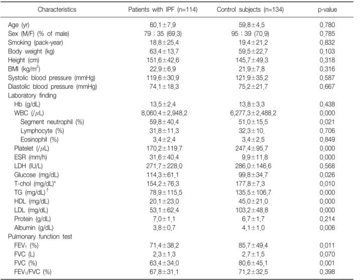
Table 1. Comparison of clinical characteristics, blood tests and pulmonary function tests between patients with IPF and control subjects
Characteristics Patients with IPF (n=114) Control subjects (n=134) p-value
Age (yr) 60.1±7.9 59.8±4.5 0.780
Sex (M/F) (% of male) 79:35 (69.3) 95:39 (70.9) 0.785
Smoking (pack-year) 18.8±25.4 19.4±21.2 0.832
Body weight (kg) 63.4±13.7 59.5±22.7 0.103
Height (cm) 151.6±42.6 145.7±49.3 0.318
BMI (kg/m2) 22.9±6.9 21.9±7.8 0.316
Systolic blood pressure (mmHg) 119.6±30.9 121.9±35.2 0.587
Diastolic blood pressure (mmHg) 74.1±18.3 75.2±21.7 0.667
Laboratory finding
Hb (g/dL) 13.5±2.4 13.8±3.3 0.438
WBC (/μL) 8,060.4±2,948.2 6,277.3±2,488.2 0.000
Segment neutrophil (%) 59.8±40.4 51.0±15.5 0.021
Lymphocyte (%) 31.8±11.3 32.3±10. 0.706
Eosinophil (%) 3.4±2.4 3.4±2.5 0.849
Platelet (/μL) 170.2±119.7 247.4±95.7 0.000
ESR (mm/h) 31.6±40.4 9.9±11.8 0.000
LDH (IU/L) 271.7±228.0 286.0±146.6 0.568
Glucose (mg/dL) 114.3±61.1 99.8±34.7 0.026
T-chol (mg/dL)* 154.2±76.3 177.8±7.3 0.010
TG (mg/dL)† 78.9±115.5 135.5±106.7 0.000
HDL (mg/dL) 20.1±23.0 45.0±21.0 0.000
LDL (mg/dL) 53.1±62.4 103.2±48.8 0.000
Protein (g/dL) 7.0±1.1 6.7±1.7 0.214
Albumin (g/dL) 3.8±0.7 4.1±1.0 0.006
Pulmonary function test
FEV1 (%) 71.4±38.2 85.7±49.4 0.011
FVC (L) 2.3±1.3 2.7±1.5 0.070
FVC (%) 63.4±34.0 80.6±45.1 0.001
FEV1/FVC (%) 67.8±31.1 71.2±32.5 0.398
IPF: idiopathic pulmonary fibrosis.
*Total cholesterol, †Triglyceride.
to assess the role of several variables as risk factors for the IPF. Differences were considered significant when the p value was less than 0.05. Statistical analyzed were performed by version 13.0 of SPSS for Windows (SPSS, Inc., Chicago, IL, USA).
Results
1. Clinical characteristics, blood tests and pulmonary function tests between two groups
One hundred fourteen patients with IPF and 134 con- trol subjects were enrolled. About 70% of both IPF pa- tients and control subjects were male (69.3% and 70.9%).
The mean age of each group was 60.1±7.9 years in IPF patients and 59.8±4.5 years in control subjects. The two groups have no significant difference in other clinical fea- tures; smoking history (18.8±25.4 pack-year vs. 19.4±
21.2 pack-year), body weight (63.4±13.7 kg vs. 59.5±
22.7 kg), height (151.6±42.6 cm vs. 145.7±49.3 cm), BMI (22.9±6.9 kg/m2 vs. 21.9±7.8 kg/m2), systolic blood pressure (119.6±30.9 mmHg vs. 121.9±35.2 mmHg), diastolic blood pressure (74.1±18.3 mmHg vs.
75.2±21.7 mmHg). The level of serum fasting glucose was significantly higher (114.3±61.1 mg/dL) in IPF pa- tients than in control subjects (99.8±34.7 mg/dL). IPF patients have significantly high level of total white blood
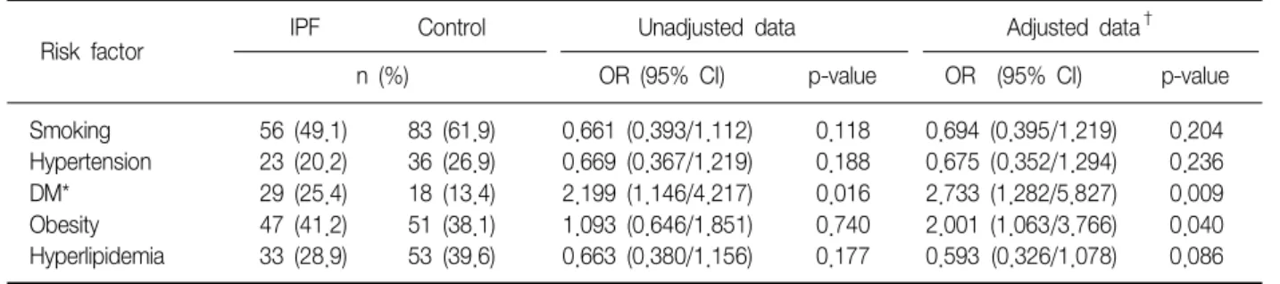
Table 2. Risk factors for IPF in relation to metabolic disorders and smoking
IPF Control Unadjusted data Adjusted data†
Risk factor
n (%) OR (95% CI) p-value OR (95% CI) p-value
Smoking 56 (49.1) 83 (61.9) 0.661 (0.393/1.112) 0.118 0.694 (0.395/1.219) 0.204 Hypertension 23 (20.2) 36 (26.9) 0.669 (0.367/1.219) 0.188 0.675 (0.352/1.294) 0.236
DM* 29 (25.4) 18 (13.4) 2.199 (1.146/4.217) 0.016 2.733 (1.282/5.827) 0.009
Obesity 47 (41.2) 51 (38.1) 1.093 (0.646/1.851) 0.740 2.001 (1.063/3.766) 0.040 Hyperlipidemia 33 (28.9) 53 (39.6) 0.663 (0.380/1.156) 0.177 0.593 (0.326/1.078) 0.086 IPF: idiopathic pulmonary fibrosis.
*Diabetes mellitus, †Adjusted for age, sex.
Table 3. Clinical characteristics, the results of blood and pulmonary function tests in IPF patients with or without diabetes mellitus
Characteristics With DM (n=29) Without DM (n=85) p-value
Age (yr) 61.5±6.9 59.8±8.1 0.255
Pack-year (py) 27.4±34.7 15.8±20.8 0.033
Body weight (kg) 64.0±9.0 63.1±15.0 0.773
Height (cm) 163.4±7.5 147.6±48.5 0.005
BMI (kg/m2) 23.9±2.6 22.5±7.8 0.178
Systolic blood pressure (mmHg) 124.5±17.2 118.0±34.2 0.329
Diastolic blood pressure (mmHg) 75.9±9.6 73.4±20.5 0.536
Laboratory findings
Hb (g/dL) 13.7±1.8 13.5±2.5 0.706
WBC (/μL) 8,522.1±3,047.0 7,902.8±2,915.4 0.331
Platelet (/μL) 122.2±112.7 186.8±118.2 0.012
ESR (mg/dL) 29.7±28.9 32.3±43.8 0.765
LDH (IU/L) 270.2±187.9 272.3±240.7 0.967
Glucose (mg/dL) 113.0±64.5 114.8±60.3 0.890
T-chol (mg/dL)* 171.3±79.9 148.3±74.6 0.163
TG (mg/dL)† 125.7±135.9 62.9±103.9 0.011
HDL (mg/dL) 32.2±22.0 16.0±21.9 0.001
LDL (mg/dL) 84.8±64.6 42.3±58.1 0.001
Protein (g/dL) 7.0±0.4 6.9±1.2 0.617
Albumin (g/dL) 3.8±0.5 3.8±0.73 0.695
Pulmonary function test
FEV1 (%) 77.4±34.8 69.4±39.3 0.332
FVC (L) 2.7±1.0 2.2±1.35 0.114
FVC (%) 68.4±31.8 61.7±34.8 0.365
FEV1/FVC (%) 79.7±17.2 63.8±33.8 0.001
IPF: idiopathic pulmonary fibrosis.
*Total cholesterol, †Triglyceride.
cell (WBC) (8,060.4±2,948.2/μL vs. 6,277.3±2,488.2/
μL), segmented neutrophil (59.8±40.4% vs. 51.0±
15.5%), and erythrocyte sediment rate (ESR) (31.6±40.4 mm/h vs. 9.9±11.8 mm/h). Total cholesterol, trigly- ceride, HDL cholesterol and LDL cholesterol were lower
in IPF patients than in control subjects (154.2±76.3 mg/dL vs. 177.8±7.3 mg/dL; 78.9±115.5 mg/dL vs.
135.5±106.7 mg/dL; 20.1±23.0 mg/dL vs. 45.0±21.0 mg/dL; 53.1±62.4 mg/dL vs. 103.2±48.8 mg/dL) res- pectively. The level of FEV1 (L), FEV1 (%), and FVC (%)
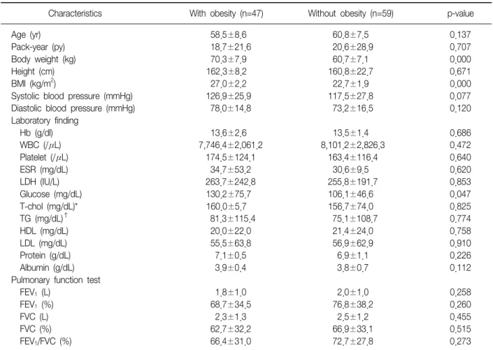
Table 4. Clinical characteristics, the results of blood and pulmonary function tests in IPF patients with or without obesity Characteristics With obesity (n=47) Without obesity (n=59) p-value
Age (yr) 58.5±8.6 60.8±7.5 0.137
Pack-year (py) 18.7±21.6 20.6±28.9 0.707
Body weight (kg) 70.3±7.9 60.7±7.1 0.000
Height (cm) 162.3±8.2 160.8±22.7 0.671
BMI (kg/m2) 27.0±2.2 22.7±1.9 0.000
Systolic blood pressure (mmHg) 126.9±25.9 117.5±27.8 0.077
Diastolic blood pressure (mmHg) 78.0±14.8 73.2±16.5 0.120
Laboratory finding
Hb (g/dl) 13.6±2.6 13.5±1.4 0.686
WBC (/μL) 7,746.4±2,061.2 8,101.2±2,826.3 0.472
Platelet (/μL) 174.5±124.1 163.4±116.4 0.640
ESR (mg/dL) 34.7±53.2 30.6±9.5 0.620
LDH (IU/L) 263.7±242.8 255.8±191.7 0.853
Glucose (mg/dL) 130.2±75.7 106.1±46.6 0.047
T-chol (mg/dL)* 160.0±5.7 156.7±74.0 0.825
TG (mg/dL)† 81.3±115.4 75.1±108.7 0.774
HDL (mg/dL) 20.0±22.0 21.4±24.0 0.758
LDL (mg/dL) 55.5±63.8 56.9±62.9 0.910
Protein (g/dL) 7.1±0.5 6.9±1.1 0.226
Albumin (g/dL) 3.9±0.4 3.8±0.7 0.112
Pulmonary function test
FEV1 (L) 1.8±1.0 2.0±1.0 0.258
FEV1 (%) 68.7±34.5 76.8±38.2 0.260
FVC (L) 2.3±1.3 2.5±1.2 0.455
FVC (%) 62.7±32.2 66.9±33.1 0.515
FEV1/FVC (%) 66.4±31.0 72.7±27.8 0.273
IPF: idiopathic pulmonary fibrosis.
*Total cholesterol, †Triglyceride.
in IPF patients was significantly lower than in control subjects (1.9±1.0 L vs. 2.2±1.3 L; 71.4±38.2% vs.
85.7±49.4%; 63.4±34.0% vs. 80.6±45.1%) respective- ly (Table 1).
2. Association of metabolic disorders with IPF
Univariate analysis was performed to evaluate non- adjusted odds ratio about risk factor between IPF pa- tients and control subjects. Odds ratio (OR) of DM was statistically different between IPF patients and control subjects. OR of DM was 2.199 (95% CI, 1.146∼4.217, p=0.016). The prevalence of DM was 25.4% (n=25) for IPF and 13.4% (n=18) for control subjects. The OR of other factors were not significant different; obesity, 1.093; smoking status, 0.661; hypertension, 0.669; hy- perlipidemia, 0.663 (Table 2). We analyzed each risk
factor by multivariate analysis in which DM and obesity were significantly associated with IPF. The adjusted OR of DM and obesity was 2.733 (95% CI, 1.282∼5.827) and 2.001 (95% CI, 1.063∼3.766). The adjusted OR of smoking status was 0.694 (95% CI, 0.395∼1.219).
Hypertension (adjusted OR, 0.675; 95% CI, 0.352∼
1.294) and hyperlipidemia (adjusted OR, 0.593; 95% CI, 0.326∼1.078) were not statistically significant (Table 2).
3. Differences between IPF patients with DM or with- out DM, and patients with obesity or without ob- esity
We analyzed IPF patients with DM or without (Table 3). Smoking history, height, triglyceride, HDL, LDL, FEV1 and FEV1/FVC were significantly higher in IPF pa- tients with DM than in IPF without DM. We also eval-
uated IPF patients with obesity or without obesity (Table 4). Glucose were significantly higher in IPF pa- tients with obesity.
Discussion
We showed that the prevalence of DM in IPF patients was significantly higher than in control subjects (25.4%
vs. 13.4%). The level of fasting glucose in IPF patients was also significantly higher than in control subjects (114.3±61.1 mg/dL vs. 99.8±34.7 mg/dL), and the ad- justed OR of DM by multivariate analysis was 2.733 (95% CI, 1.282∼5.827). These results supported a pre- vious report that the prevalence of DM was higher in patients with IPF than in control subjects4. On the other hand, there was a conflicting report that DM was not associated with IPF3. In that report, investigators had collected data by self-administered questionnaires and defined individuals who were only being in dietary or drug treatment as DM patients. They did not include latent or untreated DM. So they might have underesti- mated true association3. In this current study, obesity (OR 2.001) is also significantly associated with IPF using multivariate analysis. As the prevalence of obesity is getting increased in general, many investigators have been interested in chronic diseases which might be as- sociated with obesity. Obesity was known to be one of important risk factors in the metabolic syndrome, and associated with an increased risk of DM and heart dis- eases15,16. There have been conflict reports about rela- tionship between obesity and IPF3,4. One had reported that BMI of IPF patients was higher than in control sub- ject3. On the contrary, the other investigation showed that BMI and obesity, which was defined as BMI of >25, were not related to IPF. They showed the prevalence of obesity was not statistically different between IPF pa- tients and control subjects4. In our study, more patients were analyzed than in study of previous studies4. Ac- cordingly our study might have stronger statistical pow- er than previous studies4. Although definition of obesity in metabolic syndrome has been identified as abdominal obesity17, we used BMI as a parameter for obesity.
There are some reports that measurement of BMI as well as waist circumference has been used to identify insulin resistance and cardiovascular risk factors18,19. It is still cautious to analyze obesity-related data due to different definition of obesity. Hyperlipidemia and hy- pertension were not associated with IPF in this current study like previous studies. Smoking status was not sig- nificant related to the risk of IPF, which was incon- sistent with previous investigations that smoking history was associated with an increased risk for the develop- ment of IPF3,4,20. Pack-years of smoking was not statisti- cally associated with IPF in our study. Baumgartner et al. showed that current smoking and more than 40 pack-years of smoking in IPF patients were not signifi- cantly related to a risk of IPF3,20. Similar result was re- ported in the study of Yoshihiro et al. that there was no association with dose-response of cigarette smoking and IPF statistically3,20. We enrolled control subjects without lung diseases and abnormalities in low-dose CT. Because low-dose CT was performed to detect early lung cancer, most of control subjects had a smoking his- tory so that smoking status was not so different from patients. It might be one reason that pack-years of smoking was not associated with IPF compared to con- trol subjects in our study.
Recent studies have suggested that exogenous, en- dogenous multiple, microscopic stimuli may injure the alveolar epithelial cells, followed by an abnormal wound healing in the pathogenesis of IPF5-7. On the molecular level, a number of studies have reported that the imbalance of oxidants/antioxidants may play a sig- nificant role in the progression of pulmonary fibrosis1,2. The excess formation of reactive oxygen species (ROS) may cause tissue injuries including those involving the lung and leading to pulmonary fibrosis1,2,5-7. Many stud- ies suggested that diabetes was involved by high level of free radicals or oxidant/antioxidant imbalance and oxidative stress may play a major role in the develop- ment and progression of DM and its complication8-10,21. Oxidative stress might be produced by advanced glyca- tion end products (AGE) under hyperglycemia9,22. AGEs may inactivate enzymes and alter their structures and
functions, promote free radical formation. AGEs may be linked to influence the expression of growth factors, cy- tokines and transcription factors that are believed in me- diating the differentiation of epithelial cells to form my- ofibroblasts, such as TGF-ß1, CTGF and NF-kß23-26. AGE may activate TGF-ß-Smad signaling and influence mesangial cell hypertrophy and fibronectin synthesis22. AGEs can accumulate in alveolar macrophages and bronchial epithelial cells in idiopathic pulmonary fib- rosis27.
In the obese patients, adipose cells may play a role in endothelial dysfunction by pro-inflammatory cyto- kines, such as IL-6, TNF-α. In cultured adipocytes, ele- vated levels of fatty acids increased oxidative stress via NADPH oxidase activation16.
The lung has developed antioxidant defense mecha- nism against oxidants1. This defense mechanism may be contributed to protect against pulmonary complication by oxidative stress and show less manifestation than other organs. Many results have reported that oxidative stress induced not only IPF but also DM, including obesity. The present study appears to support previous findings that DM may be associated with increased IPF.
Moreover, our result provides an opportunity to raise interest about obesity as a risk factor of IPF. In con- clusion, we demonstrate that DM and obesity are asso- ciated with IPF. Based on these results, we need to in- vestigate more the relation between metabolic disorders such as DM, and obesity and IPF in perspective of mo- lecular basis.
Acknowledgements
I would like to thank the authors in Gachon Univer- sity Gil Hospital and Sungkyunkwan University Sam- sung Medical Center for help in recruiting patients and collecting data.
References
1. Kinnula VL, Fattman CL, Tan RJ, Oury TD. Oxidative stress in pulmonary fibrosis: a possible role for redox
modulatory therapy. Am J Respir Crit Care Med 2005;
172:417-22.
2. Teramoto S, Fukuchi Y, Uejima Y, Shu CY, Orimo H.
Superoxide anion formation and glutathione metabo- lism of blood in patients with idiopathic pulmonary fibrosis. Biochem Mol Med 1995;55:66-70.
3. Miyake Y, Sasaki S, Yokoyama T, Chida K, Azuma A, Suda T, et al. Case-control study of medical history and idiopathic pulmonary fibrosis in Japan. Respirology 2005;10:504-9.
4. Enomoto T, Usuki J, Azuma A, Nakagawa T, Kudoh S. Diabetes mellitus may increase risk for idiopathic pulmonary fibrosis. Chest 2003;123:2007-11.
5. Selman M, King TE, Pardo A. Idiopathic pulmonary fib- rosis: prevailing and evolving hypotheses about its pathogenesis and implications for therapy. Ann Intern Med 2001;134:136-51.
6. Kuwano K, Nakashima N, Inoshima I, Hagimoto N, Fujita M, Yoshimi M, et al. Oxidative stress in lung epi- thelial cells from patients with idiopathic interstitial pneumonia. Eur Respir J 2003;21:232-40.
7. Kim DS. Idiopathic pulmonary fibrosis and pulmonary fibrosis associated with collagen vascular diseases: clin- ical features, broncholaveolar lavage fluid findings and response to treatment. Korean J Med 1988;35:87-99.
8. Baynes JW, Thorpe SR. Role of oxidative stress in dia- betic complications: a new perspective on an old para- digm. Diabetes 1999;48:1-9.
9. Maritim AC, Sanders RA, Watkins JB 3rd. Diabetes, oxi- dative stress, and antioxidants: a review. J Biochem Mol Toxicol 2003;17:24-38.
10. Yu T, Robotham JL, Yoon Y. Increased production of reactive oxygen species in hyperglycemic conditions re- quires dynamic change of mitochondrial morphology.
Proc Natl Acad Sci U S A 2006;103:2653-8.
11. American Thoracic Society. Idiopathic pulmonary fib- rosis: diagnosis and treatment. International consensus statement. American Thoracic Society (ATS), and the European Respiratory Society (ERS). Am J Respir Crit Care Med 2000;161:646-64.
12. Expert Committee on the Diagnosis and Classification of Diabetes Mellitus. Report of the expert committee on the diagnosis and classification of diabetes mellitus.
Diabetes Care 2003;26 Suppl 1:S5-20.
13. Chobanian AV, Bakris GL, Black HR, Cushman WC, Green LA, Izzo JL Jr, et al. The Seventh Report of the Joint National Committee on Prevention, Detection, Evaluation, and Treatment of High Blood Pressure: the JNC 7 report. JAMA 2003;289:2560-72.
14. WHO Expert Consultation. Appropriate body-mass in- dex for Asian populations and its implications for poli- cy and intervention strategies. Lancet 2004;363:157-63.
15. National Cholesterol Education Program (NCEP) Expert Panel on Detection, Evaluation, and Treatment of High Blood Cholesterol in Adults (Adult Treatment Panel III).
Third Report of the National Cholesterol Education Program (NCEP) Expert Panel on Detection, Evaluation, and Treatment of High Blood Cholesterol in Adults (Adult Treatment Panel III) final report. Circulation 2002;106:3143-421.
16. Furukawa S, Fujita T, Shimabukuro M, Iwaki M, Yama- da Y, Nakajima Y, et al. Increased oxidative stress in obesity and its impact on metabolic syndrome. J Clin Invest 2004;114:1752-61.
17. Grundy SM, Brewer HB Jr, Cleeman JI, Smith SC Jr, Lenfant C; American Heart Association; National Heart, Lung, and Blood Institute. Definition of metabolic syn- drome: Report of the National Heart, Lung, and Blood Institute/American Heart Association conference on sci- entific issues related to definition. Circulation 2004;109:
433-8.
18. Farin HM, Abbasi F, Reaven GM. Comparison of body mass index versus waist circumference with the meta- bolic changes that increase the risk of cardiovascular disease in insulin-resistant individuals. Am J Cardiol 2006;98:1053-6.
19. Sung KC, Ryu S, Reaven GM; Health Screening Group at Kangbuk Samsung Hospital. Relationship between obesity and several cardiovascular disease risk factors in apparently healthy Korean individuals: comparison of body mass index and waist circumference. Metabo- lism 2007;56:297-303.
20. Baumgartner KB, Samet JM, Stidley CA, Colby TV, Waldron JA. Cigarette smoking: a risk factor for idio-
pathic pulmonary fibrosis. Am J Respir Crit Care Med 1997;155:242-8.
21. Choi SB, Choi EK, Ann SH, Choi MK, Park SM. The effect of continuous subcutaneous insulin infusion ther- apy on oxidative stress in Korean type 2 diabetic pa- tients. Korean J Med 2000;58:548-59.
22. Fukami K, Ueda S, Yamagishi S, Kato S, Inagaki Y, Takeuchi M, et al. AGEs activate mesangial TGF-be- ta-Smad signaling via an angiotensin II type I receptor interaction. Kidney Int 2004;66:2137-47.
23. Li JH, Wang W, Huang XR, Oldfield M, Schmidt AM, Cooper ME, et al. Advanced glycation end products in- duce tubular epithelial-myofibroblast transition through the RAGE-ERK1/2 MAP kinase signaling pathway. Am J Pathol 2004;164:1389-97.
24. Oldfield MD, Bach LA, Forbes JM, Nikolic-Paterson D, McRobert A, Thallas V, et al. Advanced glycation end products causes epithelial-myofibroblast transdifferenti- ation via the receptor for advanced glycation end prod- ucts (RAGE). J Clin Invest 2001;108:1853-63.
25. Lee CI, Guh JY, Chen HC, Hung WC, Yang YL, Chuang LY. Advanced glycation end-product-induced mitogen- esis and collagen production are dependent on angio- tensin II and connective tissue growth factor in NRK- 49F cells. J Cell Biochem 2005;95:281-92.
26. Zhou G, Li C, Cai L. Advanced glycation end-products induce connective tissue growth factor-mediated renal fibrosis predominantly through transforming growth factor beta independent pathway. Am J Pathol 2004;
165:2033-43.
27. Matsuse T, Ohga E, Teramoto S, Fukayama M, Nagai R, Horiuchi S, et al. Immunohistochemical localisation of advanced glycation end products in pulmonary fibrosis. J Clin Pathol 1998;51:515-9.