Development of PLGA Nanoparticles for Astrocyte-specific Delivery of Gene Therapy: A Review
Hyo Jung Shin1,2,4*, Ka Young Lee1,4, Kisang Kwon5, O-Yu Kwon1 and Dong Woon Kim1,2,3
1Department of Anatomy and Cell Biology, 2Brain Research Institute, Chungnam National University, Daejeon 35015, Korea
3Department of Medical Science, Chungnam National University, Daejeon 35015, Korea
4Department of Rehabilitation Medicine, Seoul National University Bundang Hospital, Seongnam 13620, Korea
5Department of Clinical Laboratory Science, Wonkwang Health Science University, Iksan 54538, Korea Received August 17, 2021 /Revised September 21, 2021 /Accepted September 23, 2021
Recently, as nanotechnology has been introduced and used in various fields, the development of new drugs has been accelerating. Nanoparticles have maintained blood drug concentration for extended periods of time with a single administration of the drug. The drug can then be selectively released only at the pathological site, thereby reducing side effects to other non-pathological sites. In addition, nanoparticles can be modified for selective target sites delivery for other specific diseases, with poly- mers being widely used in the manufacture of these nanoparticles. Poly (D,L-lactic-co-glycolic acid ) (PLGA) is one of the most extensively developed biodegradable polymers. PLGA is widely used in drug delivery for a variety of applications. It has also been approved by the FDA as a drug delivery system and is widely applied in controlled release formulations, such as in gene therapy treatments.
PLGA nanoparticles have been developed as delivery systems with high efficiency to specific cell types by using passive and active targeting methods. After the development of a drug delivery system using PLGA nanoparticles, the drug is selectively delivered to the target site, and the effective blood concentration for extended periods of time is optimized according to the disease. In this review paper, we focus on ways to improve cell-specific treatment outcomes by examining the development of as- trocyte selective nanoparticles based on PLGA nanomaterials for gene therapy.
Key words : Astrocyte, gene therapy, nanoparticle, PLGA (Poly (D,L-lactic-co-glycolic acid))
*Corresponding author
*Tel : +82-42-580-8210, Fax : +82-42-586-4800
*E-mail : [email protected]
This is an Open-Access article distributed under the terms of the Creative Commons Attribution Non-Commercial License (http://creativecommons.org/licenses/by-nc/3.0) which permits unrestricted non-commercial use, distribution, and reproduction in any medium, provided the original work is properly cited.
Introduction
A drug delivery system selectively delivers a drug to a target site and optimizes the effective blood concentration for extended periods of time according to the diseas [38], thereby maximizing the therapeutic efficacy and effect and minimizing the side effects of the drug [1, 32]. By controlling the release and absorption of drugs and targeting and deliv- ering drugs to a specific site in the body [1, 31], it is possible to retain the required amount of drug in the target site for a specified period of time. Furthermore, recent studies have focused on drug delivery systems using chemical polymers [3, 44]. The development of high molecular weight polymers that slow the release of the drug so that the drug can be effective for an extended period of time has therefore
emerged as an important topic [40].
Nanoparticles are solid spherical structures of approx- imately 100 nm, which are prepared from natural or syn- thetic polymers. A wide variety of drugs, such as hydro- philic or hydrophobic small drugs, vaccines, and biological macromolecules, can be delivered using nanoparticles.
Nanoparticles also facilitate targeted administration to spe- cific organs or cells, as well as controlled drug delivery.
Nanoparticles are broadly divided into different categories depending on their morphologies, sizes, and physical and chemical properties [17]. Nanoparticles can be carbon-based, ceramic, metal, semiconductor, polymeric, or lipid-based.
Polymeric nanoparticles have been the most extensively studied for drug delivery [20, 27]. In addition to small drug molecules, they can also be used to deliver genes and proteins. Polymeric nanoparticles can penetrate through cell membranes, have serum stability, and can be easily manu- factured. Furthermore, the surface of nanoparticles can be modified for various medical applications [24, 25].
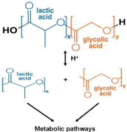
Poly (lactic-co-glycolic acid) (PLGA) is one of the most suc- cessfully used biodegradable polymers because its hydrol- ysis leads to metabolite monomers, lactic acid and glycolic
- Review -
Fig. 1. Chemical structure and hydrolysis of PLGA nanoparticles.
Fig. 2. Schematic diagram of the cellular uptake of PLGA nano- particles by type of cells in brain.
acid [23](Fig. 1). Because these two monomers are endoge- nous and easily metabolized by the body via the Krebs cycle, minimal systemic toxicity is associated with the use of PLGA for drug delivery or biomaterial applications. PLGA has been approved by the USA Food and Drug Administration (FDA) and the European Medicine Agency (EMA) for drug delivery systems in humans. These polymers are commer- cially available with different molecular weights and copoly- mer compositions. The degradation time can vary from sev- eral months to several years, depending on the molecular weight and copolymer ratios. Forms of PLGA are usually identified by the monomer ratios used. For example, PLGA 50:50 is a copolymer whose composition is 50% lactic acid and 50% glycolic acid. PLGA nanoparticles are internalized in cells partly through fluid phase pinocytosis and also through clathrin-mediated endocytosis [7]. However, the nanoparticles rapidly escape internalization into lysosomes and enter the cytoplasm within 10 min of incubation [30].
This is facilitated by the interaction of nanoparticles with vesicular membranes leading to transient and localized de- stabilization of the membrane, resulting in the escape of nanoparticles into the cytosol.
Astrocytes are the largest and most prevalent type of glial cell in the central nervous system (CNS). Astrocytes contrib- ute to the formation of the blood brain barrier (BBB), partic- ipate in the maintenance of extracellular ionic and chemical homeostasis, are involved in the response to injury, affect neuronal development and plasticity, and are critical for neuronal homeostasis [2]. Astrocytes also regulate the con- tents of the synaptic cleft and synaptic transmission as part of the tripartite synapse, control CNS metabolism, and main-
tain BBB integrity. Impairment of these functions through a disturbance in astrocyte integrity is likely to impact multi- ple aspects of brain physiology [29, 41]. Notably, astrocytes also can undergo a functional decline. Therefore, given that the primary and most important role of astrocytes in the brain is to maintain neuronal health [19, 35, 46], enhancing the efficiency of drug delivery in astrocytes is an important objective for treating brain diseases.
Currently, delivery systems for the CNS using PLGA nanoparticles rarely are specific for astrocytes (Fig. 2). Other glial cells, such as microglia, have more than 50% cellular uptake capacity, followed by neuron cells. We will therefore describe efforts to overcome these problems. In this review, we will first discuss how PLGA-based nanoparticles can be engineered to target only specific cells and then how PLGA-based nanoparticles can work by targeting only as- trocytes to deliver drugs. Finally, we will describe the poten- tial development of these nanoparticles
In vivo delivery mechanisms of PLGA nanoparticles in nanomedicine
The delivery mechanism of PLGA nanoparticles is div- ided into two main categories; the first is passive targeting by the Enhanced Permeation and Retention (EPR) effect [15, 16]. Passive targeting using the EPR effect controls the size of the nanoparticle so that it minimally penetrates into nor- mal cells but penetrates with high efficiency into the desired tissue. Nanoparticles smaller than 2 nm can easily pass through the voids in blood vessels, substances smaller than
Fig. 3. Schematic representation of PLGA nanoparticles for ac- tive targeting.
10 nm can be easily excreted through the kidneys, and nano- particles with sizes between 100-150 nm tend to accumulate in the liver [22]. Therefore, it has been determined that the optimal size of nanoparticles is 10-100 nm.
Alternatively, a nanoparticle can be passively targeted due to its charge (Fig. 3). The epidermal cells of blood vessels have many negatively charged components, so negatively charged nanoparticles tend to be repelled. Moreover, be- cause brain tissue is protected by the BBB, targeting is lim- ited by the size and surface characteristics of the nano- particles, which has been the subject of extensive research.
Passive targeting does not involve selective labeling of a spe- cific target substance but instead involves a process in which the target tissue is labeled due to a difference in the bio- logical and physical environments.
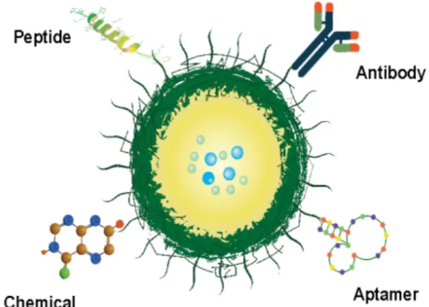
The second delivery mechanism is active targeting, in which targeting ligands are grafted at the surface of the PLGA nanoparticle [5]. The targeting ligand may be a pep- tide or an antibody that binds to a specific receptor, or devel- oped more recently, it can be in the form of an aptamer or a compound [45]. The surface of nanoparticles can also be modified in order to graft, coat, or conjugate with specific targeting moieties. For example, KB cells rapidly take up folic acid-coated PLGA nanoparticles, suggesting that they are mainly taken up by folate receptor-mediated endocytosis [18]. As another example, in glioblastoma multiformes, it was shown that PLGA nanoparticles functionalized with OX26 monoclonal antibodies against the transferrin receptor more than doubled the drug delivery [36]. Recently, ap- tamer-labeled paclitaxel using PLGA nanoparticles has been reported to increase drug targeting to cancer cells [4].
Gene therapy and PLGA nanoparticle-based deliv- ery systems
Gene-based therapy has become increasingly popular in the nanomedicine field. In past years, several studies have reported evidence of promising PLGA-based vector systems loaded with plasmid or siRNA therapeutics directed against disease-associated targets [39, 42]. This is possible because of advances in genetics and bioengineering, which enable manipulation of vectors for the delivery of genes or the in- troduction of gene editors including synthetic RNAi, miRNA, and long non-coding RNA, in addition to plasmid or CRISPR/Cas9 for silencing, enhancing, or editing of genes [10, 21]. Gene therapy, which can control gene expression, has had a major impact on diseases caused by specific gene mutations. This therapy has shown great potential for pre- cision medicine because it can treat the disease at its origin.
However, its potential still exists mainly in the laboratory, and its application is still far from being fully developed as a therapeutic agent.
Astrocyte-targeted PLGA nanoparticles for gene therapy
Astrocytes, the star-shaped glial cells in the brain, contrib- ute to formation of the BBB, and participate in the main- tenance of extracellular ionic and chemical homeostasis.
During homeostasis, astrocytes regulate neurotransmission and synaptic activity by sequestering potassium and neuro- transmitters, including glutamate. Furthermore, astrocytes secrete several neurotrophic and neuroinflammatory media- tors. Thus, astrocyte-targeted gene therapy could be used to upregulate neurotrophic factor expression and/or silenc- ing of toxic mediators. Although astrocytes play an im- portant role in neuroprotection in the brain, efficient deliv- ery of drugs to these cells is difficult. This is due to the intracranial delivery barrier called the BBB, which decreases delivery efficiency and hinders the development of drug de- livery systems targeting astrocytes.
Peptide-conjugated PLGA nanoparticles for targeting as- trocytes can be combined with an anti-Somatostatin receptor 2 (SSTR2) peptide. Furthermore, anti-SSTR2 peptide modi- fied nanoparticles could be a promising nanocarrier for glio- ma neovasculature endothelial cells and glioma cells for dual targeting [6] The method of binding compounds to the sur- face of PLGA nanoparticles is another delivery system spe- cific to astrocytes. Normal PLGA nanoparticles do not local- ize to the nucleus; however, the addition of arginine-modi-
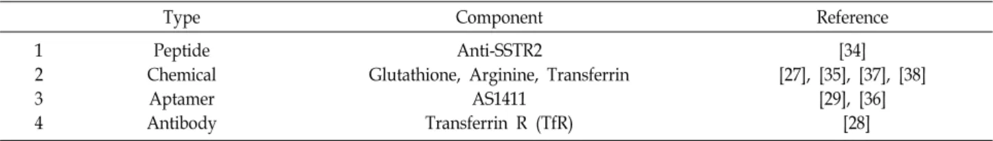
Table 1. Active targeting of PLGA nanoparticles to astrocytes
Type Component Reference
1 2 3 4
Peptide Chemical Aptamer Antibody
Anti-SSTR2
Glutathione, Arginine, Transferrin AS1411
Transferrin R (TfR)
[34]
[27], [35], [37], [38]
[29], [36]
[28]
fied polymers significantly improved nuclear localization of plasimd and successfully achieved gene expression in pri- mary human astrocytes [34]. Glutathione (GSH)-coated PLGA nanoparticles can also permeate the BBB. GSH-coated doce- taxel nanoparticles were significantly better at killing glioma cancer cells than docetaxel alone. Aptamers are a class of therapeutic oligonucleotides that bind to cell surface re- ceptors with high affinity and specificity. This property of aptamers has been exploited develop targeted drug carriers that can deliver a variety of cargo into cells. According to a recent report, the efficiency of intracellular influx of ap- tamer-conjugated PLGA nanoparticles into astrocytes was greatly improved [4, 14] compared to PLGA nanoparticles alone. Antibodies can also be used to target nanoparticles to specific cells types. For example, PLGA nanoparticles functionalized with Transferrin R monoclonal antibody-en- capsulated temozolomide increased the delivery efficiency of the drug into glioma cells.
Discussion
Nanoparticles enable effective treatment by selective tar- get site delivery to a specific disease site, while minimizing side effects that may occur in normal organs, tissues, and cells [9]. To achieve this, it is important to appropriately modify and control the characteristics of the nanomaterial polymer.
The development of nanotechnology has led to remark- able developments in drug delivery and bioimaging, and as a result, diagnosis, treatment, and methods of monitoring the progress of the treatment process have been improved, and customized treatments based to the patient’s diagnosis have now become a possibility. It is not possible to accu- rately predict how effective medical advances will be in the future by understanding the properties of materials and ap- plying them to medicine in the nanotechnology field; how- ever, it is clear that nanomaterials will play a decisive role in more accurate diagnoses and quality treatments in the near future.
Due to the enormous diversity of nanomaterials, their po- tential toxicity and environmental impact is not completely understood. Therefore, studies on the stability of these mate- rials are also being conducted at the same time. As described in this review, control of drug delivery, targeting of nano- particles, and development of cell-specific nanoparticles have developed at a rapid pace, but there are still only a few drugs that can be directly used in clinical practice. Design and targeting strategies for nanoparticles will need to be more diverse and depend on the differences in diseases, de- gree of development, and location of the lesion. Successful clinical use of nano-pharmaceuticals for therapeutic and di- agnostic purpose will then be possible.
PLGA-based nanoparticles present many advantages for drug delivery [11, 43, 47]. They can protect drugs from deg- radation and enhance their stability. Moreover, due to their size, nanoparticles can penetrate specific tissues via re- ceptors overexpressed by target cells or in the BBB. This al- lows specific delivery of drugs, proteins, peptides, or nucleic acids to their target tissues. PLGA-based nanoparticles can increase the efficacy of treatments because of the sustained release of the therapeutic agent from stable nanoparticles.
They can improve pharmacokinetic and pharmacodynamics profiles. Another major advantage of PLGA over other poly- mers is that PLGA has been approved by the FDA and EMA in various drug delivery systems, so PLGA-based nano- particles are in a good position for use in clinical trials.
However, these systems also present some disadvantages, such as a low drug loading capacity, high cost of production, and difficulty in scaling-up production. One of the most sig- nificant challenges limiting the use of drug-loaded PLGA- based nanoparticles in clinical trials is the low drug loading efficiency.
The aggregation of PLGA nanoparticles during the sol- vent evaporation processes is also a notable problem, regard- less of the specific method used. To prevent PLGA-nano- particle aggregation, polymer stabilizers are often used to coat the nanoparticle surfaces, including the use of polyvinyl alcohol, polyvinyl pyrrolidone, Tween 80, and human serum
albumin [43]. However, these stabilizers are difficult to re- move even with thorough washing protocols, and some are toxic to the BBB. In an effort to avoid this problem, a new washing method is needed, and the agglomerates must be released in a size and shape suitable for drug delivery in the body.
The brain is one of the most vital and sensitive organs in the body, which, to perform its functions in an appro- priate way, needs nutrients and gases. Due to its pivotal role and functions, it is protected in many ways, including by the skull, outer skin, three layers of meninges, and the BBB.
The BBB is a layer of endothelial cells associated with peri- cytes and astrocytes, which acts to separate blood from pa- renchymal cells, thus preventing penetration of drugs into the CNS. It therefore protects the brain from overexposure to substances such as potassium, glycine, and glutamate, which, in high levels such as those found in pathological conditions, are neurotoxic [26, 28]. The BBB is the major bar- rier for drug delivery to the brain [33, 37]. The failure of therapies administered via the intravenous or oral route is often due to their inability to cross/penetrate the brain pa- renchyma or BBB. Therefore, it is necessary to increase the efficiency of PLGA nanoparticles crossing through the BBB when developing new products. However, improving the delivery efficiency of nanoparticles is a different problem than developing nanoparticles to target specific cells.
In most cases, a single strategy does not achieve the goals of both improving delivery across the BBB and targeting as- trocytes for treatment of CNS diseases. To provide successful therapies, different strategies are needed, such as the for- mulation and construction of multifunctionally-engineered PLGA nanoparticles.
In this review, possible applications for the use of PLGA- based nanoparticles targeting astrocytes have been described.
These examples illustrate the promise of using nanoparticles for novel treatments. PLGA nanoparticles are a non-viral, biocompatible, and effective delivery system for targeting gene therapy to the human brain, specifically astrocytes. This approach may provide a powerful tool for delivering ther- apeutic genes not only to the brain, but also to other difficult to target cell types.
Acknowledgements
The authors gratefully acknowledge the financial support of the National Research Foundation of Korea (NRF) funded
by the Ministry of Science, ICT & Future Planning (NRF- 2020R1I1A1A0105309611 and NRF- 2021R1C1C2007218).
The Conflict of Interest Statement
The authors declare that they have no conflicts of interest with the contents of this article.
References
1. Al Thaher, Y., Perni, S. and Prokopovich, P. 2017. Nano-car- rier based drug delivery systems for sustained antimicrobial agent release from orthopaedic cementous material. Adv.
Colloid Interface Sci. 249, 234-247.
2. Allen, N. J. and Eroglu, C. 2017. Cell biology of astrocyte- synapse interactions. Neuron 96, 697-708.
3. Alsehli, M. 2020. Polymeric nanocarriers as stimuli-re- sponsive systems for targeted tumor (cancer) therapy: Recent advances in drug delivery. Saudi Pharm. J. 28, 255-265.
4. Aravind, A., Varghese, S. H., Veeranarayanan, S., Mathew, A., Nagaoka, Y., Iwai, S., Fukuda, T., Hasumura, T., Yoshida, Y., Maekawa, T. and Kumar, D. S. 2012. Aptamer-labeled PLGA nanoparticles for targeting cancer cells. Cancer Nano- technol. 3, 1-12.
5. Attia, M. F., Anton, N., Wallyn, J., Omran, Z. and Van- damme, T. F. 2019. An overview of active and passive tar- geting strategies to improve the nanocarriers efficiency to tumour sites. J. Pharm. Pharmacol. 71, 1185-1198.
6. Bhowmik, A., Chakravarti, S., Ghosh, A., Shaw, R., Bhandary, S., Bhattacharyya, S., Sen, P. C. and Ghosh, M. K. 2017.
Anti-SSTR2 peptide based targeted delivery of potent PLGA encapsulated 3,3'-diindolylmethane nanoparticles through blood brain barrier prevents glioma progression. Oncotarget 8, 65339-65358.
7. Chawla, J. S. and Amiji, M. M. 2003. Cellular uptake and concentrations of tamoxifen upon administration in poly (epsilon-caprolactone) nanoparticles. AAPS PharmSci. 5, E3.
8. Chang, J., Paillard, A., Passirani, C., Morille, M., Benoit, J.
P., Betbeder, D. and Garcion, E. 2012. Transferrin Adsorp- tion onto PLGA Nanoparticles governs their interaction with biological systems from blood circulation to brain can- cer cells. Pharm. Res-Dordr. 29, 1495-1505.
9. Chen, F., Shi, Y., Zhang, J. and Liu, Q. 2020. Nanoparticle- based drug delivery systems for targeted epigenetics cancer therapy. Curr. Drug Targets 21, 1084-1098.
10. Cruz, L. J., van Dijk, T., Vepris, O., Li, T., Schomann, T., Baldazzi, F., Kurita, R., Nakamura, Y., Grosveld, F., Philip- sen, S. and Eich, C. 2021. PLGA-nanoparticles for intra- cellular delivery of the CRISPR-complex to elevate fetal glo- bin expression in erythroid cells. Biomaterials 268, 120580.
11. Emerich, D. F. and Thanos, C. G. 2007. Targeted nano- particle-based drug delivery and diagnosis. J. Drug Target 15, 163-183.
12. Fricker, G. 2002. Drug transport across the blood-brain
barrier. Ernst Schering Res. Found Workshop, 139-154.
13. Grover, A., Hirani, A., Pathak, Y. and Sutariya, V. 2014 Brain-targeted delivery of docetaxel by glutathione-coated nanoparticles for brain cancer. AAPS PharmSciTech. 15, 1562- 1568.
14. Guo, J., Gao, X., Su, L., Xia, H., Gu, G., Pang, Z., Jiang, X., Yao, L., Chen, J. and Chen, H. 2011. Aptamer-functional- ized PEG-PLGA nanoparticles for enhanced anti-glioma drug delivery. Biomaterials 32, 8010-8020.
15. Jain, K. K. 2005. Nanotechnology-based drug delivery for cancer. Technol. Cancer Res. Treat 4, 407-416.
16. Jawad, Z., Xie, F. and Jiao, L. R. 2015. Applications of nano- technology in the management of cancer: miniature technol- ogy, Great Potential. JAMA Surg. 150, 1184-1185.
17. Jeevanandam, J., Barhoum, A., Chan, Y. S., Dufresne, A. and Danquah, M. K. 2018. Review on nanoparticles and nano- structured materials: history, sources, toxicity and regulat- ions. Beilstein J. Nanotechnol. 9, 1050-1074.
18. Kim, S. H., Jeong, J. H., Chun, K. W. and Park, T. G. 2005.
Target-specific cellular uptake of PLGA nanoparticles coat- ed with poly (L-lysine)-poly (ethylene glycol)-folate conju- gate. Langmuir 21, 8852-8857.
19. Liu, C. Y., Yang, Y., Ju, W. N., Wang, X. and Zhang, H.
L. 2018. Emerging roles of astrocytes in neuro-vascular unit and the tripartite synapse with emphasis on reactive gliosis in the context of alzheimer's disease. Front. Cell Neurosci.
12, 193.
20. Liu, Y., Li, K., Liu, B. and Feng, S. S. 2010. A strategy for precision engineering of nanoparticles of biodegradable co- polymers for quantitative control of targeted drug delivery.
Biomaterials 31, 9145-9155.
21. Liu, Y., Zhao, G., Xu, C. F., Luo, Y. L., Lu, Z. D. and Wang, J. 2018. Systemic delivery of CRISPR/Cas9 with PEG-PLGA nanoparticles for chronic myeloid leukemia targeted ther- apy. Biomater Sci. 6, 1592-1603.
22. Longmire, M., Choyke, P. L. and Kobayashi, H. 2008.
Clearance properties of nano-sized particles and molecules as imaging agents: considerations and caveats. Nanomedicine (Lond) 3, 703-717.
23. Makadia, H. K. and Siegel, S. J. 2011. Poly Lactic-co-Glycolic Acid (PLGA) as biodegradable controlled drug delivery carrier. Polymers (Basel) 3, 1377-1397.
24. Majewski, P. and Krysinski, P. 2008. Synthesis, surface mod- ifications, and size-sorting of mixed nickel-zinc ferrite colloi- dal magnetic nanoparticles. Chemistry 14, 7961-7968.
25. Marzaioli, V., Aguilar-Pimentel, J. A., Weichenmeier, I., Luxenhofer, G., Wiemann, M., Landsiedel, R., Wohlleben, W., Eiden, S., Mempel, M., Behrendt, H., Schmidt-Weber, C., Gutermuth, J. and Alessandrini, F. 2014. Surface mod- ifications of silica nanoparticles are crucial for their inert versus proinflammatory and immunomodulatory properties.
Int. J. Nanomedicine 9, 2815-2832.
26. Meyer, R. P., Knoth, R., Schiltz, E. and Volk, B. 2001.
Possible function of astrocyte cytochrome P450 in control of xenobiotic phenytoin in the brain: in vitro studies on mur- ine astrocyte primary cultures. Exp. Neurol. 167, 376-384.
27. Mitchell, M. J., Billingsley, M. M., Haley, R. M., Wechsler, M. E., Peppas, N. A. and Langer, R. 2021 Engineering pre- cision nanoparticles for drug delivery. Nat. Rev. Drug Discov.
20, 101-124.
28. Moore, T. L., Rodriguez-Lorenzo, L., Hirsch, V., Balog, S., Urban, D., Jud, C., Rothen-Rutishauser, B., Lattuada, M. and Petri-Fink, A. 2015 Nanoparticle colloidal stability in cell culture media and impact on cellular interactions. Chem. Soc.
Rev. 44, 6287-6305.
29. Oksanen, M., Lehtonen, S., Jaronen, M., Goldsteins, G., Hamalainen, R. H. and Koistinaho, J. 2019 Astrocyte alter- ations in neurodegenerative pathologies and their modeling in human induced pluripotent stem cell platforms. Cell Mol.
Life Sci. 76, 2739-2760.
30. Panyam, J., Zhou, W. Z., Prabha, S., Sahoo, S. K. and Labha- setwar, V. 2002 Rapid endo-lysosomal escape of poly(DL- lactide-co-glycolide) nanoparticles: implications for drug and gene delivery. FASEB J. 16, 1217-1226.
31. Patra, J. K., Das, G., Fraceto, L. F., Campos, E. V. R., Rodri- guez-Torres, M. D. P., Acosta-Torres, L. S., Diaz-Torres, L.
A., Grillo, R., Swamy, M. K., Sharma, S., Habtemariam, S.
and Shin, H. S. 2018 Nano based drug delivery systems:
recent developments and future prospects. J. Nanobiotechnol- ogy 16, 71.
32. Peng, Y., Chen, L., Ye, S., Kang, Y., Liu, J., Zeng, S. and Yu, L. 2020 Research and development of drug delivery sys- tems based on drug transporter and nano-formulation.
Asian J. Pharm. Sci. 15, 220-236.
33. Perez-Catalan, N. A., Doe, C. Q. and Ackerman, S. D. 2021 The role of astrocyte-mediated plasticity in neural circuit development and function. Neural. Dev. 16, 1.
34. Proulx, J., Joshi, C., Vijayaraghavalu, S., Saraswathy, M., Labhasetwar, V., Ghorpade, A. and Borgmann, K. 2020 Arginine-modified polymers facilitate Poly (Lactide-Co- Glycolide)-based nanoparticle gene delivery to primary hu- man astrocytes. Int. J. Nanomedicine 15, 3639-3647.
35. Radford, R. A., Morsch, M., Rayner, S. L., Cole, N. J., Pountney, D. L. and Chung, R. S. 2015 The established and emerging roles of astrocytes and microglia in amyotrophic lateral sclerosis and frontotemporal dementia. Front Cell Neurosci. 9, 414.
36. Ramalho, M. J., Sevin, E., Gosselet, F., Lima, J., Coelho, M.
A. N., Loureiro, J. A. and Pereira, M. C. 2018 Receptor-medi- ated PLGA nanoparticles for glioblastoma multiforme treat- ment. Int. J. Pharm. 545, 84-92.
37. Rochat, B. and Audus, K. L. 1999 Drug disposition and targeting. Transport across the blood-brain barrier. Pharm.
Biotechnol. 12, 181-200.
38. Saini, S., Kumar, S., Choudhary, M., Nitesh. and Budhwar, V. 2018 Microspheres as controlled drug delivery system:
an updated review. Int. J. Pharm Sci. Res. 9, 1760-1768.
39. Sajid, M. I., Moazzam, M., Kato, S., Yeseom Cho, K. and Tiwari, R. K. 2020 Overcoming barriers for siRNA ther- apeutics: from bench to bedside. Pharmaceuticals (Basel) 13, 294.
40. Senapati, S., Mahanta, A. K., Kumar, S. and Maiti, P. 2018.
Controlled drug delivery vehicles for cancer treatment and
초록:별아교세포 선택적 유전자 치료전달을 위한 PLGA 나노입자 개발
신효정1,2,4*․이가영1,4․권기상5․권오유1․김동운1,2,3
(1충남대학교 해부학교실, 2충남대학교 뇌과학연구소, 3충남대학교 의과학과, 4분당서울대병원 재활의학과, 5원광보
건대학교 임상병리학과)
최근에는 나노기술이 다양한 분야에 도입되고 활용되면서 신약개발이 가속화되고 있다. 나노입자는 약물의 단 일 투여로 장기간 동안 혈중 약물 농도를 유지하고, 병리학적 부위에만 선택적으로 방출되는 장점이 있어 비병리 주위에 대한 부작용을 줄일 수 있다. Poly (D,L-lactic-co-glycolic acid) (PLGA)는 가장 광범위하게 개발된 생분해 성 고분자 중 하나이다. PLGA는 다양한 응용분야의 약물전달에 널리 사용된다. 또한 FAD에 의해 약물전달 시스 템으로 승인되었으며, 유전자 치료제와 같은 제어방출제형에 널리 적용된다. PLGA 나노입자는 수동 및 능동 표 적화 방법을 사용하여 특정 세포 유형에 고효율의 전달 시스템으로 개발되었다. 이러한 PLGA 나노입자를 이용한 약물전달체 개발 후 표적 부위에 선택적으로 약물을 전달하고 질병에 따라 장기간 유효 혈중 농도를 최적화한다.
이 리뷰논문에서 우리는 유전자 치료를 위한 PLGA 나노 물질을 기반으로 하는 성상 세포 선택적 나노입자의 개발을 조사하여 세포 특이적으로 치료결과를 향상시키는 방법에 중점을 두고자 한다.
their performance. Signal Transduct. Target Ther. 3, 7.
41. Sofroniew, M. V. 2015 Astrocyte barriers to neurotoxic inflammation. Nat. Rev. Neurosci. 16, 249-263.
42. Singha, K., Namgung, R. and Kim, W. J. 2011 Polymers in small-interfering RNA delivery. Nucleic. Acid Ther. 21, 133- 147.
43. Singh, R. and Lillard, J. W. Jr. 2009 Nanoparticle-based tar- geted drug delivery. Exp. Mol. Pathol. 86, 215-223.
44. Sung, Y. K. and Kim, S. W. 2020 Recent advances in poly- meric drug delivery systems. Biomater Res. 24, 12.
45. Tiwari, G., Tiwari, R., Sriwastawa, B., Bhati, L., Pandey, S., Pandey, P. and Bannerjee, S. K. 2012 Drug delivery systems:
An updated review. Int. J. Pharm. Investig. 2, 2-11.
46. Vincent, A. J., Gasperini, R., Foa, L. and Small, D. H. 2010 Astrocytes in Alzheimer's disease: emerging roles in cal- cium dysregulation and synaptic plasticity. J. Alzheimers Dis.
22, 699-714.
47. Yu, X. and Pishko, M. V. 2011 Nanoparticle-based bio- compatible and targeted drug delivery: characterization and in vitro studies. Biomacromolecules 12, 3205-3212.