Objectives : The aim of this study was to evaluate the efficacy of Hwangryunhaedok-tang (HHT) and Gungangbuja-tang (GBT) on lipopolysaccharide (LPS)-induced mouse model of inflammation.
HHT and GBT are one of the representative prescriptions of cold drug and one of the representative prescriptions of hot drug, respectively. For experimental evaluation of their efficacy, we investigated
Lipopolysaccharide로 유도된 염증 mouse model에서의 黃連解毒湯과 乾薑附子湯의 효능평가
최유연1, 김미혜1, 이태희2, 양웅모1
1경희대학교 한의과대학 처방제형학교실, 2가천대학교 한의과대학 방제학교실
ABSTRACT
Evaluation of Efficacy evaluation of Hwangryunhaedok-tang and Gungangbuja-tang on lipopolysaccharide (LPS)-induced
inflammation mouse model
You-Youn Choi1, Mi-Hye Kim1, Tae-Hee Lee2, Woong-Mo Yang1
1Dept. of Prescriptionology, College of Oriental Medicine, Institute of Oriental Medicine, Kyung-Hee University
2Formulae Pharmacology Dept. School of Oriental Medicine, Ga-Chon University.
4)
•교신저자 : 양웅모
•서울시 동대문구 회기동 1번지 경희대학교 한의과대학 처방제형학교실
•Tel : +82-2-961-2209 Fax : +82-2-961-2209 E-mail : [email protected]
•교신저자 : 이태희
•경기도 성남시 수정구 성남대로 1342 가천대학교 한의과대학 방제학교실
•Tel : +82-31-750-5418 Fax : +82-31-750-5416 E-mail : [email protected]
•접수 : 2012/ 11/ 17 채택 : 2012/ 12/ 12
the anti-inflammatory effects of HHT and GBT on LPS-induced inflammation and the mechanisms of their action.
Methods : ICR mice were given a HHT (50, 500 mg/kg), GBT (100, 1000 mg/kg) extract orally on three consecutive days. On the third day, they were administered LPS intraperitoneally (35 mg/kg), 1 h after the last sample administration. Blood and liver samples were taken 6 h after the LPS challenge. Cytokine expression and inflammation-related protein factor analyses were performed by Western blotting.
Results : Oral administration of HHT significantly reduced pro-inflammatory cytokines, including interleukin (IL)-6, and interferon (IFN)-γ in the serum. While GBT inhibited an increase of IL-6, IFN-γ was not affected. Immunoblot analysis showed that LPS-induced NF-κb activation was inhibited by GBT, meanwhile HHT only inhibited NF-κb expression at high does (500 mg/kg).
In addition, HHT and GBT inhibited LPS-induced phosphorylation of Erk1/2, Jnk and p38 MAPKs. GBT also significantly inhibited i-Nos and Cox-2 expression, and HHT inhibited only i-Nos expression.
Conclusions : Both of HHT and GBT showed anti-inflammatory effects against LPS-induced endotoxemia. However, HHT significantly decreased inflammatory cytokine levels, such as IL-6 and IFN-γ more than GBT, while GBT significantly inhibited inflammatory proteins, including NF-κB, MAP Kinases, i-Nos and Cox-2, more than HHT. These results suggest that HHT and GBT regulate the different mechanisms of action and pathways, presumably by regulating cytokine levels (IL-6, IFN-γ ), NF-κB activation, and several pro-inflammatory gene expression, although both of HHT and GBT have anti-inflammatory effects.
Key word : Hwangryunhaedok-tang, Gungangbuja-tang, LPS, NF-κb, MAPKs
Ⅰ. 서 론
한의학적 이론에 따르면 처방을 여러 형태로 구분할 수 있으나 그 효능 및 기미론에 입각하여 크게 寒처방과 熱처방으로 구분할 수 있다. 黃連
解毒湯은 黃蓮, 黃芩, 黃柏, 梔子로 구성된 처방으 로, 구성약물이 모두 寒한 성질로 配伍된 대표적 인 寒처방으로 陽實證에 사용된다1). ≪肘後備急方≫
에 기록된 바에 의하면 黃蓮은 中焦에서 淸心寫胃 하고, 黃芩은 上焦에서 淸心寫肺, 黃柏은 下焦에서 는 淸心寫腎을, 梔子는 전체적인 三焦의 熱을 寫
한다고 기록되어 있다. 이와 같이 각 구성약물들 의 차가운 淸熱, 解毒 성질 배합을 이용하여 염증 관련 질환, 소화 기관 궤양, 암 그리고 심장질환에 대해 항염증 연구가 진행되고 있다2,3). 乾薑附子湯 은 辛溫한 性味로 溫中逐寒, 溫陽通脈 하는 乾薑 과 辛溫大熱한 性味로 元陽을 補租하는 主藥으로 能升能降한 성질을 가진 陽中之陽의 대표적인 약 물 附子로 구성된 처방으로 장중경의 ≪傷寒論≫에 최초로 기록되었다. 이전 연구에서, 乾薑은 Cox-2, NF-κB의 억제를 통한 항염증 효과를 보였으며, 류마티스성 관절염(rheumatoid arthritis) 환자의 무릎 통증과 부종을 완화시켰다고 보고되었다4-6).
≪本草正義≫에서는 부자의 효능을 “12經의 純陽 을 운행시키는 중요한 약으로서 밖으로는 皮毛에 이르러 表寒을 제거하고 안으로는 下元에 까지 이 르러 찬 것을 덥혀준다. 附子는 內外를 통하기 때 문에 대게 三焦, 經絡이나 모든 장부에 眞寒이 있 을 때 치료 못하는 것이 없다“라고 하여 다양한 효능을 설명하고 있다7,8). 기존 연구를 통해 附子 의 혈관확장 및 혈압상승작용을 통한 신진대사 촉 진, 국소마취작용을 가지며, 또한 진통작용 및 항 염증 작용이 보고되었다9).
염증은 감염이나 상처에 대하여 후천적인 방어기 전으로 작용한 뿐만 아니라, 많은 병리 생리학적인 중요한 역할을 한다. 염증이 진행되는 동안 다양한 감염원에 대항하는 반응을 하는데 주로 tumor necrotic factor(TNF)-α, interferon(IFN)-γ, interleukin(IL)-6 및 전염증성 유발인자(proinflammatory cytokine) 같은 염증 매개물질들이 생산된다8). 그러나 이러 한 세포활성 물질들과 전염증성 매개물질들이 과 도하게 생산되면 죽상동맥증, 류마티스 관절염, 기 관지 천식, 폐섬유종, 패혈증과 같은 많은 염증 질 병들이 나타난다. 따라서 염증반응의 억제는 염증 질환을 치료하는데 있어서 중요한 목표가 된다11).
그람 음성균의 외막성분인 lipopolysaccharide(LPS) 는 국소 염증, 항체 생산, 폐혈증과 같은 다양한 반응을 일으키는 급성유발제로서 혈류 중으로 주
입하면 급성 패혈성 쇼크와 같은 여러 가지 병리 학적 변화가 초래된다. 이러한 급성 패혈성 쇼크 는 생명과 직결되어 사망률의 증가를 초래한다10). 고농도의 LPS로 자극된 대식세포(macrophage)에서 는 염증반응으로 인해서 interleukin(IL)-6, interferon (IFN)-γ같은 cytokine 발현과 함께 전염증성 매개 물질을 분비 시킨다. 특히 endotoxin shock의 초기 단계 원인은 대식세포에서 생산하는 cytokine이라 고 보고되고 있다. 따라서 LPS 유발 급성 간 손 상모델은 현재 항염증제의 탐색을 위한 실험모델 에 다양하게 사용되고 있다13,14).
본 연구는 寒 처방과 熱 처방의 대표 처방인 黃連解毒湯과 乾薑附子湯이 LPS로 유도된 염증 모델에서 cytokine(IL-6, IFN-γ)와 전염증성 매개 물질(Nf-κb, MAPKs, i-Nos 및 Cox-2) 등의 발현 을 평가하여 어떠한 기전으로 항염증 효과를 나타 내는지를 연구하였으며, 또한 각 처방이 작용하는 기전의 차이에 대해 알아보고자 하였다.
Ⅱ. 재료 및 방법
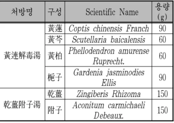
1. 黃連解毒湯, 乾薑附子湯 추출물 제조 본 실험에 사용한 黃連解毒湯과 乾薑附子湯의 처방 약물들은 경북 안동시 Omni herb 회사에서 구매하였다. 黃連解毒湯은 ≪外臺秘要≫, 乾薑附子 湯은 ≪傷寒論≫에 수록된 1濟의 기본 구성 배합 에 따라 아래의 표(Table 1)와 같이 총 용량 300g 씩 배합한 후 2 L의 증류수를 함께 100°C에서 1시 간 동안 추출한 후, 30분 동안 열을 식힌 후, Whatman #2 filter paper에 여과한 후, 3일 동안 동결건조를 하였다. 추출물은 감압 농축과 동결건 조를 통해 추출물 51.75g, 41.40g(수득률: 黃連解毒 湯: 17.25%, 乾薑附子湯: 13.80% w/w) 을 얻어 본 연구의 시료로 사용하였다. 실험 전까지는 -20 °C 에 보관을 하였으며, 매 실험 처리 전 증류수에 추 출 시료를 녹인 후 vortex하여 완전히 용해시킨 후
사용하였다.
처방명 구성 Scientific Name 용량 (g)
黃連解毒湯
黃蓮 Coptis chinensis Franch 90 黃芩 Scutellaria baicalensis 60 黃柏 Phellodendron amurense
Ruprecht. 60 梔子 Gardenia jasminodies
Ellis 90
乾薑附子湯
乾薑 Zingiberis Rhizoma 150 附子 Aconitum carmichaeli
Debeaux. 150 Table 1. Composition and Amount of HHT and GBT
2. 실험동물
실험동물은 체중 25-30g 내외의 암컷 ICR mice
(생후 5주)를 Hamamatsu, Japan으로부터 제공받 아 7일 동안 적응기를 거쳤다. 총 30마리의 mice는 실험기간 동안 물과 고형사료(labanimal, Korea)을 자유롭게 섭취하도록 하였고, 온도 23±2°C, 습도 50±10% 로 조절된 동물실에서 사육되었다. 적응 기가 끝난 후 mice 완전임의 배치로 실험군당 5마 리씩 분류하여 5마리씩 6군으로 나누었다(Table 2). 정상군 (Normal group), 대조군(LPS group), 黃連解毒湯 50 mg/kg(HHT50), 黃連解毒湯 500 mg/kg(HHT500), 乾薑附子湯 100 mg/kg(GBT100), 乾薑附子湯 1000 mg/kg(GBT1000) 으로 분류하였 다. 각 처방군의 투약량은 실제 임상에서 활용되 는 한 첩당 무게(30 g 및 16 g) 및 수율과 쥐의 대사량을 고려하여 저농도와 고농도군의 투약 농 도를 설정하였다.
Experiments groups (n=5) Treatment
Normal group The mice received the vehicle treatments as normal control LPS group
(35mg/kg) The mice received LPS (35 mg/kg) injection intraperitoneally as negative control Hwangryunhaedok-tang50
(HH50)
The mice were orally administrated HHT 50 mg/kg with intraperitoneally injected LPS
Hwangryunhaedok-tang500 (HH500)
The mice were orally administrated HHT 500 mg/kg with intraperitoneally injected LPS
Gungangbuja-tang100 (GB100)
The mice were orally administrated GBT 100 mg/kg with intraperitoneally injected LPS
Gungangbuja-tang1000 (GB1000)
The mice were orally administrated GBT 1000 mg/kg with intraperitoneally injected LPS
Table 2. Experiment Deign of Mice
3. 약물 처리 및 LPS 염증 유발
약물 투여군에는 3일 연속 일정한 시간에 각 약물을 증류수에 용해하여 200ul 경구투여 하였고, Normal군과 LPS군에는 vehicle treatments로서 생 리식염수 200 ㎕ 를 경구투여 하였다. 샘플 처리 3 일째 되는 날, 마지막 샘플 처리 1시간 후 Normal 군을 제외한 나머지군 에서는 LPS(35 mg/kg)를
복강 투여하였다. LPS 처리 6시간 이후 ether로 마취하고 희생시켜서 안와정맥으로부터 채취된 혈 액은 3000 rpm에서 20분간 원심 분리하여 분리된 혈장은 -70°C에서 냉동 보관하여 cytokine 분석에 사용되었고, 간 조직은 적출하여 간에 부착되어 있는 지방이나 근육을 깨끗이 제거한 후 차가운 생리식염수로 세척하여 혈액을 제거한 뒤 여과지
로 물기를 제거하고 -70°C에서 보관하여 염증 매 개물질들 분석에 사용되었다. 모든 동물실험 과정 은 NIH(National Institutes of Health)의 실험동 물관리 규정(Principle of Laboratory Animal Care) 과 경희 대학교의 실험동물 관리와 사용 지침의 규정에 따라 수행하였다(KHUASP(SE)-12-020).
4. Cytokine levels 측정(IL-6 and IFN-γ) 혈장 cytokine 정량용 시료는 채혈 직후, centrifuged (14000×g)에서 30분 동안 원심 분리하여 -80℃에 냉동 보관하였다. Cytokines(IL-6, IFN-γ)의 정량 은 제조업체의 지시에 따라 결정하여 Kit(BD Bioscience, San Jose, CA, USA) 를 사용하여 측 정하였다. Optical density 는 ELISA Reader (Molecular Devices, Downingtown, PA)를 사용하 여 450nm and 570nm에서 측정되었다.
5. Nuclear protein 및 whole protein 추출 Mouse의 lliver 조직은의 5배에 해당되는 cytoplasmic buffer(10mMHEPES(pH 7.9), 10 mM KCl, 0.1 mM EDTA, 0.1 mM EGTA, 1 mM DTT, 0.15% Nonidet P-40, 50 mM β-glycerophosphate, 10 mM NaF and 5 mM Na3VO4 and the protease inhibitor cocktail) 를 넣어 homogenize 하였으며, 15분 간격으로 5번 vortex 한 후 cytosolic 의 상층액을 얻고 남은 pellet에 nuclear lysis buffer(20 mM HEPES, pH 7.9, 400 mM NaCl, 1 mM EDTA, 1 mM EGTA, 1 mM DTT, 0.50%
Nonidet P-40, 50 mM β-glycerophosphate, 10 mM NaF and 5 mM Na3VO4 and the protease inhibitor cocktail)를 넣어 cytosolic lysis 방법과 동일하게 실 시하여 NF-κb의 발현을 알아보기 위해 nuclear protein 을 최종적으로 추출하였다. 또한 whole protein extracts 은 RIPA buffer (50mM Tris-HCl, pH7.4, 1%Nonidet P-40, 0.5 % sodiumdeoxycholate, 150mMNaClandtheproteaseinhibitorcocktail)에 간을 넣어 homogenize 하였으며 15분 간격으로 5번
vortex한 후 상층액을 얻어 i-Nos, Cox-2, and MAPKs(ERK1/2, JNK, p38)의 발현을 확인하였다.
6. Western blot
추출된 단백질은 Protein assay kit(Bio-Rad Laboratories, USA)의 방법으로 정량하여, 각 sample 당 30 μg의 단백질을 실험에 사용하였다. SDS 12%-polyacrylamide겔을 이용 전기영동을 통해 단 백질을 분리한 후, 분리한 단백질은 nitrocellulose 막으로 이동시켜, antibody의 비 특이적인 결합을 막기 위해 TBS-T로 용해한 5% skim milk를 이 용하여 blocking하고, TBS-T로 15분간 3번 세척 후, anti-rabbit β-actin, NF-κB, i-Nos, Cox-2, Erk1/2, Jnk, p38 MAPKs, phospho-Erk, phospho-Jnk, and phospho-p38 MAPK antibody(TBS-T에 1:1,000, Cell signaling, USA)와 이에 대한 goat-anti-rabbit 2차 항체(1:2,000, Santa cruz biotechnology Inc, USA)를 처리하였다. ECL kit의 solution을 1:1로 섞어 NC filter에 부어 1분간 흔들어 준 다음, LAS Image Gauge 프로그램을 이용하여 현상하였 다. NF-κB, i-Nos, Cox-2 밴드의 밀도를 측정은 anti-mouse β-actin 항체밴드를 대조군으로 하여 정량하였고, MAPKs phospho의 밀도 측정은 그에 상응하는 각각의 대조군으로 정량하였다.
7. 통계처리
각 data는 Mean ± SD 값으로 표시 하였으며, 각 실험 결과로부터 ANOVA(analysis of variance) 를 구한 후 Duncan’s multiple range test를 이용 하여 각 군의 평균 간의 유의성을 검정하였다. 일 반적으로 p 값이 0.005 이하인 경우 통계학적으로 유의 한 것으로 간주하였다.
Ⅲ. 결 과
1. Serum 에서의 IL-6 와 IFN-γ의 발현
LPS 대조군에서는 정상군에 비해 유의적으로 IL-6와 IFN-γ의 level이 증가됨을 확인하였다.
IL-6 발현에서 黃連解毒湯은 두 농도(50, 500 mg/kg) 모두 유의적인 감소를 보였으나, 乾薑附子湯에서
는 농도의전적인 경향은 나타나지 않았다(Fig. 1A).
IFN-γ 발현은 黃連解毒湯을 처리한 군에서 유의 적인 감소를 보였으나, 乾薑附子湯에서는 유의적 인 결과를 확인 할 수 없었다(Fig. 1B).
Fig. 1. Effects of HHT and GBT pretreatment on cytokine concentrations in serum.
(A) IL-6 (B) IFN- γ. The results are presented as the mean ± SEM.(n=5). ### indicates significance for the difference between normal control group and LPS group(### P < 0.001). * indicates significantly difference from LPS group(*** P < 0.001,
** P < 0.01).
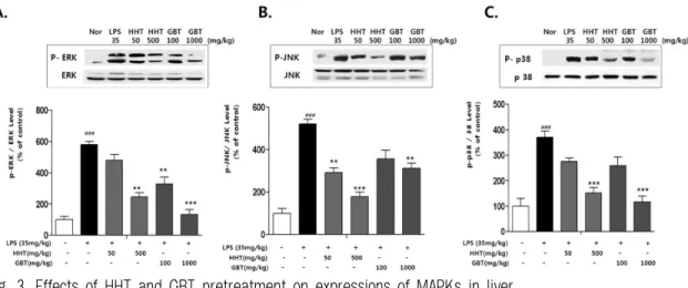
2. Liver 에서의 NF-κb, MAPKs level
LPS로 유도된 염증 모델에서 NF-κb와 MAPKs 는 전염증 매개물질들을 유도하는 중요한 역할을 하는 것으로 보고되어 있다8). 본 연구에서도 NF- κb와 MAPKs level 모두 정상군에 비해 LPS군은 과발현 됨을 확인 하였다. 반면 LPS에서 유도된 Nf-κb의 발현은 黃連解毒湯 고농도(500 mg/kg)와 乾薑附子湯 (100,1000mg/kg)에서 유의적임 감소를 확인하였다(Fig. 2). 또한 ERK1/2의 발현은 黃連 解毒湯 저농도(50 mg/kg)를 제외한 모든 군에서 유의적인 감소를 보였으며, JNK는 黃連解毒湯이 乾薑附子湯보다 유의적인 항염증효능 효과를 보였고, p38에서는 黃連解毒湯과 乾薑附子湯의 고농도(500, 1000 mg/kg)에서 유의적인 감소를 보였다(Fig. 3).
Fig. 2. Effects of HHT and GBT pretreatment on expressions of NF-κb in liver.
(A) NF-κB. ### indicates significance for the difference between normal control group and LPS group(### p < 0.001).
* indicates significantly difference from LPS group(*** p
< 0.001, ** p < 0.01).
Fig. 3. Effects of HHT and GBT pretreatment on expressions of MAPKs in liver.
(A) ERK 1/2 (B) JNK (C) p38 MAPKs. ### indicates significance for the difference between normal control group and LPS group(### p < 0.001). * indicates significantly difference from LPS group(*** p < 0.001, ** p < 0.01).
3. Liver 에서의 i-Nos와 Cox-2 발현
i-Nos와 Cox-2 단백질의 발현 억제와 NF-κb의 관련성을 확인하기 위하여 Western blot을 이용하여 단백질의 발현 정도를 비교하였다. 실험결과 LPS 군에서 i-Nos 와 Cox-2 유의적인 과발현을 보였다.
반면 乾薑附子湯을 처리한 군에서는 i-Nos와 Cox-2 모두에서 농도 의존적인 항염증 효과를 보이고 있 으며, 黃連解毒湯에서는 i-Nos의 억제는 확인할 수 있었으나, Cox-2 의 유의적인 감소는 확인할 수 없었다(Fig. 4).
Fig. 4. Effects of HHT and GBT pretreatment on expressions of I-NOS and COX-2 in liver.
(A) i-Nos (B) Cox2. ### indicates significance for the difference between normal control group and LPS group(### p <
0.001). * indicates significantly difference from LPS group(***p < 0.001, **p < 0.01).
Ⅳ. 고 찰
본 연구는 한, 열 처방의 효능 차이를 평가하기 위한 기초연구로, 黃連解毒湯과 乾薑附子湯의 항 염증 효능 및 각각의 효능 기전 차이에 대하여 알 아보고자 LPS로 급성 염증반응을 유발시켜 혈액 및 간장의 전염증성 cytokines들 및 protein 인자 들에 대하여 실험하였다.
IL-6는 monocytes/macrophages 및 간장의 Kupper cell에서 생산되는 중요한 전염증성 cytokine으로, 염증반응의 경우 급격하게 증가한다15). IFN-γ는 macrophage activating factor로 작용하며 LPS의 shock에 의해 Kuffer cell로부터 방출되어, 간장에 상처를 주고 간세포의 사멸을 일으키며, IFN-γ의 과잉 생산은 광범위의 병적상태를 유발한다. 따라 서 생체 내에서 효율적으로 IFN-γ의 생산을 억제 하는 방법에 대해 연구가 진행되고 있다16). 본 연 구에서 LPS 처리 6h 후 serum 에서의 IL-6 과발 현을 확인하였고, 黃連解毒湯을 처리한 군은 乾薑 附子湯보다 더 월등한 IL-6의 농도 감소를 보였 다. 특히 IFN-γ 농도는 黃連解毒湯군에서 LPS 대조군보다 유의적인 감소를 나타내었으나, 乾薑 附子湯군에서는 IFN-γ의 유의적인 감소를 확인하 지 못했다. 이러한 결과는 IL-6 농도의 감소 결과와 부합되며, 黃連解毒湯이 乾薑附子湯에 비해 유의성 있는 항염증 cytokine 조절 기능을 나타내었다.
NF-κb는 전사인자(transcriptional factor)로서, 염증반응과 면역 반응 등 다양한 유전자의 발현에 관여하여 종양형성, 자가 면역 질환, 염증 질환에 중요한 역할을 담당한다17,18). 본 실험에서 LPS 유 도된 그룹에서는 정상군에 비해 증가됨을 확인하 였고, 乾薑附子湯군은 NF-κb를 유의성 있게 감소 시켰rj, 黃連解毒湯의 경우 고농도(500 mg/kg)에 서만 유의적인 감소를 확인하였다.
MAP Kinases는 ERK1/2, JNK, p38 등 3가지 의 subtype으로 구성되어 있다. MAPKs의 신호경 로들은 주로 인산화 되지 않은 불활성 상태에서는
세포질에 머물다가 LPS 혹은 다른 염증인자로 유 도되어 인산화가 되어 활성화되면 핵으로 이동하 면서 i-Nos 및 전염증성 세포활성물질 발현에 관
여한다19-21). 乾薑附子湯과 黃連解毒湯 투여군 모두
LPS 투여군에 비해 MAPKs 발현을 유의성 있게 감소시켰으며, 특히 乾薑附子湯의 경우 黃連解毒 湯보다 현저한 ERK의 감소를 나타내었다.
Cox-2 및 i-Nos는 LPS에 유도된 NF-κb에 의 해 활성화 되는 대표적인 염증 유전자들에 속하 며, 이들은 이차적인 염증반응을 세포내에서 일으 키는 것으로 알려져 있다22,23). Cox는 arachidonic acid를 PG로 전환하는 효소로 혈소판 형성, 위벽 보호 및 신장 기능의 유지 등 정상 적인 생리기능 에 작용하는 Cox-1과 염증매개물질인 PGE2를 생 성하는 Cox-2로 분류된다24). 다수의 항염증제의 작용기전은 PG 합성을 억제하는 것인데 이것은 Cox-2 의 활성 저해에 의한 것이다. i-Nos는 LPS 자극에 의해 대식세포, 간세포, 심근세포 등에서 오랜 시간 동안 많은 양의 NO를 생성하는 것으로 알려져 있다25). 특히 kupper cell은 LPS 의해 i-Nos 유전자가 특징적으로 활성화 되어 i-Nos의 발현을 유도한다고 보고되고 있다26). 본 실험에서, 乾薑附子湯은 Cox-2 와 i-Nos에서 LPS 투여군에 비해 유의성 있게 발현을 감소시켰다. 黃連解毒湯 의 경우 i-Nos는 유의성 있게 감소시켰으나, Cox-2 발현에는 영향을 미치지 않았다. NF-κb와 그에 따라 활성된 i-Nos, Cox-2 억제를 종합적으로 고 찰해보면, 乾薑附子湯이 黃連解毒湯보다 더 유의 적인 감소결과를 나타내고 있다. 특히 乾薑附子湯 의 경우, NF-κb에 의한 i-Nos 및 Cox-2 발현 기 전에 영향을 미치는 것으로 유추해볼 수 있다. 이 상의 결과들을 종합해보면 黃連解毒湯과 乾薑附子 湯 모두 염증반응을 완화하는 효과를 나타내었다.
하지만 그 작용기전에 있어, 黃連解毒湯은 주로 항염증 cytokine 조절기능에 관여하고 있으며, 乾 薑附子湯 추출물에 의한 항염증 효과는 앞에서 서 술한 염증 반응의 신호 전달 체계 중 NF-κB 경
로를 차단함으로써 i-Nos와 Cox-2 의 발현의 조절 을 통해 이루어지는 것을 확인할 수 있었다.
Ⅴ. 요 약
본 연구를 통해서, 寒 처방의 대표 처방인 黃連 解毒湯과 熱 처방의 대표 처방인 乾薑附子湯 모두 항염 효능을 확인할 수 있었으나, 그 작용 기전에 있어 뚜렷한 차이를 나타내었다. 이러한 차이는 한의학의 기본 이론인 한열에 대한 개념에 대한 연구의 초석이 될 수 있기를 바라며, 각 개별 약 물의 효능 및 다른 처방들과 다른 기전적 실험이 추가적으로 필요할 것을 보인다.
감사의 글
본 연구는 한국한의학연구원 주요사업 “한의 이 론 과학화 사업 (K12274)"의 지원을 받아 수행됨.
참고문헌
1. 두호경, 박헌재. 황련해독탕의 약리학적 연구.
경희한의대논문집. 1982;5:103-14.
2. Kang H, Bang TS, Lee JW, Lew JH, Eom SH, Lee K,Choi HY. Protective effect of the methanol extract from Cryptotaeniae japonica Hassk against lipopolysaccharide-induced inflammation in vitro and in vivo. BMC Complement Altern Med. 2012;12:199.
3. Feng YB, W. Q. Luo and S. Q. Zhu, “Explore new clinical application of Huang Lian and corresponding compound prescriptions from their traditional use,” Zhongguo Zhong Yao ZaZhi.
2007;10:1221-5.
4. Srivastava KC, Mustafa T. Ginger(Zingiberofficinale) and rheumatic disorders. Medical Hypotheses.
1989;29:25-8.
5. Grzanna R, Lindmark L, Frondoza CG. Ginger an herbal medicinal product with broad anti- inflammatory actions. Journal of Medicinal Food. 2005;8:125-32.
6. Mascolo N, Jain R, Jain SC, Capasso F.
Ethnopharmacologic investigation of ginger (Zingiberofficinale). Journal of ethnopharmacology.
1989;27:129-40.
7. Sung YS, Choi HJ, Oh JM, Jee JG. The Effect of KKBT in Papain-Induced Osteoarthritis Mice Models. The Journal of Daejeon Oriental Medicine. 2012;21(1):33-52.
8. 채인식. 傷寒論譯詮. 서울:高文社. 1991:334-5.
9. Kim JH, Lee WC. A study on a paradigm of Radix Aconiti(附子) in the treatment of heart -systemic disease(心系疾患) through 'Sanghanron' (傷寒論). Journal of korean oriental internal medicine. 1999;20(1):9-32.
10. Johnson, Latoya N. Koval, Michael. Cross-talk between pulmonary injury, oxidant stress, and gap junctional communication. Antioxidants
& Redox Signaling. 2009;2:355-68.
11. van Deuren M, Dofferhoff AS, van der Meer JW. Cytokines and the response to infection.
The Journal of Pathology. 1992;168:349-56.
12. Lin BR, Yu CJ, Chen WC, Lee HS, Chang HM, Lee YC, Chien CT, Chen CF. Green tea extract supplement reduces D-galactosamine-induced acute liver injury by inhibition of apoptotic and proinflammatory signaling. Journal of Biomedical Science. 2009;25:16-35.
13. Mestre JR, Mackrell PJ, Rivadeneira DE, Stapleton PP, Tanabe T, Daly JM. Redundancy in the signaling pathways and promoter elements regulating cyclooxygenase-2 gene expression in endotoxin-treated macrophage/monocytic cells.
Journal of Biological Chemistry. 2001;276:3977-82.
14. Israf DA, Khaizurin TA, Syahida A, Lajis NH, Khozirah S. Cardamonin inhibits COXand iNOS expression via inhibition of p65 NF-κb nuclear translocation and Ik-B phosphorylation in RAW264.7 macrophage cells. Molecular Immunology.
2007;44:673-9.
15. Eduard, F.M., S.M.R. Martha, P.A. Victor and M. Pablo. Immunomodulatory effects of thalidomide analogs on LPSinduced plasma and hepatic cytokines in the rat. Biochemical pharmacology.
2004;68:1321-9.
16. Harbrecht BG, Di Silvio M, Demetris AJ, Simmons RL, Billiar TR. Tumor necrosis factor-α regulates in vivo nitric oxide synthesis and induces liver injury during endotoxemia.
Hepatology. 1994;20:1055-60.
17. Gill YG, Lee JH, Choi BT. The Serum and Immuno histochemical Analysis on the Anti-infammatory Effect of Aqueous Extract from Artemisia capillaris in the Liver of Lipopolysaccharide-injected Rat. Journal of life science. 2004;14:215-20.
18. Israf DA, Khaizurin TA, Syahida A, Lajis NH, Khozirah S. Cardamonin inhibits COXand iNOS expression via inhibition of p65NF-jB nuclear translocation and Ij-Bphosphorylation in RAW264.7 macrophage cells. Mol Immunol.
2007;44:673-9.
19. Israf DA, Khaizurin TA, Syahida A, Lajis NH, Khozirah S. Evidence for nucleotide receptor modulation of cross talk between MAP kinase and NF-kappa B signaling pathways in murine RAW264.7 macrophages. Am. J. Physiol. Cell Physiol. 286(4):C923-30.
20. Xia Z, Dickens M, Raingeaud J, Davis RJ, Greenberg ME. Opposing effects of ERK and
JNK-p38 MAP kinases on apoptosis. Science.
Science. 1995;24:1326-31.
21. DeFranco AL, Hambleton J, McMahon M, Weinstein SL. Examination of the role of MAP kinase in the response of macrophages to lipopolysaccharide. Progress in clinical and biological research. 1995;392:407-20.
22. Park HJ, Kim IT, Won JH, Jeong SH, Park EY, Nam JH, Choi J, Lee KT. Anti-inflammatory activities of ent-16alphaH,17-hydroxy-kauran-19-oic acid isolated from the roots of Siegesbeckia pubescens are due to the inhibition of iNOS and COX-2 expression in RAW 264.7 macrophages via NF-kappaB inactivation. European Journal of Pharmacology. 2007;558:185-93.
23. Qureshi AA, Tan X, Reis JC, Badr MZ, Papasian CJ, Morrison DC, Qureshi N.Suppression of nitric oxide induction and pro-inflammatory cytokines by novel proteasome inhibitors in various experimental models. Lipids in Health and Disease. 2011;10:177.
24. Lau FC, JosephJ A, McDonaldJ E. Attenuation of iNOS and COX2 by blueberry polyphenols is mediated through the suppression of NF-κ B activation. Journal of functional foods : the official journal of the International Society for Nutraceuticals & Functional Food. 2009;1:274-83.
25. Jin M, Lee HJ, Ryu JH, Chung KS. Inhibition of LPS-induced NO production and NF-kappaB activation by a sesquiterpene from Saussurea lappa. Arch Pharm Res. 2000;2:54-8.
26. Vodovotz Y, Prelich J, Lagoa C, Barclay D, Zamora R, Murase N, Gandhi CR. Augmenter of Liver Regeneration(ALR) is a Novel Biomarker of Hepatocellular Stress/Inflammation: In Vitro, In Vivo, and In Silico Studies. Journal of Molecular Medicine. 2012.