37
<원례보저>
돼지 지방 조직 및 골수 유래 성체줄기세포의 성상분석과 다능성에 관한 연구
이아영·최경임·나진주·소병재·이경우·장기윤·송재영·차상호*
농림수산검역검사본부
(접수: 2012년 9월 14일, 수정: 2013년 1월 3일, 게재승인: 2013년 1월 3일)
Characterization of multipotent mesenchymal stem cells isolated from adipose tissue and bone marrow in pigs
Ah-Young Lee, Gyeong-im Choe, Jin-Ju Nah, ByungJae So, Kyung-Woo Lee, Ki-Yoon Chang, Jae-Young Song, Sang-Ho Cha*
Animal, Plant and Fisheries Quarantine and Inspection Agency, Anyang 430-757, Korea (Received: September 14, 2012; Revised: January 3, 2013; Accepted: January 3, 2013)
Abstract : Mesenchymal stem cells (MSCs) have ability to differentiate into multi-lineage cells, which confer a great promise for regenerative medicine to the cells. The aim of this study was to establish a method for isolation and characterization of adipose tissue-derived MSC (pAD-MSC) and bone marrow-derived MSC (pBM-MSC) in pigs.
Isolated cells from all tissues were positive for CD29, CD44, CD90 and CD105, but negative for hematopoietic stem cell associated markers, CD45. In addition, the cells expressed the transcription factors, such as Oct4, Sox2, and Nanog by RT-PCR. pAD-MSC and pBM-MSC at early passage successfully differentiated into chondrocytes, osteocytes and adipocytes. Collectively, pig AD-MSC and BM-MSC with multipotency were optimized in our study.
Keywords : mesenchymal stem cells, isolation, pig, adipose tissue, bone marrow
서 론
성체줄기세포는 섬유아세포모양으로 부착해서 자라고 골, 지방, 연골세포로 분화하는 능력을 가진 세포이다. 이러한 세 포의 존재는 1970년 골수에서 처음 보고되었으나 [7], 최근 지방을 포함한 뇌, 비장, 간, 신장, 폐, 근육, 흉선, 췌장 등 다양한 조직에 존재한다고 알려져 있다 [6]. 지방 조직은 다 른 조직보다 인체 내에 양이 많고 채취 시 통증이 거의 없 으며 재충전이 가능하다는 장점을 가지고 있다. 또한, 골수 에 비해 지방 조직에는 1 g당 대략 100,000여개의 성체줄기 세포가 존재하기 때문에 [20] 단기간에 대량으로 증식하여 사용될 수 있다. 성체줄기세포는 이미 성장한 신체 조직에서 추출한다는 점에서 윤리적인 논쟁을 피할 수 있으므로 실용 적인 면에서 배아줄기세포보다 성체줄기세포 연구가 주를 이 루고 있다.
최근 생체조직공학 및 재생의학을 이용한 줄기세포 치료 방법에 관한 연구들이 활발히 이루어 지고 있다. 조직 일부 를 채취하여 필요로 하는 특정 조직으로 분화시켜 이를 이
식하는 연구 분야는 종교 및 윤리적인 부분에 제한을 받지 않고 면역거부반응과 같은 문제점을 해결할 수 있기 때문에 조직공학 분야에 활용도가 높다 [4]. 또한, 줄기세포를 원하 는 특정 세포로 분화시킬 수 있다면, 손상된 세포 및 조직의 기능을 근본적으로 재생(regeneration) 가능하다는 점에서 심 근경색, 뇌경색, 퇴행성 관절염, 골절 및 피부 등 전반적인 의학영역에서 세포치료제로서 이용 가능하며 줄기세포를 각 종 세포 및 조직으로 분화시켜 신약 후보물질의 독성 및 효 능을 빠르게 검증할 수 있기 때문에 새로운 항암제 및 진단 기술에 활용할 수 있다. 하지만, 정확한 분화 기전 및 기능 조절에 관한 기초 연구와 줄기세포의 고효율 분리, 대량 증 식, 면역거부반응, 종양형성 등 극복해야 할 과제가 남아있다.
돼지는 생명의학연구에 중요한 동물 모델로서 본 연구는 돼지의 지방, 골수에서 성체줄기세포를 분리 및 배양법을 확 립하고 골세포, 지방세포, 연골세포로 분화시켜 조직 유래 성 체줄기세포의 성상을 비교하고 분화능을 관찰하는 것에 목 적이 있다.
*Corresponding author
Tel: +82-31-463-4574, Fax: +82-31-463-4565 E-mail: [email protected]
재료 및 방법
세포 분리 및 배양
24~26개월령 모돈 유래 복부 피하 지방, 3~5일령 신생자 돈 대퇴골로부터 골수 시료를 채취하였다. 100 U/mL penicillin과 100 µg/mL streptomycin이 함유된 dulbesco’s phosphate buffered saline(DPBS; Gibco, USA)로 수 차례 세척 한 지방 조직을 가위로 잘게 자른 후, 0.1% collagenase type I solution(Gibco)을 37oC에서 1시간 반응시켰다 [14, 22]. 300× g, 5분 원심분리 후 상층액을 제거하고 DPBS를 넣고 다시 원심분리 하였다. 상층액을 제거하고 10% MSC fetal bovine serum(FBS; Gibco), 100 U/mL penicillin, 100 µg/mL streptomycin, glutamine 포함된 DMEM/F12 (Gibco) 배지를 넣고 골고루 섞은 후 100 mm dish에 분주 하여 37oC, 5% CO2, 20% O2 조건에서 배양하였다. 대퇴 골 절반을 절단 후, 총 30 mL DPBS로 골수를 채취하였다.
Ficoll(GE Healthcare, Sweden) 3 mL당 골수액 4 mL씩 분 주하여 400× g, 30분 원심분리하고 중간의 세포층을 분리하 였다 [2, 16]. DPBS를 넣고 300× g, 10분 원심분리 하여 상등액 제거 후 지방 조직과 동일한 조건으로 세포를 배양 하였다. 이 시기에는 24시간 마다 배지를 교체하며 세포가 70~80% 밀집하면 0.25% trypsin-EDTA(Gibco)을 처리하여 계대배양하였다.
세포 증식률 분석(cumulative population doubling level analysis, CPDL)
다음과 같은 공식을 이용하여 세포 증식률을 확인하였다.
CPDL = ln (Nf/Ni) ln2
Ni는 초기 세포 수, Nf는 마지막 세포 수, ln는 자연로그 를 나타내며 6-well plate에 세포를(5 × 10⁴) 세배수로 배양하 고 5일이 지나면 세포 수를 측정한 후, 다시 세포를(5 × 10⁴) 배양하여 10계대까지 반복, 계산하였다 [23].
Reverse transcription polymerase chain reaction (RT-PCR)을 이용한 초기전사표지인자 Oct4, Sox2, Nanog 발현 검사
지방 조직, 골수 유래 세포의 초기전사표지인자 Oct4, Sox2, Nanog의 발현을 확인하기 위해 RNeasy Mini kit (QIAGEN, Germany)를 이용하여 3계대 배양한 세포의 total RNA를 추출하고, cDNA를 합성하기 위해 GoScript Reverse Transcription System(Promega, USA)을 사용하였다. PCR은 GoTaq DNA Polymerase(Promega)를 이용하였다. 실험에 사용한 primer는 Table 1에 제시하였다. PCR은 pre- denaturation 95oC에서 5분, denaturation 72oC에서 30초, annealing 30초, extension 72oC에서 30초, final-extension 72oC에서 5분, 이 과정을 40 cycle 반복하였고 PCR product 를 2% agarose gel에 전기영동으로 분리한 후, 사진 촬영하 였다.
면역염색법(immunostaining)을 이용한 초기전사표지인자 Oct4, Sox2, Nanog 발현 검사
4% paraformaldehyde로 상온에서 15분 세포를 고정시켰다.
고정된 세포에 0.2% PBS-triton X-100을 상온에서 30분 처 리하여 permeabilization 시킨 후, 5% BSA로 상온에서 1시 간 blocking하였다. Primary antibody는 모두 1 : 100으로 4oC에서 24시간 처리하였고 사용한 antibody 종류는 다음과 같다. Anti-Oct4(Abcam, USA), anti-Nanog(Cell Signaling, USA), anti-SSEA-4(Millipore, USA)를 사용하였다. PBS로 세척 후 secondary antibody를 1 : 400으로 희석하여 상온에 서 1시간 처리하였다. DAPI(Invitrogen, USA)를 상온에서 10분 처리하고 간단히 세척 후, mounting solution을 처리하 고 sealing하여 형광현미경으로 촬영하였다.
유세포 분석(flow cytometric analysis)을 통한 세포표면 특이마커 발현 검사
0.25% trypsin-EDTA를 처리하여 떼어낸 세포를 PBS로 3 회 세척한 후, 성체줄기세포 표면특이마커의 발현 정도를
Table 1. Primer sequence of pluripotency markers for reverse transcription-polymerase chain reaction
Gene Primer sequence (5´-3´) PCR product size (bp) Annealing Tm (oC) GeneBank accession No.
Oct4 F-AGGTGTTCAGCCAAACGACC
335 58 NM_001113060.1
R-TGATCGTTTGCCCTTCTGGC
Sox2 F-GCCCTGCAGTACAACTCCAT
216 60 EU503117.1
R-GCTGATCATGTCCCGTAGGT
Nanog F-ATCCAGCTTGTCCCCAAAG
438 60 DQ447201.1
R-ATTTCATTCGCTGGTTCTGG
GAPDH F-TCGGAGTGAACGGATTTG
219 58 AMF017079.1
R-CCC GGA AGA TGG TGA TGG GAPDH: Glyceraldehydes-3-phosphate dehydrogenase
Flow cytometer(FACSCalibur; BD Biosciences, USA)를 이용하여 관찰하였다. 이들 항체는 형광물질이 결합된 anti- mouse CD29(BD Biosciences), anti-rat CD44(BD Bio- sciences), anti-mouse CD45(Fitzgerald, USA), anti-mouse CD90(BD Biosciences), anti-mouse CD105(Abcam)를 사용 하였다.
분화 유도 및 염색
분화를 유도하기 위해 각각의 분화 조성에 맞춘 배지에 21일간 배양하였다(Table 2). 이 때, 3일 주기로 분화 배지를 교체하였다.
결 과
돼지의 지방 조직, 골수에서 분리한 세포는 배양 일주일 내에 세포군락(colony)을 형성하였고 5일 주기로 계대 배양
하며 세포 모양을 관찰하였다. 1차 계대 시 지방, 골수 유래 세포는 섬유아세포모양으로 부착해서 자라는 것을 확인하였 고 이러한 모양은 2, 3차 계대까지 지속되었다(Fig. 1). 성체 Fig. 1. (A) Adipose tissue-derived MSC (pAD-MSC), (B) Bone marrow-derived MSC (pBM-MSC). Morphological characteristics of pMSCs at passage 1 (p1). The adherent cells exhibited a typical fibroblast-like morphology. ×40, Scale bars
= 100µm.
Table 2. Differentiation induction media
Adipogenesis Osteogenesis Chondrogenesis
Dexamethasone 100 nM Dexamethasone 10 nM Dexamethasone 100 nM
Insulin 500 ng/mL Ascorbic acid 2-phosphate 200 µM Ascorbic acid 2-phosphate 50 µg/mL
Indomethacin 100 µM β-glycerophosphate 100 mM TGF-β1 10 ng/mL
Isobutyl methyl xanthine 500 µM
Fig. 2. (A) Expression of pluripotent transcriptional marker, Oct4, Sox2 and Nanog in pAD-MSC and pBM-MSC at passage 3 by RT-PCR. GAPDH was used as a housekeeping gene. (B) Specific immuno-reactivity for Oct4, Nanog (nucleus), and SSEA-4 (cytoplasm) was observed in pMSCs. DAPI: 4’,6-diamidino-2-phenylindole, FITC: fluorescein isothiocyanate, GAPDH:
glyceraldehydes-3-phosphate dehydrogenase, RT-PCR: reverse transcription polymerase chain reaction. ×600.
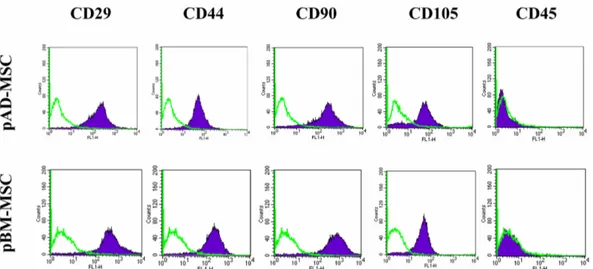
줄기세포는 미분화 상태에서 자가재생을 통해 증식할 수 있 는 능력을 가진 세포로서 이를 위해서는 다양한 특정 유전 자가 역할을 한다 [1, 9, 15, 21]. 그 중, 대표적인 초기전사 인자인 Oct4, Sox2, Nanog의 발현을 확인하기 위해 3계대 세포의 RNA를 추출하여 RT-PCR을 통해 두 가지 세포 모 두 이들 유전자의 발현을 확인하였다(Fig. 2A). 또한, 면역염 색법을 통해 핵 안에 위치한 Oct4, Nanog와 세포질에 위치 한 SSEA-4의 발현을 관찰하였다(Fig. 2B). 표면특이마커의 발현을 확인하기 위해 유세포 분석기를 통해 CD29, CD44, CD90, CD105 그리고 CD45 발현 유무를 관찰하였다. 성체
줄기세포 마커인 CD29, CD44, CD90, CD105는 발현되었 지만, 조혈줄기세포 마커인 CD45는 발현되지 않았다(Fig. 3).
세포 증식률 확인을 위해 5일 주기로 3계대부터 10계대까 지 배양하여 지방, 골수 유래 성체줄기세포의 증식률을 조사 하였다(Fig. 4). 전체적으로 지방 유래 줄기세포가 골수 유래 Fig. 3. Expression of cluster of differentiation (CD) markers in pAD-MSC and pBM-MSC at passage 3. Each sample was counted with 10,000 cells by a flow cytometer. Two kinds of cells were stained with each specific CD marker (filled histograms) and isotype matched controls (open green line). Expression level of markers represents the mean percentage of all assessed cells stained by the antibodies, respectively.
Fig. 4. pAD-MSC and pBM-MSC were seeded at 5 × 10⁴ per well, counted and passaged every 5 days. Proliferative capacities of pMSCs were expressed as cumulative population doubling level (CPDL) using the formula CPDL = ln(Nf/
Ni)ln2, where Ni and Nf are initial and final cell numbers, respectively, and ln is the natural log.
Fig. 5. Differentiation potential of pMSCs. After induction for 21 days, adipogenesis (A and B), chondrogenesis (C and D) and osteogenesis (E and F) were demonstrated by Oil red O, Alcian blue staining and Alizarin red staining, respectively.
×200, Scale bars = 100µm.
줄기세포보다 증식이 빠르다는 것을 확인할 수 있었고 골수 유래 줄기세포는 6계대 이후부터 증식률이 떨어지는 반면, 지방유래 줄기세포는 10계대까지 증식이 지속되는 것으로 보 아 골수 유래 줄기세포보다 지방유래 줄기세포의 증식률이 월등히 우수하다는 것을 확인할 수 있었다(Fig. 4).
분화는 지방, 골수 및 연골 조직으로 유도하였다. 분화배 지에서 21일간 배양 후, 각각의 분화 염색 시약으로 분화 여부를 확인하였다. 분화를 유도한 지방, 골수 유래 성체줄 기세포를 Oil red O를 이용하여 지방세포가 붉게 염색되는 것을 관찰하였고(Figs. 5A and B) 연골 조직으로 유도한 실 험은 Alcian blue로 염색하여 분화 양성을 확인하였다(Figs.
5C and D). 또한, 골 조직으로 분화한 실험은 Alizarin red 염색을 통해 정상적으로 분화하는 것을 확인하였다(Figs. 5E and F).
고 찰
줄기세포란 자가 재생 능력과 증식력, 광범위한 분화능을 가지는 상대적으로 분화되지 않은 세포이다 [5]. 줄기세포는 분화능에 따라 크게 배아줄기세포, 성체줄기세포로 나눌 수 있다. 배아줄기세포는 배아 포배(blastocysts)의 inner cell mass를 추출한 세포로 배아뿐만 아니라 모든 세포로 분화가 가능하고 성체줄기세포는 이보다 제한적으로 분화될 수 있 으나 다양한 조직에서 추출 가능하기 때문에 실용적인 면에 서 많은 연구가 이루어지고 있다. 초기전사인자로 알려진 Oct4, Sox2, Nanog는 배아줄기세포에서 강하게 발현되는 유 전자로써 자가재생과 여러 유전자의 전사를 조절한다. 또한, 줄기세포의 중요한 특성인 다능성에 관여한다고 알려져 있 기 때문에 이들 유전자를 다분화능 표지인자라고도 일컫는 다 [3, 11]. 본 연구에서는 추출, 분리, 배양한 세포의 Oct4, Sox2, Nanog 발현을 모두 확인할 수 있었고 지방 유래 줄 기세포는 계대 10, 골수 유래 줄기세포는 계대 6까지 증식능 을 보유한 결과를 종합해보면, 이는 본 연구에서 분리한 세 포가 다능성과 자가재생능을 가진 줄기세포임과 동시에 조 직 간 차이를 보임을 시사하고 있다.
성체줄기세포는 일반적으로 조혈모세포의 표식자인 CD45, CD34는 발현하지 않고 반대로 CD29, CD44, CD90, CD105는 발현한다 [8]. 이러한 표식자 발현은 종 간의 차이, 조직 유래 세포 간의 차이가 있고 또한 세포 배양 시 계대 가 후반대로 진행 될수록 그 발현이 현저히 감소한다 [18].
최근 다양하고 새로운 표면특이마커에 의해 줄기세포의 특 성 분석을 규명하는 연구가 활발히 진행되고 있다.
성체줄기세포는 대표적으로 지방세포, 연골세포, 골세포로 분화가 가능하며 분화를 유도하기 위한 분화배지의 조성은 일반적으로 알려져 있다. 분화 시약의 조성은 다양한 조직에 서 유래한 세포에 따라 차이가 있을 수 있다. 지방세포로의 분화는 lipid vacuole을 Oil red O 염색법을 이용하여 확인 하거나 adipocyte protein 2(aP2), peroxisome proliferator activated receptor gamma(PPAR
γ), lipoprotein lipase(LPL)
등 관련 유전자의 발현을 측정하여 확인할 수 있다 [17]. 골 세포의 분화는 calcium 축적과 골기질을 Alizarin red 염색 을 통해 확인하며 osteocalcin과 osteonectin의 발현을 관찰 할 수 있다 [19]. 연골세포는 Alcian blue 염색을 통해 glycosaminoglycan의 축적됨을 알 수 있고 Sox9, Aggrecan, Collagen type II의 유전자 발현 유무로 연골세포로 분화됨을 확인할 수 있다 [13]. 이 밖에 성체줄기세포는 신경세포, 췌 장내분비세포, 간세포, 혈관내피세포 및 심근세포로의 분화 또한 보고되었다 [10]. 또한, 분리된 성체줄기세포는 배지 조 건에 따라 세포의 증식능 및 분화능에 차이를 보인다고 알 려져 있다 [12].
본 연구에서는 돼지의 지방 조직, 골수에서 분리한 세포가 초기전사인자, 표면특이마커의 발현, 초기 계대한 세포의 특 정 배양조건하에서 지방세포, 연골세포, 골세포로 분화함을 확인함으로써 다능성의 특징을 지닌 세포임을 확인하였다.
성체줄기세포를 세포치료제, 재생의학 등 다양한 생명의학 연구에 적용될 경우 그 목적에 따라 원하는 세포로 정확히 분화시켜야 하기 때문에 이에 대한 배양 및 분화 조건에 대 한 추가적인 연구가 필요할 것이다. 이번 연구를 바탕으로 미래의 세포 기반 재생의학적 치료에 도움이 될 것으로 사 료된다.
감사의 글
본 연구는 농림수산검역검사본부 수의과학기술개발 연구사 업 (P-AD20-2010-12-01)에 의해 수행되었습니다.
참고문헌
1. Babaie Y, Herwig R, Greber B, Brink TC, Wruck W, Groth D, Lehrach H, Burdon T, Adjaye J. Analysis of OCT4-dependent transcriptional networks regulating self- renewal and pluripotency in human embryonic stem cells.
Stem Cells 2007, 25, 500-510.
2. Bosch P, Pratt SL, Stice SL. Isolation, characterization, gene modification, and nuclear reprogramming of porcine mesenchymal stem cells. Biol Reprod 2006, 74, 46-57.
3. Carlin R, Davis D, Weiss M, Schultz B, Troyer D.
Expression of early transcription factors Oct-4, Sox-2 and Nanog by porcine umbilical cord (PUC) matrix cells. Reprod Biol Endocrinol 2006, 4, 8.
4. Choi MJ, Byun JH, Kang EJ, Rho GJ, Kim UK, Kim JR, Park BW. Isolation of porcine multipotential skin-derived precursor cells and its multilineage differentiation. J Korean Assoc Oral Maxillofac Surg 2008, 34, 588-593.
5. Czyz J, Wiese C, Rolletschek A, Blyszczuk P, Cross M, Wobus AM. Potential of embryonic and adult stem cells in vitro. Biol Chem 2003, 384, 1391-1409.
6. da Silva Meirelles L, Chagastelles PC, Nardi NB.
Mesenchymal stem cells reside in virtually all post-natal organs and tissues. J Cell Sci 2006, 119 (Pt 11), 2204-2213.
7. Friedenstein AJ, Chailakhjan RK, Lalykina KS. The development of fibroblast colonies in monolayer cultures of
guinea-pig bone marrow and spleen cells. Cell Tissue Kinet 1970, 3, 393-403.
8. Haynesworth SE, Baber MA, Caplan AI. Cell surface antigens on human marrow-derived mesenchymal cells are detected by monoclonal antibodies. Bone 1992, 13, 69-80.
9. Jaenisch R, Young R. Stem cells, the molecular circuitry of pluripotency and nuclear reprogramming. Cell 2008, 132, 567-582.
10. Jung JS. Adipose tissue derived mesenchymal stem cells. J Korean Soc Transplant 2008, 22, 183-196.
11. Kajahn J, Gorjup E, Tiede S, von Briesen H, Paus R, Kruse C, Danner S. Skin-derived human adult stem cells surprisingly share many features with human pancreatic stem cells. Eur J Cell Biol 2008, 87, 39-46.
12. Lee RH, Kim B, Choi I, Kim H, Choi HS, Suh K, Bae YC, Jung JS. Characterization and expression analysis of mesenchymal stem cells from human bone marrow and adipose tissue. Cell Physiol Biochem 2004, 14, 311-324.
13. Mackay AM, Beck SC, Murphy JM, Barry FP, Chichester CO, Pittenger MF. Chondrogenic differentiation of cultured human mesenchymal stem cells from marrow. Tissue Eng 1998, 4, 415-428.
14. Monaco E, Bionaz M, Hollister SJ, Wheeler MB. Strategies for regeneration of the bone using porcine adult adipose- derived mesenchymal stem cells. Theriogenology 2011, 75, 1381-1399.
15. Niwa H, Miyazaki J, Smith AG. Quantitative expression of Oct-3/4 defines differentiation, dedifferentiation or self- renewal of ES cells. Nat Genet 2000, 24, 372-376.
16. Ock SA, Jeon BG, Rho GJ. Comparative characterization of porcine mesenchymal stem cells derived from bone marrow extract and skin tissues. Tissue Eng Part C Methods 2010, 16,
1481-1491.
17. Pittenger MF, Mackay AM, Beck SC, Jaiswal RK, Douglas R, Mosca JD, Moorman MA, Simonetti DW, Craig S, Marshak DR. Multilineage potential of adult human mesenchymal stem cells. Science 1999, 284, 143-147.
18. Rehman J, Traktuev D, Li J, Merfeld-Clauss S, Temm- Grove CJ, Bovenkerk JE, Pell CL, Johnstone BH, Considine RV, March KL. Secretion of angiogenic and antiapoptotic factors by human adipose stromal cells.
Circulation 2004, 109, 1292-1298.
19. Rodriguez AM, Elabd C, Amri EZ, Ailhaud G, Dani C.
The human adipose tissue is a source of multipotent stem cells. Biochimie 2005, 87, 125-128.
20. Sen A, Lea-Currie YR, Sujkowska D, Franklin DM, Wilkison WO, Halvorsen YD, Gimble JM. Adipogenic potential of human adipose derived stromal cells from multiple donors is heterogeneous. J Cell Biochem 2001, 81, 312-319.
21. Silva J, Chambers I, Pollard S, Smith A. Nanog promotes transfer of pluripotency after cell fusion. Nature 2006, 441, 997-1001.
22. Song SH, Kumar BM, Kang EJ, Lee YM, Kim TH, Ock SA, Lee SL, Jeon BG, Rho GJ. Characterization of porcine multipotent stem/stromal cells derived from skin, adipose, and ovarian tissues and their differentiation in vitro into putative oocyte-like cells. Stem Cells Dev 2011, 20, 1359-1370.
23. Yang SR, Cho SD, Ahn NS, Jung JW, Park JS, Jo EH, Hwang JW, Kim SH, Lee BH, Kang KS, Lee YS. The role of p38 MAP kinase and c-Jun N-terminal protein kinase signaling in the differentiation and apoptosis of immortalized neural stem cells. Mutat Res 2005, 579, 47-57.