두시 하태독법의 IL-4 활성 조절이 D. farinae 유도 아토피유사피부염 발병 조절에 미치는 효과
안상현1․김재규2․천진홍2,3․김기봉2,3
1세명대학교 한의과대학 해부학교실, 2부산대학교 한의학전문대학원 임상의학부, 3부산대학교한방병원 소아청소년클리닉
Received: January 17, 2017 ∙ Revised: February 8, 2017 ∙ Accepted: February 10, 2017 Corresponding Author: Kibong Kim, KMD, Ph.D.
Department of Pediatrics, Korean Medicine Hospital, Pusan National University 20, Geumo-ro, Mulgeum-eup, Yangsan-si, Gyeongsangnam-do, 50612, Republic of Korea Tel: +82-55-360-5952, Fax: +82-55-360-5952
E-mail: [email protected]
ⓒ The Association of Pediatrics of Korean Medicine. All rights reserved. This is an open-access article distributed under the tenus of the Creative Commons Attribution Non-Commercial License (http://creativecommons.org/licenses/by-nc/3.0/), which permits unrestricted non-commercial use, distribution, and reproduction in any medium, provided the original work is properly cited.
Abstract
The Effect of Douchi Hataedock Treatment for Dermatophagoides Farinae-Induced Atopic Dermatitis-like Skin Lesions by Controlling IL-4 Activity
Ahn Sang Hyun1․Kim Jae Kyu2․Cheon Jin Hong2,3․Kim Ki Bong2,3
1Department of Anatomy, College of Korean Medicine, Semyung University,
2Department of Clinical Medicine, School of Korean Medicine, Pusan National University,
3Department of Pediatrics, Korean Medicine Hospital, Pusan National University
Objectives
Hataedock method is a Korean medical therapy which removes fetal toxin by orally administering herbal decoction to neonates. This study was to observe skin damage and anti-inflammatory effect via regulating IL-4 activity in NC/Nga mice which were induced atopic dermatitis (AD)-like skin lesion by Dermatophagoides (D.) farinae after applying Douchi Hataedock method.
Methods
NC/Nga mice with 3 weeks of gestational age were used. Each 10 mice were allocated to the control group (Ctrl), the AD-induced group (AE), and the group which induced AD after administering Douchi extract (GT).
After 4 weeks from administering Douchi extract to the mice, the primary AD was induced by applying D. farinae extract 6 times per week for 3 weeks and then the secondary AD was induced by the same method after 1 week from the primary AD induction. To identify the skin damage and anti-inflammatory effect, we observed LxR, IL-4, Fc ε receptor, substance P, and NF-κB.
Results
The GT group showed alleviation of skin injury and decrease in capillary angiogenesis. Stratum corneum damage, epithelial cell hyperplasia, lymphocyte infiltration, and capillary distribution relatively decreased in the GT group.
LxR-positive reaction in the GT group were increased by 53% than that of the AE group. IL-4 production, Fc ε receptor activity, and substance P-positive reaction in the GT group were decreased by 82%, 42%, and 82%
respectively compare to those of the AE group. NF-κB-positive reaction in the GT group were decreased by 15%
compare to that of the AE group.
Conclusions
Hataedock method with Douchi extract alleviated AD via reducing inflammatory cytokines secreted at the early stage of AD. Thus, Douchi Hataedock method has a beneficial effect for the prevention and treatment of AD.
Key words: Hataedock, Douchi, Atopic dermatitis, IL-4, Fc ε recetor, Substance P, NF-k
ISSN 1226-8038(Print), 2287-9463(Online), https://doi.org/10.7778/jpkm.2017.31.1.043
특별히 정해진 치료법이 없는 난치성 질환이며, 유전 이나 환경, 약물, 심리적 요인, 면역학적 요인, 피부 장 벽 요인 등의 다양한 발병요인들이 연관되어 발병하는 것으로 알려져 있다1-2).
한의학에서는 이러한 AD의 원인을 태열 (胎熱)로 인식하였다. AD는 태열이 제대로 제거되지 못한 상태 에서 다시 풍습열사 (風濕熱邪)에 감작되어 발병하며, 이는 내인 (內因)과 외인 (外因)의 상호작용으로 인해 발생하는 것으로 이해하였다3). 이는 출산시 대부분의 영유아가 Th2 우세 상태이며, 아직 정상적인 Th1/Th2 balance로 도달하지 못한 상태에서 AD의 병인에 노출 된 결과로 볼 수 있다4).
이러한 태열은 오랫동안 영유아에서 발병하는 질환 들의 주된 원인으로 인식되어 왔다. 태열을 제거하기 위해 출생 직후 하태독법 (下胎毒法)을 시행하였으며5), 한약재를 달인 약물을 부드러운 천이나 비단에 묻혀 입안을 닦아주면서 소량 먹이는 방법이다6). 두시법 (豆 豉法), 감초법 (甘草法), 황련법 (黃連法), 주밀법 (朱蜜 法) 등이 있으며, 이 중 두시는 해표 (解表)시키는 효능 이 있어 염증 증상들을 완화시킨다7-9). 선행연구에 의 하면 발효 콩이 AD 증상 완화와 소양감 억제 효과가 있음을 보고하고 있으며10), Th2 response를 통한 Th1과 Th2 조절 효과11) 및 호산구 기도 염증 억제 효과를 보 고하고 있다8,12). 이러한 내용은 두시의 면역학적 변화 가능성을 제시한다.
AD의 발병은 house dust mite, dermatophagoides pteronissinus의 노출이 protein kinase C (PKC)의 활성을 유도함으로써 시작되며13), 이는 Th2 skewed condition induced cytokine인 IL-4, IL-5, IL-13의 생성 증가에 의 해 야기된다14). 특히, IL-4 생성은 B cell의 IgE분비를 유도하여 비만세포의 탈과립화 (degranulate)를 일으키 며, 동통과 소양증 관련 인자인 substance P 등을 생성 하여 피부손상을 가속화시킨다9).
본 연구는 두시 추출물의 AD 발병 조절 효과를 검증 하기 위해서 Nc/Nga 생쥐에게 먼저 하태독법을 실시한 후 아토피유사피부염을 유발하고 LxR, IL-4, Fc ε re- cetor, substance P, NF-kB을 관찰하였다. 이를 통해 두시 를 이용한 하태독법의 피부 손상 정도와 항염증 효과를 확인하고 AD 치료제로의 가능성을 제시하였다.
서리태 (Glycine max Merr.)를 청호 (Artemisia Apiacea Herba)와 상엽 (Mori Folium)에 발효한 두시 (duochi, fer- mameted Glycine max Merr.; 남영제약영농조합, 무주, 대한민국)의 열수추출물을 얻기 위해 100 g을 파쇄한 후 증류수 1000 ㎖에 넣고 3시간동안 전탕한 후 여과 하였다. 그 여액을 rotary evaporator에서 50 ㎖으로 농 축한 후 동결 건조하여 추출물 (수득률 15%)을 얻었다.
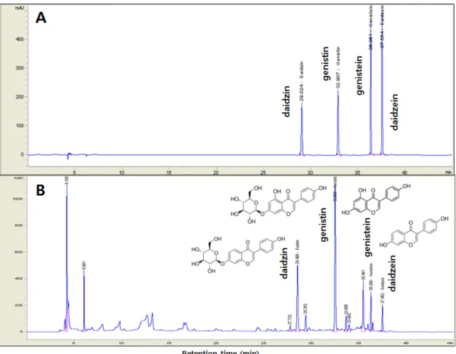
본 실험에 사용된 두시는 HPLC를 통한 isoflavone 성분 결과, genistin, genistiein, daidzine, daidzein 등이 포함 된 것으로 조사되었다 (Fig. 1).
2. Mite 항원 아토피유사피부염 동물모델
실험동물은 중앙실험동물 (서울, 대한민국)에서 분양 받은 태령 3주된 Nc/Nga 수컷 생쥐 (13~15 g)를 사용하 였다. 대조군 (Ctrl군), 아토피 피부염 유발군 (AE군), 두 시 하태독법 시행 후 아토피 피부염 유발군 (GT군)으로 나누었으며, 각 군에 각 10마리씩 배정하였다. 하태독 법은 두시추출물 (20 ㎎/㎏)이 포함된 생리식염수 0.1
㎖을 oral injection needle을 사용하여 매일 1번씩 3일간 경구투여하였다. 하태독법을 실시 4주 경과 후 아토피 유사 피부염을 유발하였다. 생쥐 등 쪽 부위 피부를 면 도한 다음 계면활성제 (surfactant)인 5% sodium dodeecyl sulfate (Sigma, USA) 1 ㎖을 면봉으로 20회 문질러서 각 질층의 lipid lamella를 제거한 후 D. farinae crude extract striper (100 ㎎, Biostir, Japan)을 3주 동안 주 6회씩 도포 하여 아토피피부염을 1차 유발하였다. 1차 유발 후 1주 일 경과 후 동일한 방법으로 2차 유발하였다. 2차 유발 72시간 후 sodium pentobarbital 용액으로 처지한 후 얻 어진 등 쪽 피부를 통상적인 방법을 통해 연속절편을 제작하였다. 본 연구과정은 부산대학교 IACUC 승인을 받아 시행되었으며 (IACUC number: PNU-2015-0924), 실험실 동물의 관리와 사용에 대해서는 NIH 가이드라 인에 따라 시행되었다 (Fig. 2).
3. 조직화학
피부 손상 변화를 관찰하기 위해 Phloxine-tartrazine 염색법을 실시하였다. Mayer's hematoxylin에 5분간 핵 염색한 후 phloxine 용액에 30분간 반응시켰다. 그런 다
음 tartrazine 용액에서 분별 후 관찰하였다.
신경 펩티드를 분비하는 비만세포의 분포와 형태 변화를 조사하기 위해 Luna's method를 실시하였다.
우선 진피 내 비만세포 과립을 aldehyde fuchsin에 30분 간 염색한 후, Weigert's iron hematoxylin과 methyl or- ange 용액에 각각 1분, 5분 동안 대조염색한 후 관찰하 였다
4. 면역조직화학
우선 피부절편을 proteinase K (20 ㎍/㎖)에 5분 동안 proteolysis 과정을 거친 후 blocking serum인 10% nor- mal goat serum에서 2시간 동안 반응시켰다. 그리고 1 차 항체인 goat anti-LxR (1:200, Santa Cruz Biotec, USA), goat anti-IL-4 (1:100, Santa Cruz Biotec), goat an- ti-Fc ε receptor (1:50, Santa Cruz Biotec), goat anti-Fc Fig. 1. The representative chromatograms of isoflavones in the standard solution (A) and Douchi (Glycine max Merr.
fermaneted by Artemisia Apiacea Herba and Mori Folium, B). Each compound was detected with AegisPak-L C18 (4.6 × 150 mm ID, 3 micro pore size) at UV 260 nm. Peak of isoflavones: daidzin, genistin, genistein, daidzein.
Fig. 2. The animal model for estimation of alleviate effects in D. farinae induced atopy like dermatitis by Douchi as Hataedock treatment.
tinylated rabbit anti-goat IgG (1:100, Santa Cruz Biotec) 에 실온에서 24시간 link 하였고, 그런 다음 avidin bio- tin complex kit (Vector Lab, USA)에 1시간동안 실온에 서 반응시켰다. 0.05% 3,3'-diaminobenzidine과 0.01%
HCl이 포함된 0.05M tris-HCl 완충용액 (pH 7.4)에서 발색시킨 후, hematoxylin으로 대조염색하였다.
5. TUNEL assay
Apoptosis 변화를 조사하기 위해 in situ apoptosis de- tection kit (Apoptag, Intergen, USA)를 이용한 TUNEL (terminal deoxynucleotid transferase-mediated dUTP-bio- tin nick-end labelling) 방법을 실시하였다. 먼저 조직 절편을 proteinase K에 5분간 proteolysis 시킨 다음 equi- libration buffer에서 20초간 처리하였다. 그런 다음 strength TdT enzyme (36 ㎕ TdT enzyme : 72 ㎕ re- action buffer)을 처리하여 37 ℃의 humidified chamber 에서 1시간 동안 반응시킨 후 strength stop/wash buffer 에서 10분 동안 처리하였다. Anti-digoxigenin-perox- idase에 1시간 동안 반응시킨 후 DAB를 처리하였다.
Light green으로 대조염색한 후 광학현미경으로 관찰하 였다.
6. 영상처리, 분석과 통계처리
면역조직화학과 TUNEL assay의 결과는 image Pro Plus (Media cybernetics, USA)를 이용한 영상분석을 통
ware (SPSS 20, SPSS Inc., USA)를 통해 이루어졌으며, one-way ANOVA 시행을 통해 유의성 (P<0.05)을 검증 하고 Duncan’s multiple range test로 사후 검증하였다.
Ⅲ. Results
1. 피부 손상 완화 효과
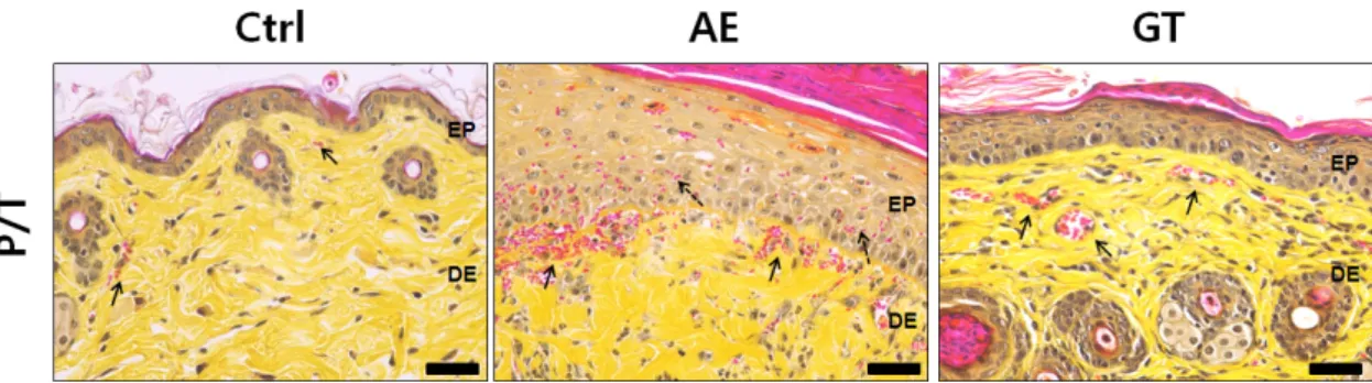
AE군의 피부 대부분의 지역에서 각질층의 탈락, 세 포과형성, 과립백혈구와 림프구의 기저층으로 침윤 증 가가 관찰되었다. 또한 손상이 심했던 상피주변의 진 피 유두에서는 많은 수의 모세혈관이 관찰되었다. 이 에 반해 GT군은 일부 지역을 제외하고는 각질층 탈락, 상피층 증가, 침윤 과립백혈구와 림프구의 출현이 감 소하였다. 진피 유두에서는 모세혈관의 수가 AE군에 비해 적었다 (Fig. 3).
2. 지방장벽 생성 효과
각질내 지방장벽인 ceramide 형성에 관여하는 Liver x receptor (LxR)의 활성을 조사하기위해 LxR 면역조직 화학을 실시하였다. AE군의 각질층에서 LxR 양성반응 은 감소하였는데, GT군에서는 LxR 양성반응이 AE군 에 비해 증가하였는데, 영상분석 결과 AE군에 비해 53
% 증가한 것으로 관찰되었다 (Fig. 4).
Fig. 3. The alleviation of skin damages by Hataedock. The damage of intercellular space of stratum corneum, hyperplasia, edema, infiltration of lymphocytes, and angiogenesis (arrow) were increased in AE, but decreased in GT (Phloxine-tartrazine stain; Bar size, 50 ㎛). Abbreviation: Ctrl, no-AE elicited group; AE, atopy dermatitis (AD) elicitated group; GT, Douchi as fermented Glycine Semen Preparata extract treated group after AD elicitation; EP, epidermis; DE, dermis.
3. IL-4 생성 조절 효과
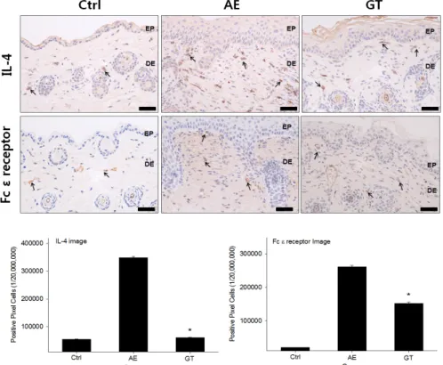
Th2 분화에 관여하는 cytokine인 IL-4의 생성증가는 아토피피부염의 과도한 염증을 유도하는데, AE군에서 는 IL-4 생성 증가가 관찰되었다. GT군에서는 IL-4 생 성이 적었는데, AE군에 비해 82% 감소한 것으로 관찰 되었다 (Fig. 5).
IL-4의 생성증가는 IgE의 분비 증가가 유도되고, 이 에 따른 Fc ε recetor 활성도 연쇄적으로 일러나는데, AE군에서는 Fc ε recetor 활성 증가가 관찰되었다. GT 군에서는 Fc ε recetor 활성이 적었는데, AE군에 비해 42% 감소한 것으로 관찰되었다 (Fig. 5).
Fig. 4. The maintain of lipid barrier by Hataedock (LxR immunohistochemistry). Abbreviation: arrow, LxR positive reaction;
EP, epithelim; DE, dermis; SC, stratum corneum; Bar size, 20 ㎛; *, p<0.05 compared with AE group.
Fig. 5. The mitigative effects of Th2 differentiation by Hataedock (IL-4 & Fc ε recetor immunohistochemistry; Bar size, 100 ㎛). Abbreviation same as Fig. 4.
type)으로 관찰되었다. 이에 반해 GT군에서는 AE군에 비해 적은 수가 관찰되었으며, 주로 과립형이었다.
한편, 진피 유두에서 관찰되는 신경펩타드인 sub- stance P 양성반응은 세포질에서 강하게 나타났으며, AE군에 비해 GT군에서 substance P 양성반응세포가 감 소된 것으로 관찰되었다. GT군에서는 AE군에 비해 82% 감소한 것으로 관찰되었다 (Fig. 6).
5. NF-kB 활성 조절 효과
염증효소 전사인자인 NF-kB 활성은 AE군에서는 증 가한 것으로 관찰되었다. 반면 GT군은 NF-kB p65 양 성반응이 감소했는데, 영상분석 결과는 AE군에 비해 15% 감소한 것으로 관찰되었다 (Fig. 7).
NF-kB의 활성은 apoptosis 유발억제를 유도하는데, GT군에서 높은 TUNEL 양성반응을 보였는데, 영상분 석 결과는 AE군에 비해 211% 증가한 것으로 관찰되었 다 (Fig. 7).
출물을 이용한 하태독법 후 D. farinae 유도 아토피유사 피부염 유발 생쥐에서 피부 손상 정도와 항염증 효과 를 확인하고자 하였다.
먼저 3주령된 어린 Nc/Nga 생쥐에 하태독법을 시행 하고 4주 후 sodium dodeecyl sulfate을 적용하여 ceram- ide 층을 붕괴해 피부장벽기능의 손상을 야기하여 AD 가 유발되기 쉬운 조건을 만들고, D. pteronissinus를 노 출시켜 AD-like skin lesion을 발생시켰다. 이는 이전 선 행연구에서처럼 항원의 도포를 통하여 Th1/Th2 균형 상태를 Th2 우세형 상태로 옮겨 한의학에서 말하는 표 열 (피부 표층의 열) 상태를 유발하기 위함이다15-6). AD 는 Th 불균형 상태의 내인 (內因)과 외부의 AD 발병인 자 (外因)의 상호반응으로 발생하게 되며, 이는 내부의 태독 (胎毒)과 외부의 사기 (邪氣)의 상호반응으로 보 는 한의학적 관점과도 일치한다16).
연구 결과, AE군에서 Ctrl군에 비해 피부 대부분의 지역에서 각질층의 탈락, 세포과형성, 과립백혈구와
Fig. 6. The regulation of itching by Hataedock (luna's stain & substance P immunohistochemistry; Bar size, 100 ㎛).
Abbreviation same as Fig. 4.
림프구의 기저층으로 침윤 증가가 관찰되었고, 각질내 지방장벽인 ceramide 형성에 관여하는 Liver x receptor (LxR)의 활성은 Ctrl군에 비해 감소하였다. 반면에 GT 군에서 피부 손상 완화와 신생모세혈관형성 감소 효과 가 관찰되었으며, 특히 각질층 손상 정도, 상피층 세포 과형성, 림프구 침윤, 모세혈관 분포 증가가 AE군에 비 해 상대적으로 감소하였다 (Fig. 3). 또한, GT군에서 AE군에 비해 각질내 지방장벽인 ceramide 형성에 관여 하는 Liver x receptor (LxR)의 양성반응은 53% 증가하 였다 (Fig. 4). 이러한 결과는 두시를 이용한 하태독법 의 시행이 피부장벽 보호효과가 있음을 보여준다.
두시를 이용한 하태독법의 피부보호 및 항염증효과 를 알아보기 위하여 아토피피부염의 과도한 염증을 유 발하는 IL-4의 생성 변화를 조사하였으며, AE군에서는 IL-4 생성 증가가 관찰되었다. GT군에서는 AE군과 비 교하여 상대적으로 IL-4 생성이 적었으며, AE군에 비 해 82 % 감소한 것으로 관찰되었다 (Fig. 5). IL-4의 생 성증가는 IgE의 분비 증가를 유도하게 되며, 이에 따라 Fc ε recetor 활성도 연쇄적으로 발생하게 된다. AE군에
서는 Fc ε recetor 활성 증가가 관찰되었으며, GT군에서 는 Fc ε recetor 활성이 AE군에 비해 42% 감소한 것으 로 관찰되었다 (Fig. 5). 이러한 결과는 하태독법이 IL-4 의 생성 변화와 Fc ε recetor 활성 변화에 영향을 줄 수 있음을 보여준다.
또한 IL-4의 증가는 비만세포의 탈과립화를 유도하 여 탈과립세포의 침착이 증가하게 된다9). 비만세포의 활성화는 각종 호산구 및 대식세포로부터 각종 proin- flammatory cytokine의 분비를 증가시켜 가려움을 유발 하는 substance P 분비를 증가시킨다. 뿐만 아니라, IL-4 의 증가는 염증반응의 전사인자인 NF-kB를 활성화시 켜 혈관 투과성 증가, 부종 등의 염증반응을 촉진한다
17). AE군는 진피 유두에서 피하 인접부까지 많은 수의 비만세포가 분포하였으며, 주로 탈과립형 (degranulated type)으로 관찰되었다. GT군에서는 AE군에 비해 적은 수가 관찰되었으며, 주로 과립형이었다. 진피 유두에 서 관찰되는 신경펩타드인 substance P 양성반응 역시 AE군에 비해 GT군에서 82% 감소한 것으로 관찰되었 다 (Fig. 6). 염증효소 전사인자인 NF-kB 활성은 AE군
Fig. 7. The regulation of NF-kB activation by Hataedock (Nf-kB p65 immunohistochemistry & TUNEL assay; Bar size, 100㎛). Abbreviation same as Fig. 4.
로 관찰되었다 (Fig. 7). 이러한 결과들은 AE군에 비해 GT군에서 염증발현이 억제되고 있음을 보여주는 것이 며, 이는 두시를 이용한 하태독법이 실제로 항염증효 과가 있음을 의미한다.
결론적으로 두시를 이용한 하태독법의 시행은 외부 의 AD 발병인자로 발생하는 Th2 우세 상태를 억제하 고 염증 발현을 억제시키는 효과를 보여주었다. 또한 LxR의 활성을 통하여 각질내 지방장벽인 ceramide 형 성을 유도하여 피부보호 효과를 보여주었다.
최근 여러 연구들을 통하여 두시나 황련, 감초를 이 용한 하태독법이 Th2 면역 조절 효과를 통하여 태열로 인한 염증성 알러지 질환에 대한 항염증효과와 피부보 호효과를 보고하고 있다9,16-9).
본 연구에서 실시한 생쥐의 하태독법의 적용 시기 는 출생 후 3주였다. 이는 실제 임상에서 시행되는 출 산 직후와는 시간적인 차이가 있다. 실험 모델 설정에 있어서 생쥐의 태령이 3주 이하는 불가하다는 실험적 한계가 있으며, 태령 3주의 생쥐는 인간에서 신생아보 다는 유소아정도의 연령으로 볼 수 있다. 저자들은 이 러한 문제점을 충분히 인지하고 있으며, 그럼에도 불 구하고 하태독법이 Th2 우세 상태 조절에 영향을 미칠 것이라고 생각되어 Th2 우세 상태가 계속 유지되고 있 는 3주령에 하태독법을 실시하였다. 하태독법 시행의 적합한 시기에 대한 부분은 추가적인 연구가 필요하다.
또한, 향후 하태독법의 아토피피부염에 대한 면역조절 과 치료기전을 설명할 수 있는 연구와 사람의 아토피 피부염 치료에 반영하기 위한 안전성 및 유효성 검증 연구가 추가적으로 필요할 것으로 보인다.
Abbreviation: LxR, liver X receptor; IL-4, inter- leukin-4; NF-κB p65, nuclear factor-κB p65; TUNEL, terminal deoxynucleotid transferase-mediated dUTP-bio- tin nick-end labelling; Ctrl, no treatment Nc/Nga mouse;
AE, atopy like dermatitis elicited Nc/Nga mouse; GT, Douchi as fermented Glycine Semen Preparata extract treat- ed group after AD elicitation; *, P<0.05 compared with AE.
하여 연구되었음.
References
1. Leung DY, Bieber T. Atopic dermatitis. Lancet.
2003;361(9352):151-60.
2. Schneider L, Tilles S, Lio P, Boguniewicz M, Beck L, LeBovidge J, Novak N, Bernstein D, Blessing-Moore J, Khan D, Lang D, Nicklas R, Oppenheimer J, Portnoy J, Randolph C, Schuller D, Spector S, Tilles S, Wallace D. Atopic dermatitis: a practice parameter update 2012.
J Allergy Clin Immunol. 2013;131(2):295-9.
3. Im GM, Joeng HW, Kim HS, Jeong WY. Oriental medical approach on the allergic disease. Korean J Orient Physiol Pathol. 2002;16(5):831-9.
4. Halonen M, Lohman IC, Stern DA, Spangenberg A, Anderson D, Mobley S, Ciano K, Peck M, Wright AL.
Th1/Th2 patterns and balance in cytokine production in the parents and infants of a large birth cohort. J Immunol. 2009;182(5):3285-93.
5. Kang MY, Jang GT, Kim JH. A study on fetal toxicosis removal therapy. J Pediatr Korean Med. 2003;17(1):
29-51.
6. Im GM, Jeong HW, Kim HS, Jeong WY. Oriental medical approach on the allergic disease. Korean J Orient Physiol Pathol. 2002;16(5):831-9.
7. Miller AK, Benson JM, Muanza DN, Smith JR, Shepherd DM. Anti-inflammatory effects of natural product for- mulations on murine dendritic cells. J Diet Suppl.
2011;8(1):19-33.
8. Yeh CY, Jung CJ, Huang CN, Huang YC, Lien HT, Wang WB, Wang LF, Chia JS. A legume product fer- mented by Saccharomyces cerevisiae modulates cutaneous atopic dermatitis-like inflammation in mice. BMC Complement Altern Med. 2014;14(1):194.
9. Jung AR, Ahn SH, Park IS, Park SY, Jeong SI, Cheon JH, Kim KB. Douchi (fermented Glycine max Merr.)
alleviates atopic dermatitis-like skin lesions in NC/Nga mice by regulation of PKC and IL-4. BMC Complement Altern Med. 2016;16:416-30.
10. Matsuda A, Tanaka A, Pan W, Okamoto N, Oida K, Kingyo N, Amagai Y, Xia Y, Jang H, Nishikawa S, Kajiwara N, Ahn G, Ohmori K, Matsuda H.
Supplementation of the fermented soy product ImmuBalance™ effectively reduces itching behavior of atopic NC/Tnd mice. J Dermatol Sci. 2012;67(2):130-9.
11. Zhang T, Pan W, Takebe M, Schofield B, Sampson H, Li XM. Therapeutic effects of a fermented soy product on peanut hypersensitivity is associated with modulation of T-helper type 1 and T-helper type 2 responses. Clin Exp Allergy. 2008;38(11):1808-18.
12. Bao ZS, Hong L, Guan Y, Dong XW, Zheng HS, Tan GL, Xie QM. Inhibition of airway inflammation, hyperresponsiveness and remodeling by soy isoflavone in a murine model of allergic asthma. Int Immunopharmacol.
2011;11(8):899-906.
13. Joo KM, Hwang JH, Bae SJ, Nahm DH, Park HS, Ye YM, Lim KM. Relationship of ceramide, and free fatty acid-cholesterol ratios in the stratum corneum with skin barrier function of normal, atopic dermatitis lesional and non-lesional skins. J Dermatol Sci. 2015;77(1):71-4.
14. Jung BG, Cho SJ, Ko JH, Lee BJ. Inhibitory effects of interleukin-10 plasmid DNA on the development
of atopic dermatitis-like skin lesions in NC/Nga mice.
J Vet Sci. 2010;11(3):213-20.
15. Matsuda H, Watanabe N, Geba GP, Sperl J, Tsudzuki M, Hiroi J, Matsumoto M, Ushio H, Saito S, Askenase PW, Ra C. Development of atopic dermatitis-like skin lesion with IgE hyperproduction in NC/Nga mice. Int Immunol. 1997;9(3):461-6.
16. Ahn SH, Kim KB. Anti-inflammatory effects of Hataedock with Douchi in atopic dermatitis-like skin lesions in house dust mite-induced NC/Nga mice. J Pediatr Korean Med. 2016;30(4):77-86.
17. Cha HY, Ahn SH, Cheon JH, Park IS, Kim JT, Kim KB. Hataedock treatment has preventive therapeutic effects in atopic dermatitis-induced NC/Nga mice under high-fat diet conditions. Evid Based Complement Alternat Med. 2016:1-13.
18. Aum SH, Ahn SH, Park SY, Cheon JH, Kim KB.
The anti-inflammatory effects of Hataedock taken Douchi extracts on atopic dermatitis-like skin lesion of NC/Nga mouse. J Pediatr Korean Med. 2016;30(2):1-9.
19. Cha HY, Ahn SH, Jeong AR, Cheon JH, Park SY, Kim KB. The effects of Hataedock on 2,4-dinitro- fluorobenzene induced atopic dermatitis like skin lesion in NC/Nga mice. J Pediatr Korean Med. 2015;29(4):
97-107.