當歸의 사람 피부아세포의 콜라게나제 활성과 프로콜라겐합성과 티로시나제 활성에 미치는 영향
이정헌1, 이세나1, 김명규1, 김명희2, 김형준1, 조학준1, 임강현1*
1 : 세명대학교 한의과대학, 2 : 세명대학교 간호학과
Effects of Angelica Gigantis Radix Extracts on the Collagenase Activity and Procollagen Synthesis in HS68 Human Fibroblasts and Tyrosinase Activity
Jung-Hun Lee
1, Sena Lee
1, Myung-Gyou Kim
1, Myoung-Hee Kim
2, Hyung-Jun Kim
1, Hak Jun Jo
1, Kang-Hyun Leem
1*1 : College of Oriental Medicine, Semyung University, Jechon 390-711, Korea, 2 : Department of Nursing, Semyung University, Jechon 390-711, Korea
ABSTRACT
Objectives:This study was designed to investigate the collagen metabolism and tyrosinase activity of Angelicae Gigantis Radix extracts (AGR).
Methods:The effect of AGR on type I procollagen production and collagenase activity in human normal fibroblasts HS68 after UVB (312 nm) irradiation was measured by ELISA method. The tyrosinase activity after treatment of AGR was measured.
Results:Type I procollagen production was recovered by AGR in UVB damaged HS68 cells. The increased collagenase activity after UVB damage was significantly recovered by AGR and the tyrosinase activity was significantly reduced. However, the L-DOPA oxidation was not changed.
Conclusion:AGR showed the anti-wrinkle effects and whitening effects
in vitro. These results suggest that AGR may have potential as an anti-aging ingredient in cosmetic herbs.
Key words:Angelicae Gigantis Radix, type I procollagen, collagenase, tyrosinase
INTRODUCTION
Angelicae Gigantis Radix (AGR) is the root of
Angelica gigasNakai
1). It's effects are to tonify blood, invigorate blood circulation, relieve pain, moisten intestines, and unblock the bowels
1,2).
It is the latest trend to be looked younger than the actual age. In these days, aging seems to be treating not as a nature to accept but as a disease or a disorder to overcome. There are two major theories of aging: the programmatic theory states that aging is an inherent genetic process, and the stochastic theory states that aging represents random environmental damage.
Processes that are associated with cellular damage
and aging are the production of free radicals (a process much enhanced after ultraviolet irradiation) and an increasing number of errors during DNA replication. Cellular manifestations of intrinsic aging include decreased life span of cells, decreased responsiveness of cells to growth signals, which may reflect loss of cellular receptors to growth factors, and increased responsiveness to growth inhibitors. All these findings are more pronounced in cells derived from photodamaged skin
3).
It has been shown that UV irradiation leads to the formation of reactive oxygen species (ROS) that activate the mitogen-activated protein (MAP) kinase pathway, which subsequently induces the expression and
*교신저자:임강현. 충북 제천시 세명대학교 한의과대학 본초학교실.
․ Tel:043-649-1341. ․ E-mail:[email protected]
․ 접수:2011년 2월 15일 ․ 수정:2011년 3월 3일 ․ 채택:2011년 3월 10일
activation of matrix metalloproteinases (MMPs) in human skin in vivo
4,5). MMPs including collagenase are considered key factors in the photoaging process.
In the present study, we investigated the effect of AGR on type I procollagen production and collagenase activity in human normal fibroblasts HS68 after UVB (312 nm) irradiation. The tyrosinase activity after treatment of AGR was measured as well.
MATERIALS AND METHODS
1. Sample preparation
Angelicae Gigantis Radix was purchased from Omniherb (Korea). Angelicae Gigantis Radix extracts (AGR) was prepared as follow. 100 g of Angelicae Gigantis Radix in 2,000 ml distilled water was heated in a heating extractor for 3 hours. The extract was filtered and concentrated by using the rotary evaporator. The extracts were lyophilized by using freeze dryer (12.2 g). The extract was dissolved in water and filtered three times through micro-filter paper and syringe filter (Whatman #2, 0.45 μm to 0.2 μm). Filtered material was placed in the disinfected vial and was sealed for further study.
2. Reagents
All reagents were purchased from Sigma-Aldrich except as mentioned below (St. Louis, MO, USA).
3. Cell culture
HS68 human fibroblasts (Health Protection Agency Culture Collections, UK) were cultured in Dulbecco's Modified Eagle's medium (Gibco, USA) containing 10%
fetal bovine serum, 1% antibiotics at 37°C in a humidified atmosphere of 5% CO
2. When cells reached above confluency, subculture was conducted at a split ratio of 1:3.
4. UVB irradiation
A UVB lamp (Vilber Lourmat, France) was used as a UVB source. In brief, HS68 cells were rinsed twice with phosphate-buffered saline (PBS), and all irradiations were performed under a thin layer of PBS (200 μl/well). Immediately after irradiation, fresh serum-free medium was added to the cells. After 24 hours incubation period, responses were measured.
Mock-irradiated blanks followed the same schedule of medium changes without UVB irradiation.
5. Cell viability
General viability of cultured cells was determined by
reduction of
3-(4,5-dimethylthiazol-2-yl)-2,5-diphenyltetrazolium bromide (MTT) to formazan. The human fibroblast cells (HS68) were seeded in 24-well plates at a density of 2×10
5/ml per well and cultured at 37°C in 5% CO
2. Cells were pretreated with the sample at a concentration of 10, 30, and 100 μg/ml for 24 hours prior to UVB irradiation. After UVB irradiation, cells were retreated with the sample and incubated for additional 24 hours, before being treated with 0.05 mg/ml (final concentration) of MTT. The blank and control group was cultivated without sample treatment. The cells were then incubated at 37°C for additional 4h. The medium containing MTT was discarded, and MTT formazan that had been produced was extracted with 200 μl of DMSO. The absorbance was read at 595 nm with a reference wavelength of 690 nm. The cell viability being calculated as follows:
Cell viability (%)
= [(OD595 of sample) / (OD595 of control)] × 100
6. Assays of collagen type I synthesis and collagenase inhibition
HS68 human fibroblasts were inoculated into 24-well plate (2×10
5cells/well) and cultured at 37°C in 5% CO
2. Cells were pretreated with the sample at a concentration of 10, 30, and 100 μg/ml for 24 hours prior to UVB irradiation. After UVB irradiation, cells were retreated with the sample and incubated for additional 24 hours. The blank and control group was cultivated without sample treatment. After culturing, the supernatant was collected from each well, and the amount of pro-collagen type I was measured with a procollagen type I C-peptide assay kit (Takara Bio, Japan). The activity of collagenase was measured with a matrix metalloproteinase-1 (MMP-1) human biotrak ELISA system (Amersham life science, USA).
7. Tyrosinase inhibition assay
Tyrosinase activity was determined essentially as
previously described
6). The reaction mixtures were
prepared by adding 40U of mushroom tyrosinase to 20
μl of AGR dissolved in distilled water (0.1, 1, and 10
mg/ml), and then adding 40 μl of 1.5 mM L-tyrosine
and 220 μl of 0.1 M sodium phosphate buffer (pH
6.5). The resulting mixture (300 μl) was incubated
for 10 min at 37°C and then absorbance at 490 nm
was measured. The same mixture, but without AGR extract, was used as a control.
8. Inhibition of L-DOPA oxidation
The inhibitory effect of AGR on L-DOPA oxidation was determined according to the method of Joshi with a slight modification
7). 50 μl of AGR dissolved in 0.1 M sodium phosphate buffer (0.1, 1, and 10 mg/ml) was added to 40 U of mushroom tyrosinase in 900 μl of 0.1 M sodium phosphate buffer (pH 6.5). After 6 min of incubation at 37°C, 3 mM of L-DOPA was added. Then the mixture was incubated at 37°C for 15 min. Activities were quantified by measuring absorbance at 475 nm. The same mixture, but without AGR extract, was used as a control.
9. Statistical analysis
The results were expressed as means ± standard error of the mean (SEM). Significances of changes were evaluated using the Students' t-test. Values of p
< 0.05 were considered significant.
RESULTS
1. Cytotoxicity on HS68 human fibroblasts
In order to evaluate the cytotoxicity of AGR, samples were prepared at various concentrations and used to treat human fibroblasts (HS68). The results of this evaluation are shown in Fig 1 at concentrations of 10, 30, 100 μg/ml. The cell viability was recalculated into 100% of control group. The cell viabilities of AGR 10 μg/ml treated, AGR 30 μg/ml treated, AGR 100 μg/ml treated are 99.4 ± 1.2%, 101.5 ± 1.2%, and 100.4 ± 0.5%, respectively. AGR showed no cytotoxicity up to the effective concentration for anti-wrinkle activity (less than 100 μg/ml).
Figure 1. Cell viability of AGR on HS68 human fibroblasts. B:
blank, distilled water treated group without UVB irradiation. C:
control, distilled water treated group with UVB irradiation. 10, 30, and 100: Angelicae Gigantis Radix extracts (AGR 10, 30, and 100 μg/ml) treated group. Data are expressed as the mean ± SEM of three experiments.
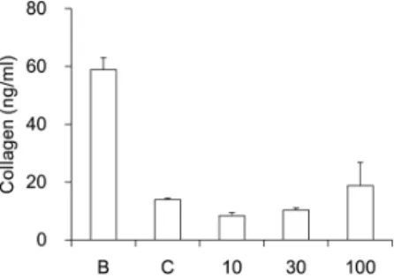
2. Assay of collagen type I synthesis
To evaluate the amount of collagen type I synthesis that occurred upon exposure to the sample, collagen type I was quantitatively detected by using the procollagen type I C-peptide assay kit previously described in methods section. Collagens are synthesized as precursor molecules, called procollagens. These molecules contain additional peptide sequences, usually referred to as 'propeptides', at both the amino-terminal end and the carboxy-terminal end. These propeptides are cleaved from the collagen triple-helix molecule during its secretion, after which the triple-helix collagens are polymerized into extracellular collagen fibrils. Thus, the amount of free propeptide stoichiometrically reflects the amount of collagen molecules synthesized
8). The amounts of type I collagen synthesis of AGR were shown in Figure 2. AGR increased the expression of type I collagen at a concentration of 100 μg/ml (18.7 ± 8.1 ng/ml).
However, there was no significant difference. The collagen amounts of AGR 30 μg/ml and 10 μg/ml treated group did not increase (10.3 ± 0.8 ng/ml and 8.4 ± 1.0 ng/ml).
Figure 2. Effect of AGR on collagen type I synthesis in human fibroblast cells. B: blank, distilled water treated group without UVB irradiation. C: control, distilled water treated group with UVB irradiation. 10, 30, and 100: Angelicae Gigantis Radix extracts (AGR 10, 30, and 100 μg/ml) treated group. Data are expressed as the mean ± SEM of three experiments.
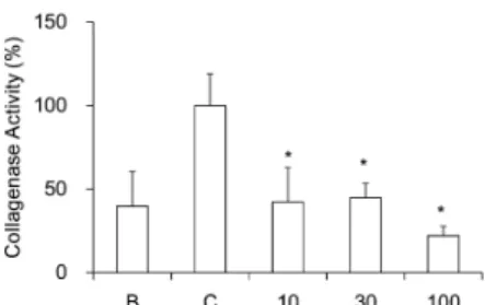
3. Assay of collagenase activity
To evaluate the collagenase activity, matrix metalloproteinase-1 (MMP-1) activity was quantitatively measured by using the previously described matrix metalloproteinase-1 assay kit. The activities of MMP-1 of AGR treatment were recalculated into 100% of control group (Figure 3).
AGR significantly reduced the MMP-1 activity at
concentrations of 10 μg/ml, 30 μg/ml, and 100 μ
g/ml in a dose dependent manner (42.2 ± 20.5%,
44.8 ± 8.5%, and 22.0 ± 5.8%, p < 0.05).
Figure 3. Effect of AGR on collagenase activity in human fibroblast cells. B: blank, distilled water treated group without UVB irradiation. C: control, distilled water treated group with UVB irradiation. 10, 30, and 100: Angelicae Gigantis Radix extracts (AGR 10, 30, and 100 μg/ml) treated group. Data are expressed as the mean ± SEM of three experiments. *: significantly different from the control, p < 0.05.
4. Inhibitory effects on tyrosinase activity
The activities of AGR on tyrosinase activity were recalculated into 100% of control group (Figure 4).
AGR significantly reduced the tyrosinase activity at concentrations of 10 and 1 mg/ml (75.9 ± 1.7% and 62.3 ± 0.5%, p < 0.05). The tyrosinase activity of AGR 0.1 mg/ml treated group did not show any significance (88.4 ± 7.1%).
Figure 4. Effect of AGR on tyrosinase activity. C: control, distilled water treated group. 0.1, 1, and 10: Angelicae Gigantis Radix extracts (AGR 0.1, 1, and 10 mg/ml) treated group. Data are expressed as the mean ± SEM of three experiments. *:
significantly different from the control, p < 0.05.
5. L-DOPA oxidation
The activities of AGR on L-DOPA oxidation were recalculated into 100% of control group (Figure 5).
Although there was no significant difference, AGR reduced the L-DOPA oxidation activity at concentrations of 10 mg/ml (89.4 ± 3.5%). AGR 1 and 0.1 mg/ml treated groups did not show any activity (105.8 ± 5.6% and 103.9 ± 19.6%
respectively).
Figure 5. Effect of AGR on L-DOPA oxidation. C: control, distilled water treated group. 0.1, 1, and 10: Angelicae Gigantis Radix extracts (AGR 0.1, 1, and 10 mg/ml) treated group. Data are expressed as the mean ± SEM of three experiments.