신경재활치료과학 제5 권 제2 호
Therapeutic Science for Neurorehabilitation Vol. 5, No. 2, 2016.
뇌졸중 환자의 상지 기능 향상을 위한 말초감각신경자극의 효과에 관한 체계적 고찰
김선호*, 박지혁**
*원주 영광병원 작업치료실
*연세대학교 작업치료학과
국문초록
목적: 본 연구는 국외 뇌졸중 환자에게 적용한 한 말초감각신경자극의 효과에 대하여 고찰하기 위한 것으 로 국외 학술지를 대상으로 체계적 문헌고찰 연구방법을 시행하였다.
연구방법: 2015년 10월 이전까지 국외 학술지에 게재된 논문을 Pubmed를 통하여 검색하였다. 주요 검색 용어로는 ‘peripheral nerve stimulation’, ‘electrical stimulation’, ‘sensory stimulation’, ‘somatosensory stimulation’, ‘stroke’, ‘hemiplegia’, ‘hemiparesis’ 와 ‘hand’, ‘arm’, ‘upper limb’를 사용하였다. 최초 검색된 논문은 501편이었으나 선정 및 배제기준을 거쳐 10편의 연구가 선정되었다.
결과: 임상적으로 널리 사용되고 있는 말초감각신경자극은 뇌졸중 환자들에게 적용이 될 때에 보다 다양 한 중재방법으로 연구에 적용이 되고 있었다. 말초감각신경자극치료는 뇌졸중 환자의 상지 기능에 효 과를 나타냈으며, 대뇌피질의 활성화에도 긍정적인 영향을 나타냈다.
결론: 본 연구는 말초감각신경자극치료의 적용에 대한 근거를 제시하며, 향후 국내연구에서는 다양한 중 재방법을 적용하여 말초감각신경자극의 효과를 더 정확하게 측정을 할 수 있는 방법에 대한 연구가 필 요하다.
주제어: 말초감각신경자극, 상지 기능, 뇌졸중
교신저자 : 박지혁([email protected]) || 접수일: 2016. 1. 18 || 심사일: 2016. 7. 19
|| 게재승인일: 2015. 8. 8
Ⅰ. 서 론
뇌졸중은 대표적인 뇌혈관 질환의 하나로써 뇌의 혈 관이 막히거나 터짐으로써 갑작스런 의식장애나 마비 현상 같은 신경학적 이상을 가져오는 급성 질환이다 (Chang, Tung, Wu, & Su, 2006) 대부분 비정상적 근 긴장(abnormalities in muscle tone)이나 운동마
비(motor paresis)로 인한 상지 기능 운동 장애를 경 험하게 되며 이는 일상생활동작을 수행하는 데 있어 가 장 큰 장애의 원인이 되기도 한다(Raghavan, 2007).
뇌졸중 환자의 손상된 측의 상지를 더욱 많이 사용 하게 하고, 보다 효율적인 움직임을 유발시키게 하기 위한 다양 한 노력들이 기울여졌었다. 오래 전부터 사용되고 있는 고 유감각 신경근 촉진법(Proprioceptive Neuromuscular
Facilitation technique: PNF), 그리고 Bobath 신경 발 달치료(Bobath’s Neurodevelopmental Technique:
NDT) 등은 마비되지 않은 쪽을 사용하여 보상기능을 획득하거나 기능을 촉진시키는 방법으로 이러한 치료 법들은 성인이 된 후에 중추신경계가 재생되거나 발 달할 수 없다는 고전적인 개념에 바탕을 둔 치료법이 다(Platz et al., 2005).
최근에는 의료공학의 획기적인 발전에 의하여 뇌의 활동을 관찰한 결과 성인의 중추신경계는 제한적이지 만 재생능력을 가지고 구조와 기능이 변화된다는 뇌 신경가소성에 대해 밝혀지고 있다(Gerloff et al., 2006). 이러한 기전을 토대로 근전도 유발 신경근 전 기자극(electromyogram-triggered neuromuscular stimulation: EMG-stim), 기능적 전기자극 (functional electronic stimulation: FES), 말초 감각신경 자극 (peripheral sensory nerve stimulation: PNS) 등과 같은 다양한 전기자극치료 기법이 개발되어 사용되고 있다(Bolton, Cauraugh, & Hausenblas, 2004;
Celnik et al., 2007; Mangold et al., 2009).
PNS는 말초 신경이 위치하는 근 피부 위에 근육의 수축이 일어나지 않는 범위의 저주파를 이용하여 전 기적 자극을 제공하는 것으로 근육의 피로와 통증이 거의 없어 위험요소가 적다(Sullivan, & Hedman, 2004). 또한, PNS는 감각운동 피질의 재조합을 유도 하는 중요한 방법 중의 하나이며, 반복적인 전기자극 은 해당 감각피질의 수용영역의 크기의 증가를 가져 온다(Deuchert et al., 2002). 최근 임상에서는 EMG-stim과 FES보다 운동·인지적 노력이 적게 요 구되는 PNS를 사용한 뇌졸중 상지 재활에 대한 연구 가 활발히 진행되고 있으며, 그 개선 효과도 보고하 고 있다(Ikuno et al., 2012).
PNS에 관한 연구들은 연구디자인과 중재적용방법 등에서 다양성을 나타내고 있으며, 자극의 강도와 기 간 등에 따라 다른 결과를 보고하고 있다(Ikuno et al., 2012). 또한, PNS가 뇌졸중 환자의 상기기능에 미치는 효과에 대한 연구는 국외에서 비교적 활발히 이루어지고 있으나 국내 연구는 미흡한 실정이다(유 인규, 박지혁, 2012). 이에 본 연구의 목적은 뇌졸중
환자의 상지 기능 향상을 위한 말초감각신경자극의 적용의 국외연구의 결과를 통합하고 연구경향을 파악 하여 이후 이루어질 국내 말초감각신경자극치료 연구 에 도움이 될 수 있는 정보를 제공하는 것이다.
Ⅱ. 연구 방법 1. 연구설계
본 연구는 국내 뇌졸중 환자의 상지기능 향상을 위 한 말초감각신경자극의 적용 연구에 관한 체계적 고 찰을 시도한 서술적 조사연구이다.
2. 연구대상
본 연구에서는 국외에서 2005년 1월 이후부터 2015년 10월까지 발표된 뇌졸중 환자의 상지 기능 향 상을 위한 말초감각신경자극 관련 최근 연구만을 대 상으로 하였으며 구체적 선정기준과 배제기준은 다음 과 같다.
1) 선정기준
(1) 말초 감각 신경 자극을 중재를 사용한 연구 (2) 상지의 기능회복과 결과를 제시한 연구 (3) 대상자가 뇌졸중 환자인 연구
(4) 실험연구인 연구
(5) 전문을 구할 수 있는 연구
2) 배제기준
(1) 외과적 수술이나 의학적 처지가 사용된 연구 (2) 영어로 저술되지 않은 연구
3. 연구 방법
1) 자료 수집
본 연구는 말초감각신경자극 치료와 관련된 연구 동 향을 분석하기 위해 PubMed에서 제공되는 연구논문
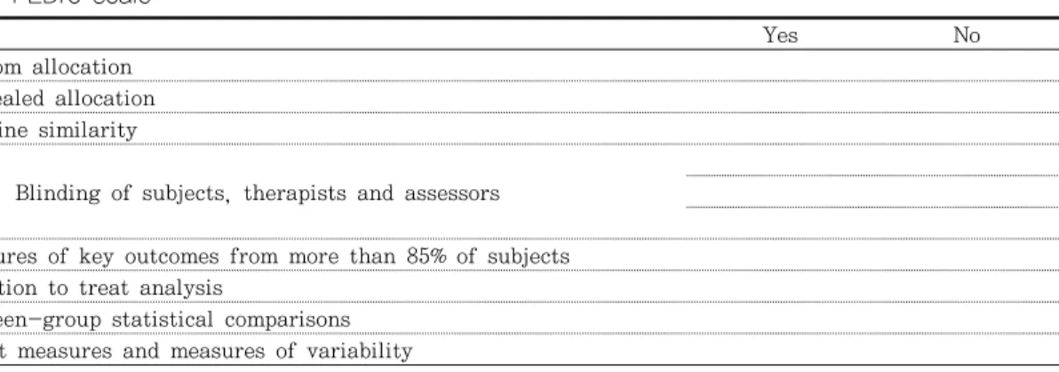
Table 1. PEDro scale
Yes No
1. Random allocation 2. Concealed allocation 3. Baseline similarity
4. 5, 6. Blinding of subjects, therapists and assessors 7. Measures of key outcomes from more than 85% of subjects 8. Intention to treat analysis
9. Between-group statistical comparisons 10. Point measures and measures of variability
들 중 2005년부터 2015년 10월까지 발표된 자료를 수집하였다. 주요 검색어는 ‘peripheral nerve stimulation’, ‘sensory stimulation’, ‘somatosensory stimulation’, ‘stroke’, ‘hemiplegia’, ‘hemiparesis’와 ‘upper extremity’를 사용하여 검색하였다. 그 결 과 총 501개의 논문이 검색되었다. 그 중 본 연구의 선정기준과 배제기준에 부합 하다고 연구자와 외부전 문가 1명에 의해 합의된 10편의 논문이 최종 선정되 었다.
2) 질적 평가방법
본 연구에서 최종적으로 선정된 10편의 연구에 대 해 질적 평가를 위해 반복적인 읽기와 분석을 통해 1 명의 연구자와 외부전문가 1명이 방법론적 평가 도구 인 PEDdro scale의 10가지 내부 타당도 항목을 사용 하여 개별적으로 근거수준을 검토하였다. PEDdro scale은 ‘예’, ‘아니오’ 답변으로 구성되면 최대점수는 10점(‘예’의 수)이다. 9점-10점은 ‘excellent’, 6점-8 점은 ‘good’, 4점에서 5점은 ‘fair’, 4점 이하는 ‘poor’
로 방법론적 질을 평가하게 된다(de Morton, 2009) (Table 1). 일치하지 않은 연구의 경우에는 연구자간 토의를 통해 합의를 하여 기재하였다.
3) 연구 분석
말초감각신경자극 치료의 효과를 알아보기 위하여 대상자수, 발병 기간, 연령 등과 같은 연구 대상자의 일반적인 특징과 연구의 질적 평가와 디자인, 중재 및 중재에 사용된 말초감각신경자극의 특징, 결과 측
정 및 측정 결과를 체계적으로 분석하였다.
Ⅲ. 연구 결과 1. 연구 대상자에 관한 일반적 특성
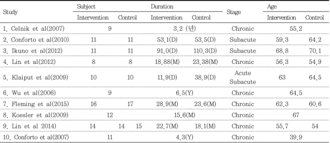
10편의 연구를 통해 총 197명의 대상자가 말초신경 감각자극치료에 대한 효과를 알아보기 위해 모집되었 다. 대상자들의 평균 연령은 39.9세에서 70.1세의 범 위를 나타냈으며, 주로 50대와 60대가 주를 이루었 다. 또한, 대상자들의 평균 발병기간은 11.9일에서 2372.5일의 범위를 나타냈다. 10편의 연구 중 7편의 연구가 만성기(>180일) 환자를 대상으로 하였으며, 2 편의 연구에서는 아급성기(30<180일), 1편의 급성기 (<30일)와 아급성기 환자를 대상으로 연구되었다 (Table 2).
2. 연구의 특징
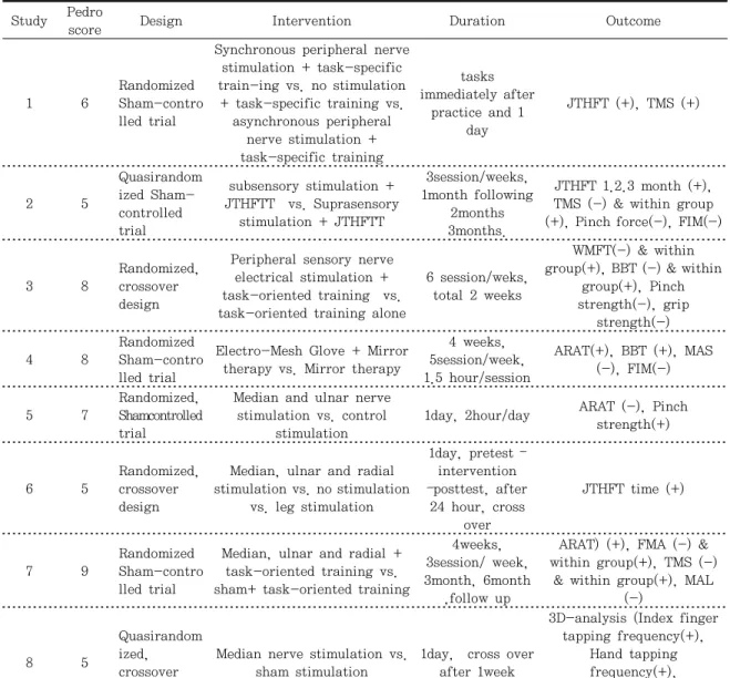
1) 연구의 질과 디자인
본 연구에 포함된 연구의 PEDro sclae에 대한 평 균 점수는 6.6 점이었다. ‘Excellent’등급의 연구 1 편, ‘Good’등급의 연구가 5편, ‘Fair’등급 연구가 4편 이며, ‘Poor’등급의 연구는 없었다. 연구의 디자인은 교차설계연구(cross-over study) 5편, 준 무작위연 구(quasi randomized study) 3편, 무작위대조실험 설계(randomized controlled trials)와 무작위 교차
Table 2. Characteristics of the Study Subjects
Study Subject Duration
Stage Age
Intervention Control Intervention Control Intervention Control
1. Celnik et al(2007) 9 3.2 (년) Chronic 55.2
2. Conforto et al(2010) 11 11 53.1(D) 53.5(D) Subacute 59.3 64.2
3. Ikuno et al(2012) 11 11 91.0(D) 110.3(D) Subacute 68.8 70.1
4. Lin et al(2012) 8 8 18.88(M) 23.38(M) Chronic 56.3 54.9
5. Klaiput et al(2009) 10 10 11.9(D) 38.9(D) Acute
Subacute 63 64.5
6. Wu et al(2006) 9 6.5(Y) Chronic 64.5
7. Fleming et al(2015) 16 17 28.9(M) 23.6(M) Chronic 62.3 60.6
8. Koesler et al(2009) 12 15.6(M) Chronic 67
9. Lin et al 2014) 14 14 15 22.7(M) 18.1(M) Chronic 55.7 54
10. Conforto et al(2007) 11 4.3(Y) Chronic 39.9
실험설계(randomized crossover trials)은 7편으로 나타났다(Table 3).
2) 중재(Intervention)
4편의 연구에서 말초감각신경자극만 단독으로 사 용되었다. 다른 4편의 연구에서는 말초감각신경자극 과 더불어 과제 지향적 훈련을 진행 하였고, 이중 2 편은 젭슨-테일러손기능검사(Jebsen-Taylor Hand Function Test: JTHFT)를 이용한 과제 훈련을 사용 하였다. 2편의 연구에서 말초감각신경자극과 함께 거 울치료를 진행한 것으로 보고하였다. 중재기간은 1일 부터 4주로 다양하게 나타났다(Table 3).
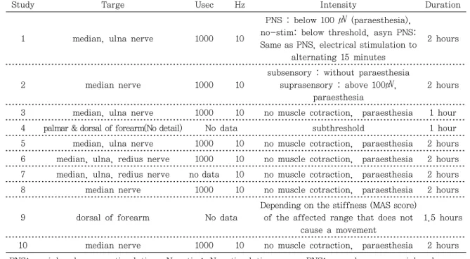
3) 전기자극
자극의 강도는 데이터가 설명되어 있지 않고 Electro-Mesh Glove를 사용한 2편의 연구를 제외한 나머지 연구에서 펄스폭(usec)/주파수(Hz)가 1000/10 으로 동일하게 사용되었다. 전극의 부착 부위는 손목 부위의 말초신경 위에 전극을 부착했다. 3편의 연구 에서 정중신경을 자극하였고, 3편의 연구가 마비 측 정중신경과 척골신경이 지나는 손목 위에 부착을 하 였다. 2편의 연구에서는 정중신경, 척골신경, 요골신 경 모두를 자극한 것으로 나타났다. 대부분의 연구에 서 사용된 전기자극의 강도는 이상감각이 약하게 느
껴지거나, 통증이 없고, 눈에 보이는 근 수축이 없는 상태로 설정하였고, PNS그룹과 비교하기 위한 허위 대조군(sham-controlled group)이나 대조군에 사용 된 전기자극의 강도는 대부분 이상감각이 느껴지지 않는 범위의 약한 강도(subsensory)로 설정하여 사 용하였다. 자극시간의 범위는 1 2시간을 적용하였 다(Table 4).
4) 결과 측정
3편의 연구가 후속평가(follow-up assessment)를 실시하였으며, 대부분의 모든 연구들이 사전 사후 중 재(pre & post intervention)에 대한 평가를 실시하 였다. 후속평가의 범위는 24 시간에서 3개월이었다.
연구에서 사용된 평가는 집기 힘(pinch strength), JTHFT, Action Research Arm Test(ARAT), 상자 블럭 검사(Box & Block test: BBT), 경두개자기자극 (transcranial magnetic stimulation: TMS)을 통한 대뇌피질의 활성화 검사, 퍼글-마이어평가(Fugl- Meyer Assessment Scale: FMA), 손과 팔의 운동역학 (movement kinematics) 평가, Wolf Motor Function Test(WMFT), Motor Activity Log(MAL), 기능적 독립 측정(Functional Independence Measure: FIM), ABILHAND Questionnaire, 근 긴장 평가 등이 사용 되었다 (Table 3).
Table 3. Characteristics of the study Study Pedro
score Design Intervention Duration Outcome
1 6
Randomized Sham-contro lled trial
Synchronous peripheral nerve stimulation + task-specific train-ing vs. no stimulation
+ task-specific training vs.
asynchronous peripheral nerve stimulation + task-specific training
tasks immediately after
practice and 1 day
JTHFT (+), TMS (+)
2 5
Quasirandom ized Sham- controlled trial
subsensory stimulation + JTHFTT vs. Suprasensory
stimulation + JTHFTT
3session/weeks, 1month following
2months 3months.
JTHFT 1.2.3 month (+), TMS (-) & within group (+), Pinch force(-), FIM(-)
3 8
Randomized, crossover design
Peripheral sensory nerve electrical stimulation + task-oriented training vs.
task-oriented training alone
6 session/weks, total 2 weeks
WMFT(-) & within group(+), BBT (-) & within
group(+), Pinch strength(-), grip
strength(-)
4 8
Randomized Sham-contro lled trial
Electro-Mesh Glove + Mirror therapy vs. Mirror therapy
4 weeks, 5session/week, 1.5 hour/session
ARAT(+), BBT (+), MAS (-), FIM(-)
5 7
Randomized, Shamcontrolled trial
Median and ulnar nerve stimulation vs. control
stimulation
1day, 2hour/day ARAT (-), Pinch strength(+)
6 5
Randomized, crossover design
Median, ulnar and radial stimulation vs. no stimulation
vs. leg stimulation
1day, pretest – intervention –posttest, after
24 hour, cross over
JTHFT time (+)
7 9
Randomized Sham-contro lled trial
Median, ulnar and radial + task-oriented training vs.
sham+ task-oriented training
4weeks, 3session/ week, 3month, 6month
,follow up
ARAT) (+), FMA (-) &
within group(+), TMS (-)
& within group(+), MAL (-)
8 5
Quasirandom ized, crossover design
Median nerve stimulation vs.
sham stimulation
1day, cross over after 1week
3D-analysis (Index finger tapping frequency(+),
Hand tapping frequency(+), Reach-to-grasp
movements(+)
9 8
Randomized Sham-contro lled trial
Electro-Mesh Glove + Mirror therapy vs. Mirror therapy
vs.control treatment
4weeks, 5 session/weeks, 1.5hour / session
FMA (+), BBT (+), kinematic parameters(+),
MAS (-), MAL (-), ABILHAND Questionnaire(-)
10 5
Quasirandom ized Sham-contro lled trial
median nerve stimulation vs.
control stimulation (sub-sensory median nerve
stimulation)
1day, cross over
after 30day JTHFT (+)
JTHFT: Jebsen-Taylor Hand Function Test TMS: Transcranial Magnetic Stimulation, FIM: Functional
Independence Measure , WMFT: Wolf Motor Function Test, BBT: Box and Block Test, ARAT: Action
Research Arm Test, MAS : Modified Ashworth scale of muscle spasticity, FMA: Fugl-Meyer
Assessment Scale, MAL: Motor Activity Log, 10MWT: 10-Meter Walk Test
Table 4. Characteristics of electrical stimulation
Study Targe Usec Hz Intensity Duration
1 median, ulna nerve 1000 10
PNS : below 100 ㎶ (paraesthesia), no-stim: below threshold, asyn PNS:
Same as PNS, electrical stimulation to alternating 15 minutes
2 hours
2 median nerve 1000 10
subsensory : without paraesthesia suprasensory : above 100㎶,
paraesthesia
2 hours
3 median, ulna nerve 1000 10 no muscle cotraction, paraesthesia 1 hour 4 palmar & dorsal of forearm(No detail) No data subthreshold 1 hour 5 median, ulna nerve 1000 10 no muscle cotraction, paraesthesia 2 hours 6 median, ulna, redius nerve 1000 10 no muscle cotraction, paraesthesia 2 hours 7 median, ulna, redius nerve no data 10 no muscle cotraction, paraesthesia 2 hours
8 median nerve 1000 10 no muscle cotraction, paraesthesia 2 hours
9 dorsal of forearm No data
Depending on the stiffness (MAS score) of the affected range that does not
cause a movement
1.5 hours
10 median nerve 1000 10 no muscle cotraction, paraesthesia 2 hours
PNS: peripheral nerve stimulation, No-stim: No stimulation, asyn PNS: asynchronous peripheral nerve stimulation
5) 측정 결과
집기 힘(pinch strength)은 3편의 연구 중 1편에서 유 의하게 개선된 것으로 나타났다. 운동 역학(movement kinematics) 평가를 실시한 2편의 연구에서는 동작 의 효율성 개선과 운동빈도의 향상을 보고하였다.
JTHFT를 실시한 연구는 모두 유의한 개선을 나타냈 다. 특히, 거울치료와 과제지향적 훈련을 병행하였던 연구에서 시행된 상지기능평가(JTHFT, BBT, ARAT, FMA, WMFT)는 모두 유의한 향상을 보였다. 이중 3 편은 대뇌피질의 활성화에 대한 연구로 유의한 향상을 보고하였다. 한편, 근 긴장 평가, MAL, ABILHAND Questionnaire, FIM을 사용한 연구에서 모두 대조군과 비교하여 유의한 개선을 나타내지 않았다(Table 3).
Ⅳ. 고찰
본 연구에서는 뇌졸중 환자에서 말초감각신경자극 치료에 관한 중재효과의 체계적 고찰을 통해 연구대
상자의 특성과 연구의 중재특성 및 말초감각신경자극 의 효과에 대해 알아보았다. 최근 임상적으로 널리 사용되고 있는 말초감각신경자극은 뇌졸중 환자들에 게 적용이 될 때에 보다 다양한 중재방법을 활용하여 연구에 적용이 되고 있었다.
말초감각신경자극은 거울치료 및 과제지향적 훈련 과 함께 적용되어 상지기능의 개선효과를 극대화시킬 수 있는 것을 확인하였다. 거울치료는 마비되지 않은 사지의 움직임이 거울 반영을 통해 마비된 사지의 움 직임으로 투영되고, 이러한 시각 정보가 주는 움직임 의 영상이 손상된 뇌를 자극 시킨다는 이론으로 환자 의 능동적인 참여를 유도하여 상지 기능 회복에 기여 하는 치료법이며, 과제지향적 훈련은 신경학적 손상 을 가진 환자의 기능회복과 개선에 대한 운동 조절과 운동 학습을 접목하여 접근하는 치료법으로 두 중재 법 모두 대뇌활성화를 통한 뇌신경가소성의 원리를 기초로 임상에서 널리 사용되어져 왔다(Thieme et al., 2012; Yang, 2011). 감각신경에 대한 반복적인 전기자극 역시 해당 감각피질(somatosensory cor-
tex)의 수용영역(receptive field)의 크기를 확대시킨 다는 것이 밝혀졌다(Deuchert et al., 2002). 무엇보 다 말초감각신경자극은 무엇보다 자극 동안 참여자의 자발적인 참여나 집중을 요구하지 않기 때문에 다른 작업과 동시에 적용하기 용이하다(Kattenstroth, et al., 2012). 말초 감각신경 자극과 자발적이고 능동적 인 훈련을 병행하였을 때, 감각 신경이 분지하는 피 부에서 입력되는 말초 구심성 입력이 자극되면서 피 질척수로의 활성화를 강화시키는데 시너지 효과가 있 다(Lin et al., 2012; Saito et al.,2013). 이는 말초 감각신경자극과 거울치료, 과제지향적 훈련을 병행 한 연구들의 결과 비교를 통해 상지기능회복에 긍정적 효과를 보고한 결과와 일치 하는 것이라 할 수 있다.
반면, 같은 평가에 대해 서로 다른 결과를 보고하기 도 하였다. 집기 힘(pinch strength)은 3편의 연구 중 1편에서 유의하게 개선된 것으로 나타났는데, 2시간의 말초 감각신경 자극을 제공한 직후에 바로 장측 잡기와 손끝잡기를 평가하여 대뇌 운동피질의 흥분성이 증가 된 상태에서 평가하였기 때문에 더 유의한 결과를 얻은 것으로 판단되며(Klaiput, & Kitisomprayoonkul, 2009), 또한, 다른 2편의 연구는 과제훈련이 대뇌 피 질의 가소성에 영향을 주지만 거울치료 동안 손의 근 력 향상을 위한 직접적인 과제는 제공하지 않았기 때 문에 유의하지 않은 결과가 나온 것으로 사료된다 (Fleming et al., 2015; Lin et al., 2012). 또한, ARAT에서는 3편의 연구 중 2편에서 유의한 개선을 보인 것으로 나타났는데 중재 기간이 길고, 과제지향 적 훈련을 병행한 연구에서 긍정적인 영향을 주었던 것으로 사료된다(Fleming et al., 2015; Lin et al., 2012). ARAT에서의 유의미한 개선을 나타내지 않은 이유로 짧은 중재기간과 대상자들의 상지 기능에 대 한 천장효과(ceiling effect)의 영향을 제언하고 있다 (Klaiput, & Kitisomprayoonkul, 2009). 선행연구 에서 말초감각신경자극은 움직임의 조절과 관련이 있 는 근 긴장에 대한 감소를 보고하고 있지만, 근 긴장 도(muscle tone)에 관한 평가는 2편의 연구 모두 유 의미한 개선을 이끌어내지 못하였다(Lin et al., 2012; Lin et al., 2014). 이는 사전 검사에서 대상자
들의 강직수준이 경미하거나 중간 정도(MAS 2)로 높은 선정기준이 영향을 미쳤거나, 중재 전 전기자극 의 설정을 위한 시도로 인해 강직을 미리 줄여주는 효과를 나타냈을 수도 있다고 제언하고 있다.
특히, 말초감각신경자극과 거울치료 및 과제지향적 훈련을 병행한 연구에서 근긴장도, MAL, ABILHAND Questionnaire, FIM 모두 대조군과 비교하여 긍정적 인 효과를 이끌어 내지 못하였다. 일상생활기능 (Daily function) 평가 도구인 운동활동지표(Motor Activity Log), ABILHAND Questionnaire이 유의미 한 개선을 보이지 않은 것과 몇 몇의 연구에서 대조 군과의 유의한 차이가 없었던 이유로 작은 연구대상 자 수와 교차설계연구(cross over design)였던 점 그 리고 대상자들의 천장효과(ceiling effect)와 대조군 의 중재 영향력을 이유로 설명하였다. 향후 손상 수 준 또는 병변 부위에 따라 계층화를 통한 연구대상자 수를 증가 시키는 것이 중요하며 명확한 자극의 강도와 자극을 제공하는 타이밍을 제공을 통한 말초감각신경 자극의 정확한 프로토콜이 필요할 것이라 판단된다.
몇 몇 소규모 연구들은 손 기능 향상은 감각전기자 극에 의해 단기 이월 효과(short-term carry-over effect)로 야기되었다는 것을 보고하였다. 감각전기 자극 후에 30일 이상 동안 효과가 유지됨을 보고하였 고(Conforto et al., 2007), 저자는 이후 연구에서 감 각전기자극과 과제훈련을 1개월 동안 병행한 환자는 2-3개월 동안 그 효과가 지속되는 것을 보고하였다 (Conforto et al., 2010). 또한, 신체훈련의 효과를 향상시키기 위해 과제훈련을 함께 병행하는 것이 감 각전기자극을 단독으로 사용하는 것보다 낫다고 보고 하고 있다. 척추손상환자의 연구뿐 아니라 뇌졸중 환 자를 대상으로 한 연구도 감각전기자극은 집중적인 운동훈련의 효과를 강화 할 수 있다(Beekhuizen, &
Field-Fote, 2008). 과제훈련은 대뇌운동피질의 흥 분성을 증가시키고 영구적인 뇌 신경가소성의 변화를 유도한다(Nudo et al.,1996). 또한, 감각전기자극은 장기적인 상승작용 메카니즘에 의해 지속적인 가소성 의 변화를 강화 할 수 있다(Sawaki et al., 2006). 장 기적인 이월효과를(carry-over effect)를 얻기 위해
서는 감각전기자극과 과제훈련을 병행하는 것이 중요 한 요소일 수 있을 것이다.
감각전기자극(sensory electrical stimulation)에 서 전기자극의 파라미터(parameter)는 중요한 역할 을 한다. 정중신경에 150Hz의 고주파 신경 자극은 대칭측 대뇌반구의 운동피질(motor cortex)의 흥분 성을 줄일 수 있다고 보고하였다(Mang, Lagerquist,
& Collins, 2010). 인구근육에 5Hz의 감각전기자극 을 적용하였을 때 피질의 흥분성의 증가를 유도해냈 으며, 반면에 20 Hz -40Hz를 적용했을 때는 피질의 흥분성이 감소했다고 보고하고 있다(Kaelin-Lang, 2002) 또한, 자극 되어지는 주파수에 따라 피질의 흥 분성이 달라지며, 100Hz의 자극은 10, 50, 200Hz일 때 보다 더 효과적이었다고 보고하였다(McKay et al., 2002). 그러나 감각전기자극을 사용한 연구를 근거로 하여 대부분 10Hz가 사용되었고(Fraser et al., 2002), 이 연구들은 긍정적인 효과를 보여주었 다. 이러한 결과는 최적의 주파수는 감각전기자극이 전달되는 위치에 따라 변화 될 수 있음을 나타낸다.
추후 연구는 모든 치료적 대상을 위한 최적의 주파수 를 결정하는 것이 필요할 것이라 사료된다. 전기자극 의 자극시간 또한 중요하다. 연구들을 비교한 결과 대부분 연구에서 2시간의 감각전기자극시간을 실시 했다. 2시간의 자극은 손과 팔의 기능 개선에 긍정적 인 효과를 보였다. 그러나 중재 프로토콜은 임상재활 세팅에서 수행되어 현실적이지 못했다(Ikuno et al., 2012). 이후 입원 재활 환자를 대상으로 1시간의 전 기자극과 과제훈련을 병행하여 그 효과를 조사한 결 과 피로의 정도에 큰 차이는 없었고, 부작용도 나타 나지 않았다. 게다가, 그룹 내에서 손과 팔의 기능에 유의한 개선을 보였다(Ikuno et al., 2012). 한 연구는 대뇌피질의 흥분의 증가가 45분-60분이 전기자극 후 정점을 나타냄을 보고하였다(Langhorne, Bernhardt,
& Kwakkel, 2011). 따라서 전기적 자극의 최대효과 를 생성하기 위해서는 적어도 30분의 자극이 필요 할 수 있다고 판단된다.
자극의 강도(stimulation intensity)의 측면에서 모순된 결과가 있었다. 1개월 간의subsensory자극이
suprasensory자극보다 JTHFT에서 유의한 향상을 보였다(Conforto, 2010). 이 결과는 저자의 가설과 일치하지 않았다. 그룹간 유의미한 차이가 있었지만 두 그룹은 비슷한 수준으로 모두 JTHFT에서 개선되 었다. 향후 연구에서는 무작위 거짓통제임상시험 (randomized sham-controlled clinical trial)에서 전기자극의 최적의 강도를 결정하는 것이 필요할 것 이라 판단된다. 이전 연구에 따르면, 기능적 전기자 극(functional electrical stimulation: FES)과 같은 전기자극의 운동을 만들어 내는 진폭(motor amplitude) 은 감각성 자극의 진폭보다 더 클 수 있다(Langhorne, Bernhardt, & Kwakkel, 2011). 그러나, 감각전기자극 은 전기의 운동성 자극에 의하여 생길 수 있는 피로 또 는 통증을 발생시키지 않는 이점을 갖는다. 또한, 감각 전기자극은 장기간 동안 과제지향적 훈련의 동시수행 을 가능하게 해준다. 여러 연구들에서 나타난 바와 같 이, 감각전기자극은 강제유도운동치료(constraint-in- duced movement therapy : CIMT)를 필요로 하는 뇌 졸중 환자의 상지재활의 효과를 강화 시킬 수 있는 보조 도구로서 유용하게 사용될 수 있을 것이다(Beekhuizen,
& Field-Fote, 2008; Conforto, 2010).
감각전기자극은 팔의 마비가 있는 뇌졸중 환자가 기능적인 과제훈련과 결합하여 사용하면 손과 팔의 기능을 향상 시킬 수가 있다. 그러나 연구들을 검토 한 결과 적절한 무작위 대조군 연구(Randomized controlled trials: RCT)가 부족하기 때문에 아직 명 확한 결론에 이르지 못하고 있다. 향후 연구는 가장 효과적인 전기자극의 주파수(frequency), 기간 (duration) 및 강도(intensity)에 대하여 명확히 해결 해야 하며, 또한, 어떤 뇌졸중 단계(stage)에서 가장 효과를 나타낼 수 있는지 명확히 해결해야 할 것이 다. 또한, 뇌졸중 환자를 대상으로 한 감각전기자극 의 효과성를 밝히기 위해 장기추적연구와 큰 모집단, 무작위 다기관 시험이 필요할 것으로 사료되며, 다양 한 중재방법을 적용한 연구로 말초감각신경자극의 효 과를 극대화시킬 수 있는 방법을 적용할 필요가 있 다. 국외에 비해 국내의 말초감각신경자극의 연구는 활발하게 이루어지지 못하고 있다. 앞으로 국내 작업
치료 연구에서도 이러한 상지기능의 향상을 위해 효 과성이 입증된 다양한 중재방법을 통한 말초감각신경 자극의 활용이 필요하다.
Ⅴ. 결론
본 연구는 뇌졸중 환자에서 말초감각신경자극에 관 한 상지 기능에 대한 중재효과의 체계적 고찰을 통해 연구대상자의 특성과 연구의 중재특성 및 말초감각신 경자극의 효과에 대해 알아보았다 최근에는 임상적으 로 널리 사용되고 있는 말초감각신경자극은 뇌졸중 환자들에게 적용이 될 때에 보다 다양한 중재방법으 로 연구에 적용이 되었다. 말초감각신경자극치료는 뇌졸중 환자의 상지 기능에 효과를 나타냈으며, 대뇌 피질의 활성화에도 긍정적인 영향을 나타냈다. 특히, 특정과제와 함께 적용되는 말초감각신경자극은 전반 적인 상태를 촉진하고 치료효과를 극대화 시키는 것 을 확인하였다. 하지만, 연구 디자인의 한계와 적은 수의 대상자는 명확한 말초감각신경자극의 주파수 (frequency), 기간(duration) 및 강도(intensity)를 해결하는데 문제를 가지고 있었다. 향후 연구에서는 무작위 대조군 연구(Randomized controlled trials:
RCT)와 큰 모집단, 장기추적연구와 무작위 다기관 시험이 필요할 것으로 판단되며, 다양한 중재방법을 적용한 연구로 말초감각신경자극의 효과를 극대화시 킬 수 있는 방법을 적용할 필요가 있다.
Reference
유인규, 박지혁, (2012) 뇌졸중을 경험하는 대상자를 위한 체성감각자극 중심치료의 효과 및 방법 제 언, 신경재활치료과학, 1(2), 5-13.
Beekhuizen, K.S., Field-Fote, E.C. (2008). Sensory stimulation augments the effects of massed practice training in persons with tetraplegia.
Archives of Physical Medicine and Rehabilitation.
89, 602-608. doi:10.1016/j.apmr.2007.11.021
Bolton, D. A., Cauraugh, J. H., & Hausenblas, H.
A. (2004). Electromyogram-triggered neuro- muscular stimulation and stroke motor recovery of arm/hand function : A meta-analysis.
Journal of Neurological Sciences, 223(2), 121-127. doi:10.1016/j.jns
Celnik, P., Hummel, F., Harris-Love, M., Wolk, R., & Cohen, L. G. (2007). Somatosensory stimulation enhances the effects of training functional hand tasks in patients with chronic stroke. Archives of Physical Medicine and Rehabilitation, 88(11), 1369-1376. doi:10.
1016/j.apmr.2007.08.001
Chang, J. J., Tung, W. L., Wu, W. L., & Su, F. C.
(2006). Effect of bilateral reaching on af- fected arm motor control in stroke-with and without loading on unaffected arm. Disability and Rehabilitation, 28(24), 1507-1516.
doi:10.1080/09638280600646060
Conforto, A. B., Cohen, L. G., dos Santos, R. L., Scaff, M., & Marie, S. K.N.(2007). Effects of somatosensory stimulation on motor function in chronic cortico-subcortical strokes. Journal of Neurology, 254(3), 333-339. doi:10.
1007/s00415-006-0364-z
Conforto, A. B., Ferreiro, K. N., Tomasi, C., dos Santos, R. L., Moreira, V. L., Nagahashi Marie, S. K. N., … Cohen, L. G. (2010). Effects of somatosensory stimulation on motor function after subacute stroke. Neurorehabilitation and Neural repair, 24(3), 263-272. doi:10.
1177/1545968309349946
de Morton, N. A. (2009). The PEDro scale is a valid measure of the methodological quality of clinical trials: a demographic study. Aust J Physiother, 55(2), 129-133, 2009. doi:
10.1016/S0004-9514(09)70043-1
Deuchert, M., Ruben, J., Schwiemann, J.,
Meyer, R., Thees, S., Krause, T., Blankenburg, F., Villringer K., Kurth, R., Curio, G., & Villringer, A. (2002). Event-related fMRI of the somatosensory system using electrical finger stimulation. Brain Imaging, 13, 365-369.
Fleming, M.K., Sorinila, I.O., Roberts-Lewis, S.F., Wolfe, C.D., Wellwood, I., & Newham, D.J. (2015). The Effect of combined somato- sensory stimulation and task-specific train- ing on upper limb function in chronic stroke:
A double-blind randomized controlled trial.
Neurorehabilitation and Neural repair, 29(2), 143-152. doi: 10.1177/1545968314533613 Fraser, C., Power, M., Hamdy, S., John, R., David,
H., Lgor, H., … David, T. (2002). Driving plasticity in human adult motor cortex is associated with improved motor function after brain injury. Neuron, 34(5), 831–840.
Gerloff, C., Bushara, K., Sailer A , Wassermann, E.M., Chen, R., Matsuoka, T., Waldvogel, D., Wittenberg, G.F., Ishii, K, Cohen, L. G.,
& Hallett, M. (2006). Multimodal imaging of brain reorganization in motor areas of the contralesional hemisphere of well recovered patients after capsular stroke. Brain, 129(Pt 3), 791-808.
Ikuno, K., Kawaguchi, S., Kitabeppu, S., Kitaura, M., Tokuhisa, K., Morimoto, S.,… Shomoto, K. (2012). Effects of peripheral sensory nerve stimulation plus task-oriented train- ing on upper extremity function in patients with subacute stroke: A pilot randomized crossover trial. Clinical Rehabilitation, 26(11), 999-1009. doi: 10.1177/0269215512441476 Ikuno, K., Kawaguchi, S., Kitabeppu, S., Kitaura,
M., Tokuhisa, K., Morimoto, S.,… Shomoto, K. (2012). Effects of peripheral sensory
nerve stimulation plus task-oriented train- ing on upper extremity function in patients with subacute stroke: A pilot randomized crossover trial. Clinical Rehabilitation, 26(11), 999-1009. doi: 10.1177/0269215512441476 Kaelin-Lang, A., Luft, A.R., Sawaki, L.,
Burstein, A.H., Sohn, Y.H., Cohen , L.G.
(2002). Modulation of human corticomotor excitability by somatosensory input. The Journal of Physiology. 540(2), 623-633.
Kattenstroth, J. C., Kalisch, T., Peters, S., Tegenthoff, M., & Dinse, H. R. (2012).
Long-term sensory stimulation therapy im- proves hand function and restores cortical responsiveness in patients with chronic cer- ebral lesions: Three single case studies.
Human Neuroseience, 6(244), 1-13. doi: 10.3389/
fnhum.2012.00244
Klaiput, A., & Kitisomprayoonkul, W. (2009).
Increased pinch strength in acute and subacute stroke patients after simultaneous median and ulnar sensory stimulation. Neurorehabilitation and Neural repair, 23(4), 351-356. doi: 10.1177 /1545968308324227
Koesler, .IB., Dafotakis, M., Ameli, M., Fink, G.R., & Nowak, D.A. (2009) Electrical soma- tosensory stimulation improves movement kinematics of the affected hand following stroke. Journal of Neurology Neurosurgery and Psychiatry. 80(6), 614-619. doi:10.1136/
jnnp.2008.161117
Langhorne, P., Bernhardt, J., & Kwakkel, G.
(2011). Stroke rehabilitation. Lancet 377 (9778) 1693-1702.
Lin, K. C., Chen, Y. T., Huang, P. C., Wu, C.
Y., Huang, W. L., Yang, H. W., … Lu, H. J.
(2012). Effect of mirror therapy combined with somatosensory stimulation on motor
recovery and daily function in stroke pa- tients: A pilot study. Journal of Formosan Medical Association, 113(7), 422-428. doi:10.
1016/j.jfma.2012.08.008
Lin, K.C., Huang, P.C., Chen, Y.T., Wu, C.Y.,
& Huang, W.L. (2014).Combining afferent stimulation and mirror therapy for re- habilitating motor function, motor control, ambulation, and daily functions after stroke.
Neurorehabilitation and Neural Repair, 28(2), 153-162. doi: 10.1177/1545968313508468 Mang, C.S., Lagerquist, O., & Collins, D.F.
(2010). Changes in corticospinal excitability evoked by common peroneal nerve stim- ulation depend on stimulation frequency.
Experimental Brain Research, 203(1), 11-20.
Mangold, S., Schuster, C., Keller, T., Zimmermann- Schlatter, A., & Ettlin, T. (2009). Motor Training of Upper Extremity With Functional Electrical Stimulation in Early Stroke Rehabilitation. Neurorehabilitation and Neural Repair, 23(2), 184-190. doi: 10.1177/154596 8308324548
McKay, D., Brooker, R., Giacomin, P., Ridding, M., Miles, T. (2002). Time course of induction of increased human motor cortex excitability by nerve stimulation. Neuroreport 13(10), 1271-1273.
Nudo, R.J., Wise, B.M., SiFuentes, F., &
Milliken, G.W. (1996). Neural substrates for the effects of rehabilitative training on motor recovery after ischemic infarct. Science. 272, 1791-1794.
Platz, T., Eickhof, C., Van-Kaick, S., Engel, U., Pinkowaki, C., Kalok, S., & Pause, M.
(2005) Impairment-oriented training or Bobath therapy for severe arm paresis after stroke: A single-blind, multicentre randomized
controlled trial, Clin Rehabil, 19(7), 714-721.
Raghavan, P. (2007). The nature of hand motor impairment after stroke and its treatment.
Current Treatment Options in Cardiovascular Medicine, 9(3), 221-228. doi: 10.1007/s11936 -007-0016-3
Saito, K., Yamaguchi, T., Yoshida, N., Tanabe, S., Kondo, K., & Sugawara, K. (2013). Combined effect of motor imagery and peripheral nerve electrical stimulation on the motor cortex.
Experimental Brain Research, 227(3), 333- 342. doi:10.1007/s00221-013-3513-5
Sawaki, L., Wu, C.W., Kaelin-Lang, A., & Cohen, L.G. (2006). Effects of somatosensory stimulation on use-dependent plasticity in chronic stroke.
Stroke. 37, 246-247.
Sullivan, J. E., & Hedman, L. D. (2004). A home program of sensory and neuromuscular electrical stimulation with upper-limb task practice in a patient 5 years after a stroke. Physical Therapy, 84(11), 1045-1054.
Thieme, H., Bayn, M., Wurg, M., Zange, C., Pohl, M., & Behrens, J. (2012). Mirror ther- apy for patients with severe arm paresis af- ter stroke-A randomized controlled trial.
Clinical Rehabilitation, 27(4), 314-324. doi:
10.1177/0269215512455651
Wu, C.W., Seo, H.J., & Cohen, L.G. (2006).
Influence of electric somatosensory stim- ulation on paretic-hand function in chronic stroke. Archives of Physical Medicine and Rehabilitation, 87, 351-357.
Yang DY (2011). Effects of biofeedback with task-related training on motor function and neural plasticity in subjects with stroke (Doctor’s thesis). Dongshin University of Korea, Naju.
Abstract
Systematic Review on Effect of Peripheral Sensory Nerve Stimulation on Upper Extremity Function for Stroke Patients
Kim, Sun-Ho, M.S, O.T*, Park, Ji-Hyuk, Ph.D., O.T.**
*Dept. of occupational therapy, Young Kwang Rehabilitation Hospital
**Dept. of occupational therapy, Yonsei University
Objective: This study is executed systematic review targeted at international journals intended to investigate on effect of peripheral sensory nerve stimulation on upper extremity function for stroke patients.
Method: After literature search, researchers selected for 10 studies registered up to October 2015 based on PubMed database, using the following search terms: peripheral nerve stimulation, electrical stimulation, sensory stimulation, somatosensory stimulation, stroke, hemiplegia, hemiparesis and hand, arm, upper limb.
Result: There were significant improves of upper extremity function and positive effect on the cortical activation in the use of peripheral sensory nerve stimulation.
Conclusion: domestic studies in future requires a study of the method for measuring more accurately the effect of peripheral sensory nerve stimulation in RCT studies applying various intervention.
Keywords: Peripheral nerve stimulation, Stroke, Upper-limb