* Corresponding author
Phone: +82-51-606-2335, Fax: +82-51-891-0004 E-mail: [email protected]
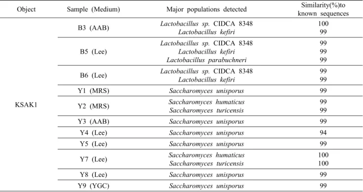
PCR-based Identification of Microorganisms in a Kefir Grain
Won Hoe Koo
1, Min-gook Seo
1and Jung Hoon Ahn
2*1
Korea Science Academy, Busan, Korea
2
전체 글
1
2
수치


관련 문서
(1) PCR monitoring of bphC gene of Ralstonia eutropha H850 in soil Amount of bphC gene amplification of H850 cells was not generally proportional to the cell density
For confirmation of results of direct real-time melting curve analysis, we also performed an in-house JAK2 V617F ASP and a BsaXI-treated nested PCR-direct
Comparison of genotypes using REP-PCR and Integron-IS26 PCR results among 78 IRAB strains isolated from 2 university hospitals.. The result of REP-PCR genotypes and
Sequencing results of groEL gene of Anaplasma phagocytophilum detected in blood, kidney and spleen of wild rodents captured in Jeollanam-do area using a
The object of this study is to analyse the bacteria of periapical bascess and fascial space abscess using multiplex real-time polymerase chain reaction (MRT-PCR) before
In the present study, 10 study stations were established, and seasonal changes for various ecological aspects of the community such as species composition, number of
To assess the clinical usefulness of performing Q-PCR in practice as a diagnostic technique, we compared blindly the Q-PCR results using blood samples of the
As a result of pyrG N-PCR sequence analysis, the positive rate of Borrelia in 27 wild mice captured in October and November was 29.63% (8 positive / 27 total) and only
