슬관절 관절경 검사 시 초음파 검사의 유용성
조인병원, 대전한국병원1, 충남대학교 의과대학 정형외과학교실2
변기용∙이광진1∙김경천2∙김동규2∙김보건2
서 론
슬관절 병변에 대한 진단으로 현재 자기 공명 영 상 촬영이 선택적 진단법으로 자리매김하고 있으나, 고가의 비용으로 인하여 환자들이 겪는 부담은 크다
통신저자: 김 경 천
대전광역시 중구 문화로 33
충남대학교 의과대학 정형외과학교실 Tel: 042-280-8485, Fax: 042-252-7098 E-mail: [email protected]
Ultrasonographic Utility for Arthroscopic Examination of Knee
Ki-Yong Byun, M.D., Ph.D., Kwang-Jin Rhee, M.D.
1, Ph.D, Kyung-Cheon Kim, M.D.
2, Ph.D, Dong- Kyu Kim, M.D.
2, Bo-Kun Kim, M.D.
2Join Hospital, Daejeon Hankook General Hospital
1, Department of Orthopedic Surgery, School of Medicine,Chungnam National University, Daejeon, Korea
2Purpose: To evaluate the relationship between the real pathology & abnormal finding found by ultrasonography.
Without an MRI test being done beforehand, an arthroscopy is done after an ultrasonography to show abnormal lesions during a knee abnormality.
Materials and Methods: The subjects were 42 patients out of 49 cases, excluding those with rheumatoid arthritis, septic arthritis and patients suspected with a ligament tear, which were examined by ultrasonography alone before receiving a knee arthroscopy in our hospital from July 2007 to July 2008. In every case, a physical examination, sim- ple X- ray and knee ultrasonography was done. An arthroscopy was performed when there was ultrasonographic abnormal finding. Before the procedure, a MRI test was not performed and when abnormal findings were found by an arthroscopy, an appropriate surgery was done.
Results: During the ultrasonographic examination, there were various sized effusions in the suprapatellar pouch.
Also, in addition there were eleven cases of medial meniscus abnormalities, sixteen cases of lateral meniscus abnor- malities, and two cases of cystic lesions. Throughout the arthroscopic examination, there were 14 cases of medial meniscus abnormalities, 20 cases of lateral meniscus abnormalities, 15 cases of cartilage damages, 9 cases of medi- al pathologic plica, 2 cases of intra-articular loose bodies, 5 cases of chondromalacia, 2 cases of cyst, and 2 cases of synovitis. When an effusion abnormality was found by the ultrasonography in a suprapatellar pouch, there was a 100% probability of knee pathology. When a medial meniscus abnormality was found with an ultrasonography, there was a 90.9% probability of a real pathology. When a lateral meniscus abnormality was found there was 81.2% proba- bility of a real pathology. Ultrasonography was 100% accurate when it came to cystic lesions.
Conclusion: Knee ultrasonography performed before an arthroscopy seems to be a very useful examination method when suspecting intra-articular lesions.
Key Words: Knee, Arthroscopy, Sonography
는 단점이 있다. 본 연구에서는 슬관절 병변에 대한 다른 검사법으로 초음파의 유용성에 대하여 알아보 기 위하여, 초음파 검사상 이상 소견과 실제 병변과 의 관계를 연구하였다.
대상 및 방법
2007년 7월부터 2008년 7월까지 본원에서 슬관 절 관절경 시술을 받았던 환자 중 류마티스 관절염 이나 화농성 관절염과 인대 손상이 의심 되는 환자 는 제외하고 시술 전 초음파 검사만을 시행한 42명 의 환자 49례를 대상으로 하였으며 남자 20명, 여자 22명으로 평균나이는 42.4세(15세~76세)이였다.
전례에서 이학적 검사 및 단순 방사선 검사와 슬 관절의 초음파 검사를 시행 후 초음파 검사 상 이상 소견이 있는 환자에 대해 관절경 검사를 시행하였 고, 시술 전 자기 공명 영상 촬영 검사는 시행하지 않았으며, 관절경 검사는 수면마취 하에서 시행하였 고, 관절경 검사 상 이상소견 시에는 이에 적절한 수 술 을 시 행 하 였 다 . 초 음 파 장 비 는 메 디 슨 사 의 ACCUVIX-V10�을 사용하였고 6 MHz~12 MHz 의 Linear probe를 사용하여 검사하였다.
초음파를 이용하여 슬개상 주머니 삼출(Supra- patellar effusion) 및 낭종과 내, 외측 반월상 연골판 의 이상소견 유무를 검사하였다. 초음파상 반월상 연 골판 이상소견은 반월상 연골판 자체 내에 저신호 강 도 영역(hypoechogenic area)과 역동적 검사
(dynamic examination)시 반월상 연골판의 외측 돌 출(outward extrusion) 이 있는 경우로 정하였으며, 그 유무를 반대측 슬관절과 비교하여 결정하였다.
결 과
관절경 검사 시 내측 반월상 연골판 파열 14례, 외측 반월상 연골판 파열 20례, 연골손상 15례, 내 측 활액막 추벽 9례, 관절 내 유리체 2례, 연골 연화 증 5례, 낭종 2례, 활액막염 2례 였다(Fig. 1, 2).
수술 소견으로는 반월상 연골판 절제술 31례, 반 월상 연골판 봉합술 3례, 연골성형술 15례, 낭종 제 거술 2례, 활액막 제거술 2례, 유리체 제거술 2례, 활액막 추벽 제거술 9례, 관절경 검사만 시행한 경 우 5례 였다.
관절경적 치료는 내측 반월상 연골판 절제술 13 례, 외측 반월상 연골판 절제술 18례, 내측 반월상 연골판 봉합술 1례, 외측 반월상 연골판 봉합술 2 례, 연골성형술 15례, 낭종 절제술 15례, 활액막 절 제술 2례, 관절 내 유리체 제거술 2례, 추벽 절제술 9례, 관절경적 검사 5례 였다.
초음파 검사상 슬개상 주머니에서 삼출액 소견이 있을 경우에 그 크기는 4 mm이상으로 다양하였으 며, 어떤 병변이든지 실제 슬관절 병변이 있을 확률 은 100% 이었고, 초음파 검사상 내측 반월상 연골 판 이상 시 양성 예측율 90.9%, 음성 예측율 89.4%, 민감도 71.4%, 특이도 97.1%이었으며, 외
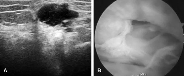
Fig. 1. A 50-year-old female diagnosed with a Medical meniscus radial tear. (A) Sonographic finding. (B) Arthroscopic finding.
A B
측 반월상 연골판 이상 시 양성 예측율 81.2%, 음 성 예측율 78.7%, 민감도 65.0%, 특이도 89.6%이 었다(Table1). 낭종 소견은 100%에서 정확도가 있었다. 수술 중이나 수술 후에 감염 등 다른 합병증 은 발생하지 않았다.
고 찰
슬관절 반월상 연골판 병증의 진단을 위하여 현재 자기 공명 영상 촬영이 널리 보급되면서 고가임에도 불구하고, 선택적 진단법으로 사용되고 있다. 그러
나 슬관절 증상을 호소하는 많은 환자들이 금전적인 문제로 진단 및 치료에 어려움을 겪고 있어, 최근에 는 비교적 저가이고, 사용이 간편하며 역동적 검사 가 가능한 초음파에 대한 관심이 높아지고 있다.
반월상 연골판 병증에 대하여, Tomasella 등7) 에 의하면 슬관절 내 초음파가 1차적인 진단법이라고 하였고, Friedl 등1)에 의하면 슬관절의 인대와 반월 상 연골판 손상에 대하여 이학적 검사 후 1차적 진 단 검사로 초음파를 사용해야 한다고 하였다. 또한 Najafi 등4) 에 의하면 슬관절 병증에 대한 초음파의 민감도는 100%, 특이도는 95%, 양성 예측율은 내
Table 1A. This table shows comparative relationships of positive or negative results of ultrasonographic and arthroscopic finding on medial meniscus and lateral meniscus pathology of our study
Arthroscopy (+) Arthroscopy (-)
Medial meniscus Ultrasonography (+) 10 01
Ultrasonography (-) 04 34
Lateral meniscus Ultrasonography (+) 13 03
Ultrasonography (-) 07 26
Table 1B. This table shows sensitivity, specificity, positive predictive value, negative predictive value from results of ultrasonographic and arthroscopic finding on medial meniscus and lateral meniscus pathology of our study
Ultrasonography
Lateral Meniscus Medial Meniscus Average
Sensitivity 65.0% 71.4% 67.6%
Specificity 89.6% 97.1% 93.8%
Positive Predictive value 81.2% 90.9% 85.4%
Negative Predictive value 78.7% 89.4% 84.4%
Fig. 2. A 41-year-old female diagnosed with a meniscal cyst. (A) Sonographic finding. (B) Arthroscopic finding.
A B
측 반월상 연골판 파열 95%, 외측 반월상 연골판 파열 93%, 음성 예측율은 내, 외측 반월상 연골판 파열 모두 100%라 보고하여 슬부 병변의 진단에 있어 초음파가 매우 유용하다 하였으며, Grifka 등2) 은 반월상 연골판 손상에 대하여 임상적인 이학적 검사와 초음파 검사의 결과를 비교하였는데, 민감도 는 비슷한 결과를 얻었으나, 특이도에서는 초음파가 훨씬 우수한 결과를 얻었다. 그의 연구에서 초음파 검사상 특이도는 외측 반월상 연골판이 98%, 내측 반월상 연골판이 90%의 결과를 보였고, 이학적 검 사상 특이도는 외측 반월상 연골판 47%, 내측 반월 상 연골판 81%의 결과를 보였는데, 이것은 이학적 검사가 위양성이 많았기 때문이며, 특히 젊은 환자 에서 근육 경련, 급성 외상 시 위양성이 많아 초음파 검사가 유용하다고 하였다. 그러나 Holzach 등3) 에 의하면 초음파는 아직 임상적인 중요성을 갖지 못하 고 실험적인 사용에 한정되는 진단방법이라 하였으
며, Sandhu 등5)에 의하면 최근에 고해상 초음파가 개발되어 반월상 연골판 손상에 대해 높은 민감도를 나타내는 결과를 보였으나, 정확한 파열의 위치, 정 도에 대한 정보를 얻는 것은 자기공명영상 촬영과 비교하여 덜 우수한 결과를 보였다고 하는 등 아직 슬관절 병증의 진단에 있어 초음파의 유용성에는 다 양한 의견이 있는 것이 현실이다.
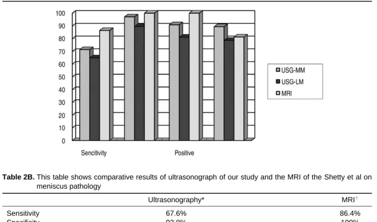
본 연구에서는 슬관절 병변에 대한 초음파의 결과 와 Shetty 등6) 의 자기 공명 영상 촬영 결과를 비교 한 결과에 의하면, 초음파상 반월상 연골판 이상소 견은 자기 공명 영상 촬영보다 민감도는 상당히 낮 았으나(초음파 평균 67.6%, 자기 공명 영상 촬영 86.4%), 특이도는 비슷한 결과를 보였다(초음파 평균 93.8%, 자기 공명 영상 촬영 100%)(Table 2). Shetty 등6)의 연구에서는 초음파가 자기공명영 상 촬영과 비교하여 초음파의 특이도가 86.4%, 자 기공명영상 촬영의 특이도가 100%의 결과를 보여
Table 2A. This graph shows comparative results of ultrasonograph of our study and the MRI of the Shetty et al on meniscus pathology. (USG=Ultrasonography, MM=medial meniscus, LM=lateral meniscus)
Table 2B. This table shows comparative results of ultrasonograph of our study and the MRI of the Shetty et al on meniscus pathology
Ultrasonography* MRI
�Sensitivity 67.6% 86.4%
Specificity 93.8% 100%
Positive Predictive value 85.4% 100%
Negative Predictive value 84.4% 81.3%
* The average results of our study about the sonography on knee joint (Including MM and LM).
�
The average results of the Shetty et al
6)about the MRI on knee
초음파가 열등한 결과를 얻었으나, 본 연구 결과에 서는 특이도에서도 초음파와 자기공명영상 촬영이 대등한 결과를 보여 Shetty 등6)과는 다른 결과를 보였다. 특히 외측 반월상 연골판은 내측 반월상 연 골판보다 민감도와 특이도가 낮았는데, 이는 외측 반월상 연골판은 슬와공(popliteal hiatus)이 있는 관계로 파열과 구별이 힘들기 때문으로 사료된다.
본 연구 결과에서 보여주듯 초음파 검사의 특이도 가 우수하여 자기공명영상 촬영의 특이도와 비슷한 결과를 보였다. 이것은 관절경 검사 전에 초음파검 사를 시행하는 것이 위양성 관절경 시술을 줄일 수 있다는 것을 의미하므로, 필요 없는 수술적 치료를 피할 수 있고, 비용이 저렴하고, 간편하게 검사가 가 능하며, 역동적 검사가 가능하다는 점에서 우수한 진단법으로 사료된다. 그러나, 한계점으로 검사자에 따라 다양한 결과가 나올 수 있어, 초음파에 대한 충 분한 지식과 숙달이 필요하다고 사료된다.
결 론
슬관절의 자기 공명 영상 촬영과 비교하여 초음파 검사는 비용, 간편성 측면에서 관절경 시술을 시행하 기 전에 관절 내 병변을 확인하여 시술여부 결정에 도움을 주는 아주 유용한 검사방법으로 사료된다.
참고문헌
01)
Friedl W, Glaser F: Dynamic sonography in the diagnosis of ligament and meniscal injuries of the knee, Arch Orthop Trauma Surg, 110: 132-8, 1991.
02) Grifka J, Richter J, Gumtau M: Clinical and
sonographic meniscus diagnosis. Orthopade, 23:
102-11, 1994.
03) Holzach P, Mattli J, Benz K, Streicher U and
Matter P: Ultrasonography of meniscus lesions.
Sportverletz Sportschaden, 4: 135-8, 1990.
04) Najafi J, Baqheri S and Lahiji FA: The value
of sonography with micro convex probes in diag- nosing meniscal tears compared with arthroscopy. J Ultrasound Med, 25: 593-7, 2006.
05) Sandhu MS, Dhillon MS, Katariva S, Gopal
V, Nagi ON: High resolution sonography for analysis of meniscal injuries. J Indian Med Assoc, 105: 49-50, 2007.
06) Shetty AA, Tindall AJ, Jajmes KD, Relwani
J and Fernando KW: Accuracy of hand-held ultrasound scanning in detecting meniscal tears.
J Bone Joint Surg Br, 90: 1045-8, 2008.
07) Tomasella G, Turra S, Olmeda A, Soliman A