Received: Nov, 6. 2014./Accepted for Publication: Dec. 30.
2014.
Corresponding Author: 정재호
주 소 서울시 동대문구 회기동 1번지, 경희의료원 PACS팀 Tel: 02-958-9484
E-mail: [email protected]
◈ 원 저 ◈
PACS환경에서 디지털유방엑스선 영상 화질에 관한 연구
정재호 경희의료원 PACS팀
The research on Full Field Digital Mammography Image Quality in PACS Environment
Jae-Ho Jung
Department of PACS, Kyung Hee University Hospital
4)
Abstract
The full-field digital mammography (FFDM), which has been known as a digital breast imaging system, carries out more outstanding performance than the screen-film mammography in overall image quality, skin & nipple, description of pectoral muscle and expression of micro-calcification.
Thus, in this thesis, I perform experiments for both the enhancement of image quality and accurate estimation of the result in question, when detecting the very tiny-sized lesions in mammography. The image of digital breast X-rays is the important diagnostic tool for detecting early breast cancer and micro calcification lesion.
The experiment of how much compression rate has an effect on the result of diagnosis in the case of microcalcification lesion, with JPEG2000 40:1 compression and over 50% enlargement led to obscure or definitely unacceptable diagnostic results is performed. And in another study of assessment of PSNR degree. I recognized the importance of standardized management system in mammography, where not to mention the accurate reading of the image has the most crucial role in diagnosis
Keyword : PACS, JPEG2000, Full field digital mammography
Ⅰ. 서 론
유방촬영술(Digital mammography)은 유방암의 조기 진단에서 매우 중요한 진단방법으로서 비 촉지성 유방 암의 조기 발견율을 높여 유방암에 의한 여성의 사망률 을 감소시키고 있다1). 종전의 Screen-film 시스템에서 Digital mammography 시스템으로 대체되어 관용도가 넓고, 노이즈가 적으며, 신호 대 잡음비가 높은 장점을 가지고 있어 유방암을 진단하는데 많은 도움을 주고 있 다2,3). 2009년 미국의 Cancer statistics에 따르면 유방 암이 여성의 암 발병률 1위, 사망률 2위로 여성에 있어 가장 흔한 악성종양으로서 점차 증가하는 추세이다4). 유방암 조기 진단을 위해 자가 촉진 이외에 Screening 검사로 Mammography가 널리 사용되고 있다. Full- Field Digital Mammography (FFDM)는 전반적인 화 질, 피부와 유두, 대흉근의 묘사, 미세석회화 표현등 기존의 screen-film mammography(SFM) 타입보다 뛰 어나다5-6). Picture Achieving and Communication System(PACS) 운영 환경의 도입으로 SFM 타입의 아 날로그 환경은 급속도로 막을 내리고 FFDM 영상장비 가 널리 사용되고 있다. 기존의 전통적인 SFM 타입 환경에서는 유방촬영술 팬텀을 촬영하여 방사선장비, 필름과 카세트의 조합, 현상기에 의해 얻어진 필름의 농도, 대조도, 균일성등 영상의 질이 적정 수준으로 유 지 되는지를 관리하였다7). 디지털유방 엑스선 영상검출 기에 있어서는 디텍터의 종류, 선질, 영상압축방법에 따른 다양한 요소들이 영상의 화질에 관여한다. 따라서 본 논문에서는 영상의 화질에 관여되는 영상의 압축율, 확대율에 따른 영상의 화질변화를 연구하고자 하였다.
유방촬영영상에 대한 진단 시 영상의 배율을 조정하 여 판독하는 경우가 많다. 이에 영상 압축 적용 시 미 세한 모양의 병변 진단에 영향이 있을 것으로 판단되어 압축률 및 압축방법에 따른 임상적 영향을 정성적, 정 량적 측정법을 통해 의료영상의 화질의 변화에 대한 데 이터 정보를 구축하여 의료영상의 압축 영향 평가기준 을 제시 하고자 하였다. 실제 초미세병변 검출영상을 선택하여 확대율(0%, 50%, 100%)에 따른 영상의 화질 비교를 실시하였다. 배율변화에 따른 JPEG 2K, JPEG 압축률의 변화를 비교 평가하였다.
Ⅱ. 대상 및 방법
1. 실험대상
본 연구에서 사용한 유방촬영용 검사 장치는 2011년 도입된 Senographe DS(GE healthcare, Milwaukee, Wisconsin, USA)이며 실험 전 장치의 품질관리기준을 이수 하였다. 본 실험에 사용된 phantom은 Fig 1.의 지방조직과 유선조직 50:50의 비율로 구성된 두께 4.2cm ACR Accreditation image quality phantom (FLUKE Biomedical, 18-220)을 이용하여 촬영하였다.
Fig 1. ACR Accreditation image quality control phantom
2. 실험방법
정량적 화질평가 방법으로서 PSNR(Peak Signal to Noise Ratio), RMSE(Root Mean Squared Error), MAE(Mean Absolute Error) SSIM(Structural SIMilarity)등을 측정하여 상대적 평가를 실시한다.
Raw데이터와 압축영상간의 왜곡측정을 위해 Image J 를 이용하여 평가하였으며 먼저 JPEG2000알고리즘을 이용한 압축영상의 화질이 Raw데이터와 노이즈발생 정 도를 평가하기위해 Signal to Noise Ratio(SNR)를 구 하였다. Raw데이터와 압축영상간의 같은 양의 데이터 에 대해 동일한 위치에 대해 표준편차를 계산하기 위해 RMSE(Root Mean Square Error)를 측정 하였으며 Raw데이터와 압축영상간의 오차들의 절대값의 평균 등 을 측정하기 위해 MAE(Mean Absolute Error)를 측정
하였다. Full Reference 영상화질평가 방법 중 luminance change, contrast change, structural change를 감지할 수 있는 구조적 유사도(Structural SIMIlarity: SSIM)를 측정하였다.
정성적 화질평가방법에서 본 연구에 사용된 Radiation Quality는 공칭 초점값(nominal focal spot) 0.4를 넘지 않는 조건에서 IEC 61267에서 제정 된 선질을 이용하였다. Fig 1 과 같은 모양의 ACR Accreditation image quality phantom을 이용하여 측 정하였다. 팬텀영상의 분석은 각각의 요소들(Fibers, Specks, Masses)의 크기가 큰 것부터 순서대로 평가하 며 Fiber의 모든 길이가 다 보이고 위치와 방향이 올바 르면 1점을 부여하고, 다 보이진 않지만 1/2이상 보이 고 위치와 방향이 올바르면 Fig 2. 섬유소 검출 점수의 예와 같이 0.5점을 부여한다.
Fig 2. Example for fiber detection score
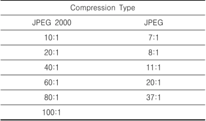
촬영모드는 AEC Standard를 선택하였으며 target 모드는 Molybdenum/Rhodium (Mo/Rh)선질을 선택하 였다. 이렇게 획득되어진 영상에 JPEG 2K, JPEG 압축알고리즘 JPEG2000 영상 압축은 페가수스 키트 (PICTools 32-Bit Imaging Development Kits, Pegasus Imaging Corporation, Tampa, FL. U.S.A) 을 적용하여 [Table 1]에서와 같은 비율로 압축을 실시 한다. 판독 전문의 3명이 각각 압축된 영상의 Detection score를 작성하였다. 또한, 실제 미세질환 영상 5장을 선택하여 관찰자 5명이 각 영상에 대해 5회 씩 관찰하여 그 평균값을 도출하였으며 실험방법은 압 축률(10:1, 20:1, 40:1, 60:1, 80:1, 100:1)과 확대율 (50%, 100%)의 차이에 대해 5단계의 평가 (1:
definitely unacceptable for diagnosis, 2: probably unacceptable, 3:equivocal, 4: probably acceptable, 5: definitely acceptable for diagnosis)를 실시하였 다. 압축률에 따른 정량적 화질평가를 위해 디지털 mammography 영상장치를 대상으로 [Table 1]과 같이 Raw데이터를 각각 압축하였다.
Table 1. Apply compression ratio
Compression Type
JPEG 2000 JPEG
10:1 7:1
20:1 8:1
40:1 11:1
60:1 20:1
80:1 37:1
100:1
Ⅲ. 결과
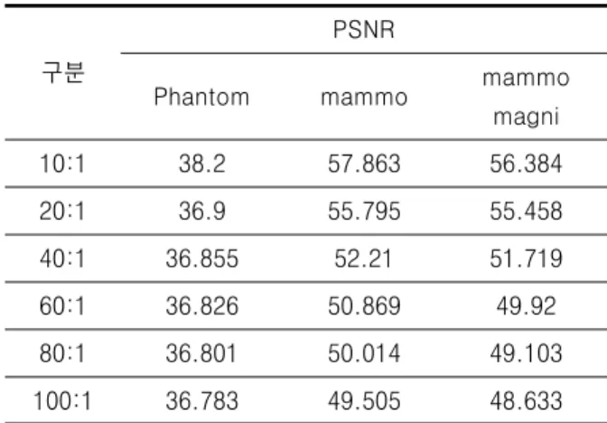
본 연구에서 영상의 종류별 압축률의 영향을 알아보 기 위해 Mammo ACR Phanatom, 일반 Digital mammography, magnification mammography 영상을 대상으로 각각의 압축률별 PSNR, RAMSE, MAE, SSIM등을 측정하였다. 그 결과 JPEG 2000, 20:1 압축 에서 PSNR값이 ACR Phantom 36.9db로 mammo 55.8db, mammo magni 55.5db,보다 낮았으며 RMSE 값은 ACR Phantom 이 55.2로서 5.02, 6.9를 나타낸 mammo, mammo magni보다 현저히 높게 나타났 다.ACR Phantom영상이 JPEG 2000 압축에서 mammography영상보다 수학적 왜곡도가 상대적으로 심한 것을 알 수 있었다. 하지만 그에 반해 JPEG 압축 알고리즘은 Phantom영상, mammography, magnification mammography 영상 모두에서 균일한 값을 나타내었다. SSIM 측정값은 압축률에 따른 유의 미한 변화값은 도출되지 않았다.
Table 2. PSNR according to Compression ratio
구분
PSNR
Phantom mammo mammo
magni
10:1 38.2 57.863 56.384
20:1 36.9 55.795 55.458
40:1 36.855 52.21 51.719
60:1 36.826 50.869 49.92
80:1 36.801 50.014 49.103
100:1 36.783 49.505 48.633
Table 3. RMSE according to Compression ratio
구분
RMSE
Phantom mammo mammo
magni
10:1 54.1 4.356 6.209
20:1 55.2 5.052 6.908
40:1 55.483 7.633 10.624
60:1 55.67 8.906 13.069
80:1 55.834 9.828 14.358
100:1 55.944 10.421 15.156
Table 4. MAE according to Compression ratio
구분
MAE
Phantom mammo mammo
magni
10:1 26.4 2.802 2.602
20:1 27.789 3.771 3.065
40:1 28.264 5.113 5.212
60:1 28.547 5.762 6.41
80:1 28.745 6.189 7.026
100:1 28.891 6.466 7.445
압축률별 모의병소 실험에서는 미세석회화(speck), 섬유소(fiber), 종괴(mass) 검출능력은 압축률에 따른 미세한 변경은 있었으나 최대 JPEG 2K 100:1 압축, JPEG 41:1 압축에서도 허용기준 10점 이상을 유지하였 다.
0 10 20 30 40 50 60 70
10대1 20대1 40대1 60대1 80대1 100대1
dbPSNR(JPEG 2K)
phantom
mammo
mammo magni
Fig 3. PSNR according to Compression ratio (JPEG2000)
0 10 20 30 40 50 60 70
8 10 13 23 41
DB
PSNR(JPEG)
phantom mammo
mammo magni
Fig 4. PSNR according to Compression ratio(JPEG)
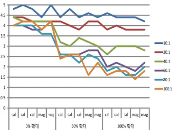
실제 미세병변영상 검출능력 실험에서는 확대하지 않 은 원본영상일 때 진단능 평가 에서 100:1까지도 평균 4.1 (probably acceptable)이상을 유지하였다. 50%확대 영상에서는 20:1 압축에서 4.1(probably acceptable) 유지하였으나 40:1이상 압축에서는 측정값 평균 3.2(equivocal) 판독 모호 함으로 나타났다.
0 0.5 1 1.5 2 2.5 3 3.5 4 4.5 5
cal cal cal mag mag cal cal cal mag mag cal cal cal mag mag
0% 확대 50% 확대 100% 확대
10:1 20:1 40:1 60:1 80:1 100:1
Fig 5. Detection Score for the actual micro resions mammography
Ⅳ. 고찰 및 결론
디지털 유방엑스선 영상의 정량적 화질평가에서 JPEG 2000압축에서는 그림5에서처럼 압축률에 의한 변화보다는 영상의 특성이 고주파영역이 많이 포함되며 미세병소 부분이 많이 포함될수록 PSNR, RMSE, MAE 값에서 영상의 수치적 왜곡이 많이 발생된다는 것을 알 수 있었다. 실제 미세병변영상의 평가에서는 JPEG 2000 20:1 영역에서는 100%확대율까지 진단 가능한 허 용범위이나 40:1압축 이후부터는 50%영역부터 판독 모호 하거나 명확히 판독 불가한 영역으로 나타났다.
따라서 압축률에 따른 진단능의 변화에서는 확대율 1:1 영상일때는 판독에 별다른 지장이 없다고 추정되었으나 50% 이상 확대 시 판독이 모호하거나 판독불가능 영상 의 양상을 보여 이에 대한 고려가 있어야 할 것으로 사 료된다. 일반적인 고유의 mammography 영상과 magnification mammography 영상에서는 JPEG2k 압 축에서 PSNR수치는 최대 압축인 100대1에서도 비교적 우수한 49.5 dB, 48.6dB를 나타내어 압축에 따른 영상 의 열화는 크지 않은 것으로 나타났으며 JPEG압축에서 40:1 압축에서도 51.9dB로 측정되어 압축알고리즘에 의한 변인은 크지 않음을 알 수 있었다. 이상과 같은 내용으로 ACR Phantom을 이용한 모의병소 실험을 통 해 확대율에 따른 모의병소 검출능력은 거의 변화가 없 음을 인지하였고 압축률에 따른 정량적 평가 수치 또한 허용범위 내였으나 다만, 고주파영역의 영상 또는 미세 한 병변영상 진단 시 노이즈발생 또는 왜곡의 발생 가
능성이 있음을 알 수 있었다.
실제 미세병변영상의 확대율에 따른 진단능의 변화에 서는 압축률이 미치는 영향이 매우 크며 의료기관에서 압축률 지정 시 전문판독의의 판독 시 확대 판독에 대 한 정보를 숙지하고 이에 대한 신중한 압축률을 설정하 여야 하겠다.
Ⅴ. 참고문헌
1. Belbachir. A. and Goebel. P., "Medical Image Compression: Study of the Influence of Noise on the JPEG 2000 Compression Performance,"
International Conference on Pattern Recognition, vol. -, pp. 893-896, 2006.
2. Hamamoto. K., "Study on Medical Ultrasonic Echo Image Compression by Jpeg2000 - Optimization and the Subjective Assessment of the Quality -," Proceedings of the Annual International Conference of the IEEE Engineering in Medicine and Biology Society, vol. 25, pp. 833-836, 2003.
3. Haseeb. S. and Khalifa. O. O., "MEDICAL IMAGE COMPRESSION EVALUATION BY JPEG AND JPEG 2000," Proceedings of the International Conference on Computer and Communication Engineering -Cd-Rom Edition-, vol. -, pp. 1049-1053, 2006.
4. Karssemeijer. N., Frieling. J. T. M. and Hendriks. J. H. C. L., "Spatial Resolution in Digital Mammography," Invest. Radiol., vol.
28, pp. 413, 1993.
5. Harvey. J. A. and Bovbjerg. V. E., "Quantitative Assessment of Mammographic Breast Density:
Relationship with Breast Cancer Risk,"
Radiology, vol. 230, pp. 29-41, 2004.
6. Elmore. J. G., Wells. C. K., Lee. C. H. and Howard. D. H., "Variability in radiologists' interpretations of mammograms," ACP J. Club, vol. 122, pp. 74, 1995.
7. G. Gennaro, G. Gennaro and C. di Maggio,
"Dose comparison between screen/ film and full-field digital mammography ," Eur. Radiol., vol. 16, pp. 2559-2566, 2006.