INTRODUCTION
According to the report of the National Cancer Information Center of Korea in 2001, the occurrence rates of bladder cancer in Korea are 7.67 and 2.00 per
100,000 in men and women, respectively. Urothelial carcinoma was the most common histologic subtype in this report, and it comprised 81.8% of the newly diag- nosed bladder tumors. Urothelial carcinoma can easily be managed by local excision when it is diagnosed
형태 계측학적 분석과 ThinPrep ® 액상 소변세포검사를 이용한 악성 요로상피 세포 검출
고려대학교 구로병원 병리과,1 고려대학교 구로병원 동결폐조직은행2
신봉경
1,2·이 영석
1·정회선
1,2·이상호
1,2·김현철
1·김애리
1,2·김인선
1·김한겸
1,2Urothelial carcinoma accounts for 90% of all the cases of bladder cancer. Although many cases can be easily managed by local exci- sion, urothelial carcinoma rather frequently recurs, tends to progress to muscle invasion, and requires regular follow-ups. Urine cytology is a main approach for the follow-up of bladder tumors. It is noninvasive, but it has low sensitivity of around 50% with using the conventional cytospin preparation. Liquid-based cytology (LBC) has been devel- oped as a replacement for the conventional technique. We compared the cytomorphometric parameters of ThinPrep® and cytospin prepa- ration urine cytology to see whether there are definite differences between the two methods and which technique allows malignant cells to be more effectively discriminated from benign cells. The nuclear-to- cytoplasmic ratio value, as measured by digital image analysis, was efficient for differentiating malignant and benign urothelial cells, and this was irrespective of the preparation method and the tumor grade.
Neither the ThinPrep ® nor the conventional preparation cytology was definitely superior for distinguishing malignant cells from benign cells by cytomorphometric analysis of the adequately preserved cells.
However, the ThinPrep® preparation showed significant advantages when considering the better preservation and cellularity with a clear background.
(Korean J Cytopathol 2008;19(2):136-143)
Key Words : Liquid-based urine cytology, Urothelial carcinoma, Cytomorphometric analysis, Digital image analysis, Nuclear-to-cytoplasmic ratio.
Detecting Malignant Urothelial Cells by Morphometric Analysis of ThinPrep ® Liquid-based Urine Cytology Specimens
Bong Kyung Shin, M.D.,
1,2Young Suk Lee, M.D.,
1Hoiseon Jeong, M.D.,
1,2Sang Ho Lee, M.D.,
1,2Hyunchul Kim, M.D.,
1Aree Kim, M.D.,
1,2, Insun Kim, M.D.,
1and Han Kyeom Kim., M.D.,
1,2Department of Pathology, Guro Hospital,
Korea University Medical College
1and Korea Lung Tissue Bank (KLTB)
2, Seoul, Korea
논문접수 : 2007년 7월 18일 논문수정 : 1차 : 2007년 11월 20일,
2차 : 2008년 2월 22일, 3차 : 2008년 7월 14일 게재승인 : 2008년 7월 28일
책임저자 : 김 한 겸
주 소 : (152-703) 서울 구로구 구로동 80번지, 고려대학교 구로병원 병리과
전 화 : 02-818-6872 팩 스 : 02-818-6239
E-mail address : [email protected]
*이 연구는 2006년 6월 대한세포병리학회로부터 연구비 지원을 받아 진행되었습니다.
원 저
early; 1 however, these tumors recur rather frequently.
According to Saad A et al., about 70% of superficial bladder urothelial carcinomas will recur in the first five years following transurethral resection (TUR), and 10- 20% will progress to muscle invasion. 2 Therefore, those patients treated for bladder urothelial carcinoma should be closely followed up to detect recurrent diseases.
Although cystoscopy with biopsy is the gold stan- dard for the diagnosis and follow-up of bladder can- cers, this process requires the combined use of other diagnostic approaches because of the invasiveness of the cystoscopy procedure, the considerable discomfort to the patients and the inability to detect all carcinoma in situ cases and lesions of the upper urinary tract. 3,4 Therefore, urinary cytology is another important approach for making the diagnosis and follow-up of bladder cancer. This procedure is noninvasive and quite sensitive for high grade urothelial lesions irrespec- tive of their location in the urinary tract. 3,4 However, urinary cytology has a mean sensitivity of just around 50% for low grade tumors urinary cytology, and this procedure is hampered by the large number of non- diagnostic samples. 5, 6
Liquid-based cytology (LBC) using a filtration process and thin layer deposition of cells has been developed to replace the conventional cytospin preparation tech- nique, and it is expected to improve the cell recovery capabilities and offer better cell preservation. 3 Many studies have shown that preparation with the Cystic ThinPrep ® 2000(Cystic Corp, Boxborough, Massachusetts, USA) resulted in increased cellularity and a pronounced reduction of inflammatory debris, red blood cells and crystals. 7-11 However, the advantages of LBC have been questioned by other studies that compared LBC with the cytospin preparation. For example, Nassar et al., in their analysis of seventy-nine urine samples, calculated that although the ThinPrep ® method was simpler to perform and it produced clean, easier-to-read slides, the cytospin preparation was more efficient for detect- ing malignant cells with less technical artifacts. 12 Piaton et al. also proposed that the cytocentrifugation method
still offered the highest quality for the current treatment of urinary samples, when considering both the diagnos- tic performance and the cost. 3
For urine cytology, further investigation is needed to determine the efficiency of liquid-based cytology to detect urothelial carcinoma cells. Besides the change in preparation technique, cytomorphometric analysis of urothelial cells that includes the DNA contents, the nuclear area, the cytoplasmic area and the nuclear-to- cytoplasmic ratio has been proposed as another sensi- tive and specific tool for detecting malignant cells in urine cytology. 13,14 Van der Poel et al., in their study that compared digital image analysis and conventional microscopic examination of urinary washing cytology, they measured the DNA contents and nuclear morphol- ogy of Feulgen-stained urothelial cells with using an automated image analysis system, i.e. QUANTICYT, and they concluded that cytomorphometric analysis was superior to conventional urine cytology for detect- ing malignant cells. 14 However, van der Poel et al.'s study used bladder washing specimens instead of the conventional voided urine, and this required Feulgen- stained slides rather than the usual Papanicolaou stain, and they used the DNA content and nuclear morpholo- gy as measured by a specific karyometric system QUANTICYT as the major parameters for detecting malignant cells. When considering the invasiveness of acquiring the specimens, the requirement for specific equipment and the complex process for measuring and interpreting the morphometric parameters, van der Poel et al.'s approach may not be easily applied to the pres- ent clinical environment. Another study by Bishop and Sims evaluated the feasibility and utility of a thin-layer cytology preparation for morphometric analysis of non- gynecologic specimens, including urine. They pro- posed that the thin-layer preparation cytology had sig- nificant advantages for morphometric studies over the conventional preparation, although any differences of the morphologic measures were not observed between the two cytology procedures. 13
In this study we investigated whether the cytomor-
phometric analysis of urothelial cells can adequately discriminate malignant urothelial cells from benign urothelial cells. We also compared the cytomorphomet- ric parameters of ThinPrep® with those of cytospin preparation urine cytology to determine whether there are definite differences between the two techniques and which technique is better for discriminating benign cells from malignant cells.
MATERIAL AND METHODS
Case Selection
Thirty-four ThinPrep® urine cytology specimens and 50 conventional cytospin preparation urine cytology specimens were selected from the pathology files of Korea University Guro Hospital, and all of these were cytologically and histologically confirmed as urothelial carcinoma. The urine cytology specimens of 100 benign cases, (50 ThinPrep® and 50 conventional preparations) were also selected for comparison. Of the 34 ThinPrep® preparation cases of urothelial carcino- ma, 17 were high-grade and 17 were low-grade tumors, while the 50 cytospin preparation cases of carcinoma were composed of 28 high and 22 low grade tumors.
All the slides were the same Papanicolaou-stained ones that were examined at the time of the original diagnos- tic process with conventional light microscopy.
Digital Image Analysis
All the slides were submitted to digital image analysis with using ImagePro® 6 software. Morphometric parameters such as the cell size, the nuclear area, the cytoplasmic area and the nuclear/cytoplasmic area ratio were measured for both the benign and malignant urothelial cells. Well-preserved, non-overlapping and/or individually scattered cells were usually select- ed, and some loosely clustered, easily discernable cells were also accepted and measured. Tight three-dimen-
sional clusters or papillae were excluded; squamous cells, either contamination or metaplastic, were not sub- mitted to digital image analysis. In the ThinPrep®
preparation group, the number of malignant cells, both in the high- and low-grade tumors that were adequate for image analysis, was usually between 20 and 50 observed per slide, and only rarely did they exceed 100. In the benign group, 10 to 30 urothelial cells were usually observed per slide, including both the superfi- cial and intermediate cells, except for a few inflamma- tory conditions that showed more than 50 or rarely 100 cells per slide, including numerous severely degenerat- ed ones. In the cytospin group, the number of ade- quately preserved cells was markedly smaller. The malignant group still showed more than 20 cells; how- ever, the benign group usually produced less than 10 sufficiently well-preserved cells, including both the superficial and intermediate cells. Up to 50 adequate, superficial or intermediate urothelial cells were ana- lyzed from one slide.
In each of the ThinPrep® and cytospin groups, the differences in morphometric parameters were separate- ly compared between the benign and malignant cells, with and without classifying the malignant tumors into high and low grade. Those parameters that were signifi- cantly different between the benign and malignant cells were again compared between the ThinPrep® and cytospin groups to investigate which preparation pro- duced more distinct differences between the benign and malignant cells.
Statistics
T-tests were used to determine the p values, and p values ≤ 0.05 were considered significant.
RESULTS
Both the liquid-based and cytospin preparation tech-
niques yielded individual cells that were suitable for
morphometric analysis. In some of the malignant cases, the urine cytology showed cell clusters of papillae with irregular borders, variable sized and shaped nuclei with fine granular chromatin and small nucleoli (Fig. 1A). In the higher grade malignant cases, the cells were signifi- cantly pleomorphic and individually scattered or they formed small, loose clusters or syncytials; the cells dis- played coarse chromatin, prominent nucleoli and fre- quent apoptosis in a rather dirty background of inflam- mation, hemorrhage and/or necrosis(Fig. 1C). The ThinPrep ® liquid-based preparation generally present- ed well-preserved cells with a relatively clean back- ground in both the malignant and benign cases (Fig. 1 and Fig. 3). In the malignant cases, the cytospin prepa- ration showed an adequate number of atypical cells for making the diagnosis in spite of the occasional cases with a dirty background and cellular degeneration (Fig.
2). However, in the benign cases, the cytospin prepara- tion generally displayed lower cellularity and more cel- lular degeneration than did the ThinPrep ® preparation (Fig. 3).
On the digital image analysis, the nuclear-to-cyto- plasmic area ratio showed significantly distinct values between the benign and malignant urothelial cells in both the ThinPrep ® and conventional urine cytology.
In the cytospin preparation, the mean nuclear-to-cyto- plasmic area ratio was higher for the malignant urothe- lial cells than that for the benign urothelial cells (29.2%
versus 16.4%, respectively (p<0.05)) (Table 1). In the
ThinPrep ® , the mean value of the nuclear-to-cytoplas- mic area ratio was also significantly higher for the malignant urothelial cells (29.5%) than that for the benign (17.2%) urothelial cells (p<0.05) (Table 1).
However, on comparing the value of the nuclear-to- cytoplasmic area ratio of the ThinPrep ® with that of the conventional cytospin urine cytology for the malignant urothelial carcinoma group, they were very similar to each other (29.5% versus 29.2%, respectively). Further, the values of the nuclear-to-cytoplasmic area ratio for the benign group were not significantly different between the ThinPrep ® and conventional cytospin urine cytology (17.2% versus 16.4%, respectively) (Table 2).
On classifying the urothelial carcinoma into the high- and low-grade groups, the mean nuclear-to-cytoplas- mic area ratios of the malignant cells of the two groups were 32.3% and 28.7% in the ThinPrep ® preparation, and these values were 31.7% and 26.5% in the conven- tional preparation cytology, respectively. In both prepa- rations, the mean nuclear-to-cytoplasmic area ratio of the high-grade tumor cells was higher than that of the low-grade cells, and the latter value was still significant- ly higher than that of the benign urothelial cells.
However, on comparing the nuclear-to-cytoplasmic area ratios of the malignant cells between the two preparation techniques, there were still no significant differences regardless of the grade of malignant cells.
As for the cellularity, the degree of degeneration and
Table 2. Comparison of malignant with benign urothelial lesions by ThinPrep® and conventional cytospin technique
Malignant Benign
ThinPrep® Conventional ThinPrep® Conventional
Nuclear-to-cytoplasmic area ratio (%) P value
29.5 29.2 17.2 16.4
>0.05 >0.05
Table 1. Comparison of ThinPrep® with cytospin preparation urine cytology in malignant and benign urothelial lesions
ThinPrep® Conventional
Malignant Benign Malignant Benign
Nuclear-to-cytoplasmic area ratio (%) P value
29.5 17.2 29.2 16.4
<0.05 <0.05
the background, the ThinPrep® preparation technique seemed to have some advantages. Although both the ThinPrep® and conventional cytology produced suffi- cient numbers of cells for making a diagnosis, preserva- tion of the individual cells seemed to be better with the ThinPrep® preparation, which demonstrated more eas- ily measurable single cells. The lack of an interfering background inflammation, blood and/or necrosis was also helpful in selecting interesting cells (Fig. 1 and 2).
Moreover, in the benign urine specimens, the cellularity and preservation were significantly superior in the ThinPrep® cytology (Fig. 3).
In summary, the value of the nuclear-to-cytoplasmic
area ratio, as measured by digital image analysis, was efficient for differentiating malignant urothelial cells from benign urothelial cells irrespective of the prepara- tion method and the tumor grade. Neither the ThinPrep® nor conventional cytospin preparation cytology was definitely superior to the other technique for distinguishing malignant cells from benign cells by cytomorphometric analysis of the adequately preserved cells. However, upon considering the better preserva- tion and cellularity with a clear background, the ThinPrep® preparation seemed to have some advan- tage.
A B
Fig. 1. ThinPrep® preparation cytology of urothelial carcinomas. (A) The cytological findings of a low grade urothelial carcinoma include cell clusters of papillae with irregular borders, variability in size and shape or nuclei with fine granular chromatin and small nucleoli (Papanicolaou stain). (C) In a higher grade tumor, the cells are significantly pleomorphic, individually scattered, or form small, loose clusters or syncytia in rather dirty background of inflammation, hemorrhage and/or necrosis (Papanicolaou stain). B and D are the correspondant histologic findings of A and C, respectively (H&E).
C D
DISCUSSION
Although liquid-based urine cytology, with it's defini- tively superior cellularity and cell preservation, was once expected to replace the conventional cytology, the results of many studies that compared the efficiency of the two preparation techniques didn't agree one another. 3, 7-12 Van der Poel et al. proposed in one study that image analysis was superior to cytological analysis for predicting tumor recurrence after obtaining normal
findings via cystoscopic examination. 14 Bishop and Sims measured similar morphometric parameters of var- ious non-gynecological specimens, including body flu- ids, urine, respiratory aspirates and fine needle aspi- rates, and they compared the ThinPrep ® and conven- tional preparation techniques, but they didn't focused on urine specimens. 13 In this study we attempt, with using digital image analysis(ImagePro ® 6 software) and measuring the cytomorphometric parameters, to com- pare the effectiveness of the ThinPrep ® and cytospin preparation urine cytology for detecting urothelial cell carcinoma.
In the definitely confirmed urothelial carcinomas, the malignant cells were clearly discriminated by a cyto- morphometric parameter (the nuclear-to-cytoplasmic area ratio) from the benign urothelial cells in both the ThinPrep ® and cytospin preparations. The nuclear-to- cytoplasmic area ratio by itself may be a sensitive and specific discriminator to detect malignant cells in the urine cytology of any preparation technique. Yet when comparing the malignant urothelial cells observed in the ThinPrep ® and the cytospin preparation, there were no significant differences in the cytomorphometric parameters measured by digital image analysis.
Therefore, it cannot be claimed that for detecting malig- nant cells by cytomorphometric analysis, one prepara- tion is definitively superior to the other if an adequate
Fig. 2. Conventionally prepared cytology of urothelial carcino- ma. In malignant cases, cytospin preparation shows an ade- quate number of atypical cells for diagnosis in spite of the dirty background and cellular degeneration (Papanicolaou stain).
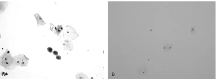
Fig. 3. ThinPrep® and conventional preparation cytology of a benign condition ("negative for malignancy"). ThinPrep® preparation shows well-preserved cells with relatively clean background in benign cases as well as in malignant ones (A) while the cytospin preparation presents a lower cellularity and more degeneration (B). (Papanicolaou stain).