돼지 관상동맥 스텐트 재협착에 대한 Paclitaxel Nanoparticle 국소전달요법의 효과
13
0
0
전체 글
(2) inhibits neointima formation after stent overdilation injury in porcine coronary arteries without systemic toxicity. (Korean Circulation J 2000;30( (2) ):208-220) ) KEY WORDS:Stent·Restenosis·Paclitaxel·Nanoparticle.. clitaxel과 같은 항microtubule 제제는 세포주기의 세. 서. 론. 포분열기 뿐만 아니라, 간기(interphase)에 있는 암세 포 및 정상세포 모두에 영향을 미치게 된다.. 관상동맥질환에 대한 치료법의 하나로서 관상동맥. 최근 쥐의 대동맥을 이용한 생체 외 연구에서 극소량. 중재술은 1977년 Gruentzig1)가 소개한 이후 다양한. (30 pmol~100 nmol)의 paclitaxel로 전처치한 경우. 첨단 의공학의 발달과 함께 시술 경험 축적으로 초기에. 에 혈관 평활근 세포의 이동과 증식, 혈소판 유도 성장. 는 단순한 병변만을 치료하였으나, 최근에는 심한 병변. 인자(platelet-derived growth factor;PDGF)의 자. 이 있는 관상동맥까지 시술이 가능하게 되었다. 또한,. 극에 의한 혈관 평활근 세포의 이동이 억제되었고, 쥐. 시술 성공률도 점차 높아져 관상동맥 우회술을 대신하. 의 경동맥을 이용한 생체 내 연구에서 풍선확장으로 내. 2)3). 다. 피세포를 탈락시키는 손상을 가한 후 종양을 치료하는. 양한 관상동맥 중재술 방법 중에서도 1987년부터 임. 용량보다 100배가 적은 혈중 농도(50~60 nmol)가. 상에서 이용되고 있는 관상동맥내 스텐트 시술은 풍선. 되도록 paclitaxel을 전신 투여(2 mg/kg)하여 중막의. 확장술에 의한 내막 박리를 효과적으로 치료할 수 있고. 혈관 평활근세포 증식과 신생내막의 혈관 평활근 세포. 항혈소판 제제의 사용에 따라 스텐트 혈전 형성이 감소. 축적을 예방하였다.9) Paclitaxel은 인간의 관상동맥 스. 되어 임상에서 널리 사용되고 있으며, 풍선확장술에 비. 텐트 재협착의 예방에 치료적 가치가 있으리라 여겨지. 해 재협착율도 감소시키는 경향이 있다. 그러나, 관상. 며, 인체에서의 생체 내 연구에 앞서서 이러한 효과에. 동맥 스텐트 시술 환자의 20~30%에서 발생하는 후기. 대하여 인체와 유사한 동물 모형을 이용한 생체내 연구. 스텐트 재협착이 여전히 중요한 임상적 문제로 남아있. 가 중요하리라 생각된다.. 여 관상동맥질환의 치료에 널리 이용되고 있다.. 다.4)5). 본 연구는 최근 쥐9)와 토끼10)의 경동맥을 이용한 연. 관상동맥 스텐트 재협착의 주요 기전은 신생내막. 구에서 혈관 평활근세포의 증식과 이동을 억제하는 효. (neointima)의 형성으로서 신생내막 형성에 의하여 후. 과가 있는 paclitaxel을 nanoparticle로 제조한 다음,. 기 내강소실(late lumen loss)이 발생한다고 알려져 있. 국소 전달요법(local drug delivery)을 이용하여 인간. 6)7). 다.. 최근에 스텐트 재협착의 예방 및 치료에 관하여. 과 유사한 관상동맥 재협착이 유도될 수 있는 동물 모. 새로운 시술 기구, 새로운 방법인 국소 약물, 유전자 혹. 형은 돼지로서,6)11) 관상동맥 내로 국소 투여하여 관상. 은 방사선 전달요법 등 다양한 시도가 이루어지고 있으. 동맥 스텐트 재협착에 대한 paclitaxel의 예방 효과를. 나, 그 효과는 아직 확실하지 않다.. 알아보고자 하였다.. Paclitaxel(Taxol)은 최근에 발견되어 항microtu-. 대상 및 방법. bule 제제로 분류되는 새로운 항암제로서 1970년대 중반 이후 항암 효과를 인정받고 1990년도에 Food & Drug Administration(FDA)으로부터 불응성 유방암과 8). 난소암에 사용 허가를 받은 약제이다.. 연구대상. Paclitaxel은. 돼지 관상동맥에 paclitaxel의 국소전달없이 스텐트. 세포 분열에 필요한 방추체(mitotic spindle)의 주요. 과확장 손상만을 가하였고(Ⅰ군;n=10), 다른 관상동. 구성 요소인 microtubule을 안정화시키고 micro-. 맥내로 paclitaxel nanoparticle 국소전달요법을 시행. tubule이 tubulin으로 depolymerization이 되는 것을. 한 후 스텐트 과확장 손상을 가하였다(Ⅱ군;n=10).. 차단함으로써, tubulin-microtubule dynamics를 파괴. 실험에 사용되었고 추적관상동맥 조영술 및 조직검사. 하여 일종의 세포고사를 일으키게 된다. 그러므로 pa-. 가 가능하였던 돼지는 모두 14마리이었고 스텐트는 근 209.
(3) 위부 좌전하행지 혹은 근위부 우관상동맥에 시술하였. 방 법. 고 좌전하행지와 우관상동맥이 3.0 mm 이상 컸던 돼 12). 지 6 마리에서는 두 개의 관상동맥에 각각 하나씩 두. 돼지 관상동맥 스텐트 재협착 모형 과 Paclitaxel의 국소. 개의 스텐트를 시술하였고 하나의 혈관이 작았던 돼지. 전달. 8마리에서는 좌전하행지 혹은 우관상동맥에 하나의 스. 동물실험은 전남대학교 의과학연구소 윤리위원회의. 텐트를 시술하였으며, Ⅰ군은 좌전하행지 6예, 우관상. 심의를 거쳐서 시행되었으며, 실험동물은 25~35 kg의. 동맥 4예,Ⅱ군은 좌전하행지 6예, 우관상동맥 4예로서. 암퇘지를 이용하였고 실험 종료 후 동물 사육사에서 4. 양 군간에 차이가 없었다.. 주 동안 관찰한 후 관상동맥 조영술 등의 추적 실험을 하고 희생시켜 심장을 적출한 다음 관상동맥 병변에 대. A. B. C. Fig. 1. Local delivery of Paclitaxel nanoparticle and stent overdilation injury of porcine left anterior descending coronary artery. After initial coronary angiogram (A), balloon and stent were chosen (A). After local delivery of Paclitaxel nanoparticle using the Dispatch Catheter, stent was inflated with stent to artery ratio (B). After stenting, overdilated stent and no edge dissection or flow limitation were proved (C).. A. B. Fig. 2. Porcine right coronary angiogram 4 weeks after stent overdilation injury with local delivery of paclitaxel nanoparticle. Overdilated stent was visualized in right coronary artery (A) and angiographic restenosis was not developed (B).. 210. Korean Circulation J 2000;30(2):208-220.
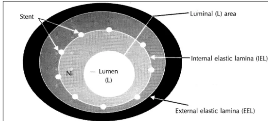
(4) 한 조직병리검사를 실시하였다. 관상동맥 손상 3일 전. 록공중합체(분자량:28,900, PEO 함:91.8 mol%). 부터 일일 aspirin 100 mg, ticlopidine 250 mg을 투. 10 mg과 Paclitaxel 2.5 mg을 10 ml의 DMF/THF. 여하였으며 실험돼지는 ketamine 12 mg/kg와 xyla-. (1/7:v/v)에 녹인 후에 이것을 molecular cut-off. zine 8 mg/kg를 근육 주사하여 마취하였다. Atropine. 12,000의 투석막안에 넣어서 멸균된 증류수를 이용하. 1 mg을 정맥주사하고 lidocaine으로 국소마취한 후 경. 여 투석하였다. 세 시간 간격으로 증류수를 교환하면서. 동맥을 절개하여 8 French 동맥관을 위치시켰다. 관상. 48시간동안 투석을 행하였다. 투석이 끝난 후의 nan-. 동맥 조영술 시술 직전에 동맥내로 Heparin 10,000. oparticle의 입자 크기는 dynamic light scattering으. unit를 주사하였고 목표한 관상동맥을 유도도자로 선별. 로 측정하였더니 평균 150~200 nm이었고 입자모양. 한 후, 스텐트(Wiktor stent, Medtronics, U.S.A.)가. 은 전자현미경으로 관찰하였더니 원형이었다. 블록공중. 감긴 풍선도자를 이용하여 스텐트:동맥 내경 비율을. 합체에 담지된 paclitaxel nanoparticle의 농도는 블록. 1.3 :1로 하여 관상동맥에 과확장 손상을 가하였다. 공중합체의 무게에 대하여 약 8~9 wt.%이었다.. (Fig. 1). 국소전달요법은 치료군에서 관상동맥 스텐트 과확장 손상 전에 실시하였으며, spiral perfusion inTM. 관찰 지표. fusion catheter인 Dispatch Catheter (Boston Sc-. 시술 직후와 4주 후에 시행한 관상동맥 조영술에 대. ientific, USA)를 이용하여 시행하였으며, 대조군과 비. 한 정량적 분석은 비디오 기록을 Kontron사의 Cardio. 교를 위하여 시술 전후로 관상동맥 조영술을 시행하여. 500 컴퓨터를 이용하여 하였고, 스텐트 시술을 한 근. 비디오에 기록한 후 정량적 관상동맥 조영술 분석을 하. 위부와 원위부 기준혈관의 내경, 스텐트 시술부위의 내경. 였다. 국소전달을 위한 주입압력(infusion pressure)은. 을 측정한 후 근위부와 원위부 기준혈관 내경의 평균값에. 13). 평균 4기압으로 하였다.. 추적 관상동맥 조영술과 조직. 병리검사는 스텐트 시술 후 28일에 실시하였다(Fig. 2).. 대하여 스텐트 시술부위 내경의 비율을 제외한 백분율로 내경협착(diameter stenosis) 정도를 평가하였다.. Paclitaxel nanoparticle은 30 μmol/L농도로 하여. 추적 관상동맥 조영술을 시행한 후 돼지는 potas-. 10 ml를 분당 1 ml 씩 10분 동안 국소에 전달하였다.. sium chloride 정맥주사로 희생시켰으며, 추출한 돼지. Paclitaxel nanoparticle의 국소 전달요법을 시행한 직. 심장은 10% 중성 buffered formalin으로 관류 고정하. 후 90% 알코올 용액에 관상동맥을 담근 후 자외선 활. 였다. 스텐트를 함유하고 있는 돼지의 관상동맥은 심장. 성도(ultraviolet activity)를 측정한 결과, 전체 전달된. 으로부터 추출한 후에 powerful light source를 이용한. 양의 약 10% 정도의 자외선 활성도가 관찰되었다. Pa-. 투시에 의해 쉽게 확인되었고, 조직 절개시에 돼지 관. clitaxel nanoparticle의 제조 방법은 이미 보고된 방법. 상동맥 스텐트는 쉽게 촉지할 수 있었다. 관상동맥 절. 14)15). 과 유사하게 투석방법으로 제조되었다. 즉, 소수성. 편은 스텐트의 상, 하부 1 cm까지 적출하였고 stent fi-. 인 poly γ-benzyl L-benzyl L-glutamate(PBLG). lament는 혈관을 2~3 mm 간격으로 절단한 후 혈관. 와 친수성인 poly ethylene oxide(PEO)로 구성된 블. 이 변형되거나 손상되지 않도록 현미경 시야에서 제거. Fig. 3. Calculation methods of neointima and histopathologic area stenosis in a porcine stent restenosis model. Neointimal area was calculated as:Internal elastic lamina area subtracted by luminal area and histo-pathologic area stenosis: (internal elastic lamina areaneointimal area)×100/(internal elastic lamina area).. 211.
(5) 하였다. 각 동맥 분절의 절단편은 Hematoxylin/Eosin. 통계학적 분석. 염색을 실시하였다. 또한 신생내막의 증식을 비교하기. 모든 자료는 평균±표준편차로 표시하였으며, pacli-. 위하여 증식세포 핵항원(proliferating cell nuclear an-. taxel 국소전달없이 스텐트 과확장 손상만을 시킨 대조. tigen;PCNA)에 대하여 면역조직화학법으로 염색을. 군(Ⅰ군)과 paclitaxel 국소전달후 스텐트 과확장 손상. 하였다. 모든 조직병리학적 측정 및 관찰은 병리학자와. 을 시킨 치료군(Ⅱ군)사이의 비교는 paired t test를 이. 함께 하였고 calibrated microscope(Leitz CBA 8000). 용하여 검정하였고 p 값이 0.05 미만인 경우에 통계학. 을 이용하여 확립된 방법16-18)에 따라 시행하였다(Fig.. 적 유의성을 두었다.. 3). 각 내경의 단면부위는 calibrated digital micros-. 결. copic planimetry로 측정하였으며, 스텐트 시술 혈관의. 과. 신생내막 두께(neointimal thickness), 중막 두께(medial thickness), 신생내막과 중막의 두께 비율(neoi-. 돼지 관상동맥내에 paclitaxel 국소전달없이 스텐트. ntima/media thickness ratio), 내강 면적(luminal area),. 과확장 손상을 시킨 대조군(n=10, Ⅰ군)과 paclitaxel. 신생내막 면적(neointimal area), 내탄력층 면적(internal. 국소전달후 스텐트 과확장 손상을 시킨 치료군(n=10,. elasticlamina area:IEL area), 중막 면적, 외탄력층. Ⅱ군)의 관상동맥 조영술 소견, 신생내막 형성 정도, 조. 면적(external elastic lamina area:EEL area) 등을. 직병리학적 협착 정도, 신생내막내 세포증식 정도는 다. 측정하였다. 스텐트 과확장 손상 방법에 의한 혈관 손. 음과 같았다.. 상의 평가는 Schneider 등19)이 중막의 파괴 정도에 의. 1) 정량적 관상동맥 조영술로 측정한 근위부 기준혈. 하여 분류한 조직학적 손상지수(histological injury. 관의 내경은 Ⅰ군 3.18±0.12 mm, 군 3.16±0.15 mm,. score)법을 변형하여 사용하였으며, IEL이 손상되지. 원위부 기준혈관의 내경은 Ⅰ군 2.93±0.26 mm,Ⅱ군. 않고 내피세포만 손상된 경우를 0점, IEL이 손상되고. 2.91±0.18 mm로서 두 군 사이에 유의한 차이는 없. 중막이 눌린 경우 1점, IEL과 중막이 파괴된 경우 2점,. 었다. 시술 직후 스텐트 과확장 손상에 의하여 스텐트. 중막과 EEL가 파괴된 경우를 3점으로 하였다. 스텐트. 시술부위의 확장된 내경은 Ⅰ군 3.29±0.17 mm,Ⅱ군. 근위부와 원위부 혈관을 기준혈관으로 하여 두 혈관의. 3.39±0.21 mm, 내경협착은 Ⅰ군 -7.94±2.58%, Ⅱ. 평균값을 기준혈관으로 하였다. 신생내막 면적은 혈관. 군 -11.90±7.74%로서 스텐트 과확장 정도는 두 군. 의 내탄력층 면적에서 내강 면적을 빼서 계산하였다.. 사이에 유의한 차이가 없었다. 스텐트 시술 4주후 근위. 스텐트를 시술한 혈관의 조직병리학적 면적협착(area. 부 기준혈관의 내경은 Ⅰ군 3.42±0.12 mm, Ⅱ군 3.44. stenosis)은 100×(1-내강 면적/IEL 면적)으로 계산. ±0.23 mm, 원위부 기준혈관의 내경은 Ⅰ군 3.15±. 하였다.. 0.10 mm, Ⅱ군 3.24±0.19 mm, 스텐트 시술부위의 감소한 내경은 Ⅰ군 2.72±0.09 mm, Ⅱ군 2.52±0.09. Table 1. Quantitative coronary angiographic findings of stented porcine coronary arteries without local delivery of paclitaxel nanoparticle (GroupⅠ) and with the local delivery (Group Ⅱ) Group Ⅰ. Group Ⅱ. p value. Immediately after stenting Proximal reference diameter (mm). 3.18±0.12. 3.16±0.15. 0.58. Distal reference diameter (mm). 2.93±0.26. 2.91±0.18. 0.51. 3.29±0.17. 3.39±0.21. 0.70. -7.94±2.58. -11.90±7.74. 0.63. Stented artery diameter (mm) Diameter stenosis (%) Four weeks after stenting Proximal reference diameter (mm). 3.42±0.12. 3.44±0.23. 0.30. Distal reference diameter (mm). 3.15±0.10. 3.24±0.19. 0.08. 2.72±0.09. 2.52±0.09. 0.08. 20.66±2.12. 26.44±7.00. 0.98. Stented artery diameter (mm) Diameter stenosis (%). 212. Korean Circulation J 2000;30(2):208-220.
(6) mm, 내경협착은 Ⅰ군 20.66±2.12%, Ⅱ군 26.44±. 내막 면적과 조직병리학적 면적협착이 유의하게 적었. 7.00%로서 두 군 사이에 유의한 차이는 없었다(Table 1).. 다(p=0.04, 0.03, respectively, Table 2).. 2) 스텐트 시술 4주후 실험돼지를 희생시켜 관찰한. 3) 면역조직화학검사에서 PCNA 양성 세포분율은Ⅰ. 조직병리검사에서 스텐트를 시술한 관상동맥내에 신생. 군 46.80±5.20%, Ⅱ군 31.10±3.70%로. 내막이 형성되었음을 알 수 있었다(Fig. 4). 손상지수. 서, paclita-xel 국소전달 없이 스텐트 과확장 손상만. 는 Ⅰ군 1.13±0.09, Ⅱ군 1.15±0.13, 외탄력층 면적. 을 시킨 Ⅰ군보다 국소전달후 스텐트 과확장 손상을 시. 은 Ⅰ군 11.54±2.33 mm2, Ⅱ군 12.13±1.84 mm2,. 킨 Ⅱ군에서 신생내막내 세포증식이 유의하게 적었다(p. 2. 내탄력층 면적은 Ⅰ군 9.10±2.13 mm , Ⅱ군 8.58± 1.73 mm2, 내강 면적은 Ⅰ군 5.32±0.76 mm2, Ⅱ군 2. 6.44±0.98 mm 로서 두 군 사이에 유의한 차이는 없 2. =0.04, Fig. 5). 4) 실험후 관찰한 4주 동안 사망한 돼지는 없었으며 약제의 알러지 또는 독성 부작용은 관찰되지 않았다.. 었다. 그러나, 신생내막 면적은 Ⅰ군 3.77±1.40 mm ,. Paclitaxel 국소전달후 전후에 각각 검사한 백혈구는. Ⅱ군 2.39±0.33 mm2, 조직병리학적 면적협착은 Ⅰ군. 17,960±3,330/mm3, 21,760±5,920/mm3, 혈소판. 31.13±7.15%, Ⅱ군 22.24±19.26%로서, paclitaxel. 176,750±128,200/mm3, 224,330±108,330/mm3,. 국소전달없이 스텐트 과확장 손상만을 시킨 Ⅰ군보다. Blood urea nitrogen 14.3±3.2 mg/dl, 13.7±4.7. 국소전달후 스텐트 과확장 손상을 시킨 Ⅱ군에서 신생. mg/dl, creatinine 1.3±0.5 mg/dl, 1.1±0.2 mg/dl,. A. B. Fig. 4. Histopathologic findings. Neointimal area and histopathologic area stenosis were smaller in stented porcine coronary artery with local delivery of paclitaxel nanoparticle (B) than without the local delivery (A). Table 2. Histopathologic assessment of stented porcine coronary arteries without local delivery of paclitaxel nanoparticle (Group Ⅰ) and with the local delivery (Group Ⅱ) Group Ⅰ Injury score External elastic lamina area (mm2). Group Ⅱ. p value. 1.13±0.09. 1.15± 0.13. 0.47. 11.54±2.33. 12.13± 1.84. 0.66. Internal elastic lamina area (mm2). 9.10±2.13. 8.58± 1.73. 0.69. Luminal area (mm2). 5.32±0.76. 6.44± 0.98. 0.06. Neointimal area (mm2) Histopathologic area stenosis (%) PCNA (%) PCNA:Proliferating cell nuclear antigen. 3.77±1.40. 2.39± 0.33. 0.04. 31.13±7.15. 22.24±19.26. 0.03. 46.80±5.20. 31.10± 3.70. 0.04. 213.
(7) A. B. Fig. 5. Immunohistochemical staining of Proliferating cell nuclear antigen (PCNA) in the stented porcine coronary arteries. PCNA index was lower in stented porcine coronary artery with local delivery of paclitaxel nanoparticle (B) than without the local delivery (A).. Aspartate aminotransferae 51.0±11.5 U/L, 53.6±. 다 6개월 후 관상상 재협착율을 유의하게 감소시킬 수. 32.1 U/L, Alanine aminotrasferase 46.6±7.5 U/L,. 있다고 알려져 있다.23)24) 최근에 관상동맥 스텐트는. 41.0±18.4 U/L, prothrombin time은 9.7±1.0 sec,. 풍선확장술에 의해 발생할 수 있는 내막 박리를 효과적. 9.9±0.3 sec, activated partial thromboplastin time. 으로 치료할 수 있고 ticlopidine 및 platelet glyco-. 은 17.3±2.9 sec, 19.1±1.8 sec, 총 콜레스테롤은. protein Ⅱb/Ⅲa receptor blocker와 같은 새로운 항. 81.5±10.5 mg/dl, 88.3±10.3 mg/dl, 중성지방 35.8. 혈소판제의 개발로 인하여 스텐트 혈전을 예방할 수 있. ±10.4 mg/dl, 35.1±16.1 mg/dl, 고밀도 지단백 콜레. 게 되었으나, 아직도 스텐트 재협착율은 20~30%로서. 스테롤은 38.3±14.7 mg/dl, 43.5±4.6 mg/dl, 저밀도. 비교적 높은 편이다. 특히, 구제적 스텐트 시술(bail-. 지단백 콜레스테롤 37.0±7.4 mg/dl, 36.6±8.7 mg/dl,. out stenting)을 하거나, 직경이 3.0 mm 이하인 작은. 리포프로테인 리틀 에이 [lipoprotein (a)] 9.5±2.7. 혈관 또는 당뇨병 환자, 혈전 형성능(thrombogenic. mg/dl, 10.3±0.0mg/dl로서, paclitaxel 국소전달요법. activity)이 증가되어 있는 불안정형 협심증이나 급성. 전과 4주후에 유의한 차이는 없었다(Table 3).. 심근경색증 환자에서 스텐트를 시술하는 경우, 20 mm. 최근에 관상동맥 질환의 치료에 많이 이용되는 경피. 이상의 긴 스텐트나 2개 이상의 스텐트를 중첩시켜 시. 적 관상동맥 성형술(percutaneous transluminal cor-. 술한 경우 등은 스텐트 재협착율이 비교적 높게 보고되. onary angioplasty;PTCA)은 일차적 성공률이 향상. 고 있다.. 되고 급성 합병증의 발생이 감소함에 따라 관상동맥질. 관상동맥 스텐트 시술은 풍선확장술에 비하여 형태. 환의 내과적 치료법으로서 중요성이 점차 증가하고 있. 학적으로 관상동맥 혈관벽을 확고하게 확장시키는 효. 으나, 시술을 받은 환자의 30~50%에서 발생하는 후. 과가 있어서, 시술 직후 최소혈관내경(minimal luminal. 20). diameter;MLD)을 극대화하고 오랫동안 양호한 상태. 재협착 예방을 위하여 최근에 도입된 다양한 중재적. 로 유지할 수가 있으므로 혈관의 재구성(remodeling). 시술 방법3)21)22)중에서 스텐트 시술만이 풍선확장술보. 에 영향을 주어 재협착의 예방에 기여하게 된다고 알려. 기 재협착이 가장 중요한 제한점으로 알려지고 있다.. 214. Korean Circulation J 2000;30(2):208-220.
(8) Table 3. Laboratory findings before and four weeks after local delivery of paclitaxel nanoparticle Before local delivery of paclitaxel. 4 weeks after local delivery of paclitaxel. CBC. p value. WBC 17.9 ±. (103/mm3) Platelet (103/mm3). 3.3. 21.7±. 176.7 ±128.2. Hb (g/dl). 10.7 ±. 1.0. 5.9. 0.13. 224.3±108.3. 0.41. 9.9±. 0.6. 0.09. 81.5 ± 10.5. 88.3± 10.3. 0.25 0.92. Lipid profile (mg/dl) Total cholesterol Triglyceride. 35.8 ± 10.4. 35.1± 16.1. HDL-C. 38.3 ± 14.7. 43.5±. 4.6. 0.36. LDL-C. 37.7 ±. 7.4. 36.6±. 8.7. 0.81. Apo A1. 37.3 ±. 7.7. 31.1±. 4.8. 0.11. Apo B. 27.3 ±. 0.0. 27.3±. 0.0. 1.00. Lp (a). 9.59±. 2.76. 10.3±. 0.0. 0.08. 14.3 ±. 3.2. 13.7±. 4.7. 0.87. 1.3 ±. 0.5. 1.1±. 0.2. 0.49. Renal function test BUN (mg/dl) Creatinine (mg/dl) Liver function test AST (U/L). 51.0 ± 11.5. 53.6± 32.1. 0.90. ALT (U/L). 46.6 ±. 7.5. 41.0± 18.4. 0.64. 9.7 ±. 1.0. 9.9±. 0.3. 0.53. 17.3 ±. 2.9. 19.1±. 1.8. 0.39. PT (sec) APTT (sec). Fibrinogen (mg/dL) 279.0 ± 14.1 272.3± 14.9 0.65 WBC:White blood cell, Hb:hemoglobin, HDL-C:high density lipoprotein-cholesterol, LDL-C:low density lipoprotein-cholesterol, Lp (a):lipoprotein (a), BUN: blood urea nitrogen, AST: aspartate am/inotransferase, ALT:alanine aminotransferase, PT:prothrombin time, aPTT:activated partial thromboplastin time. 져 있다.25-27) 또한 최근의 연구26)27)에서 풍선확장술. 억제제인 cilazapril,43)44) 그리고 antigrowth factor인. 후 관상동맥의 외탄력층이 수축하여 후기 혈관내경의. platelet-derived growth factor antibody,45-47) c-. 감소에 66% 정도 기여한다는 사실이 밝혀졌으며, 이러. myc, c-myb, c-fos 등의 protooncogene에 대한. 한 혈관의 탄력 반동이 풍선확장술후 재협착의 중요한. anti-sense nucleotide48)49) 및 nitric oxide synth-. 기전의 하나로 여겨지고 있다. 관상동맥 스텐트의 지지. ase gene 등에 의한 gene transfection50)51)에 의한. 력(radial force)은 외탄력층의 수축을 억제할 수 있어. 치료 등의 유전자 요법에 이르기까지 많은 약물요법이. 후기 혈관내경의 감소를 줄여 풍선확장술후 발생하는. 시도되어 왔으나, 신생내막의 생성을 억제하여 재협착. 재협착을 줄일 수가 있다. 그러나, 관상동맥 스텐트 시. 을 예방할 수 있는 확실한 방법은 아직까지 알려지지. 술은 풍선확장술에 비하여 오히려 신생내막의 형성이. 않은 실정이다.. 촉진되어 후기 내강 손실이 더 크게 되는 경우도 있 다.27). 본 연구에서 사용한 paclitaxel(Taxol)은 최근에 발 견된 taxane의 일종이며 항microtubule 제제로 분류. 스텐트 시술후 발생하는 재협착의 주요 기전은 신생. 되는 새로운 항암제로서, 1963년 Wani 등이 태평양. 내막의 형성이며, 이의 발생을 예방하기 위하여 nitric. 주목(Pacific yew, Taxus brevifolia)의 껍질에서 추출. oxide,30) antithrombin 인 hirudin, hirulog,31)32) 지질. 되었고 1971년에 화학 구조가 밝혀졌다. 즉, 하나의. 32). 19). 34-36). 세. 15-member taxane ring system이 하나의 4-mem-. 포증식 억제제인 colchicine,37-39) angiopeptin,40)41). ber oxetan ring과 연결되어 복합 에스테르인 taxane. 강하제인 lovastatin,. probucol 과 fish oil, 42). 세포파괴제인 methotrexate,. angiotensin 전환효소. 을 형성하며, 이렇게 형성된 taxane ring의 C-13 부 215.
(9) mics에 영향을 끼치는 것이 항암 효과의 기본이다.53) 그러나, tubulin이 microtubule로 중합(polymerization) 되는 것을 차단하는 vinca alkaloids와는 달리, paclitaxel은 microtubule을 안정화시키고 microtubule이 tubulin으로 depolymerization이 되는 것을 차단한다 (Fig. 6). 이 기전에 의하여 tubulin-microtubule dynamics를 파괴하여 분열하는 세포를 분열 중기(metaphase)에 정지하도록 하여 일종의 계획적인 세포사망 회로인 세포고사가 초래된다. 또한 paclitaxel을 이용한 최근의 생체 내 연구에서는 PDGF에 의한 혈관 평활근 세포의 이동과 증식을 억제한다는 사실이 밝혀진 후 동 물 실험에 의한 생체내 모형에서도 손상 받은 경동맥에 서 신생내막의 형성을 억제하는 것으로 알려져 있다.9) 국소 전달에 의한 약물요법은 전신적으로 투여하는 것에 비하여 소량으로 최대의 효과를 얻을 수 있기 때 문에 전신적 부작용이 거의 없으며 혈관 내에 약물이 Fig. 6. Structure of paclitaxel and its action on tubulinmicrotubule dynamics. Taxane ring, complex esters consisting of a 15-member taxane ring system linked to an unusual four-member oxetan ring, is linked to an ester side chain attached at the C-13 position of the ring, which is essential for anti-tumor activity (A). The taxanes and vinca alkaloids affect microtubule dynamics through the binding to tubulin subunits. The taxanes stabilize microtubules and block their depolymerization into tubulins (B). The consequent disruption of tubulinmicrotubule dynamics causes metaphase arrest in dividing cells, a likely consequence of which is activation of a programmed cell death pathway, or apoptosis.. 위에 항암 효과를 나타내는 ester side chain이 연결되 52). 오랫동안 잔존할 수 있어 더욱 효과적일 것으로 생각되 어,52)53) 재협착 예방을 위한 임상적 연구와 동물 모형 에서 재협착의 새로운 치료법 연구에 활용되고 있으나 국내에서는 아직 이를 이용한 생체내 연구가 적은 실정 이다.56-58) Paclitaxel은 풍선확장술 또는 스텐트 시술후 재협착 의 과도한 혈관 평활근 세포 증식에 대한 국소 전달 치 료제로서 이용하기 좋은 장점을 지니고 있다.10) 첫째, paclitaxel은 지용성(lipophilic character)이 커서 세포 벽의 공수성 장벽(hydrophobic barrier)을 쉽게 통과. Microtubule은 세포 분열에 필요. 하여 세포 흡수(cellular uptake)가 촉진된다.59) 이러. 한 방추체(mitotic spindle)의 주요한 구성 요소로서. 한 성질로 transfilter coculture system을 이용한 생. 세포 증식에 중요한 조절 기능을 하며, 다양한 세포의. 체 외 연구10)에서 내피세포에 짧은 시간(20분) 동안. 기능과 활동, 즉, 모양유지, 이동과 고정(anchorage),. 한 번 노출시켜 인간 장골동맥의 평활근 세포의 증식과. 세포외 분비기능, 세포 기관사이의 물질 이동, 성장 인. 이동을 14일까지 억제할 수 있었다. 둘째, 독특한 작용. 자들과 세포 표면 수용체와의 상호작용과 세포내 신호. 기전으로 소량의 농도를 짧은 시간 동안 한번 사용하여. 전달의 조절 등에 필요하다. 그러므로 항microtubule. 도 장기간 지속하는 항증식 작용을 얻을 수 있다.40) 즉,. 제제는 세포주기의 세포 분열기 뿐만 아니라 간기(in-. paclitaxel은 β-tubulin에 강력히 결합하여 microtu-. terphase)에 있는 암세포와 정상세포 모두에 영향을 미. bule을 안정화시키고 microtubule이 tubulin으로 depoly-. 치게 된다. Microtubule은 soluble protein subunit인. merization이 되는 것을 차단하여 microtubule assem-. α-및 β-tubulin heterodimer와 동적인 평형 상태를. bly 쪽으로 평형을 이동시켜 세포질 내에 unorganized,. 유지하고 있으며, 정상적인 평형 상태는 free tubulin의. decentralized microtubule이 많이 형성되게 하여 세포. 농도와 다양한 세포 및 화학 중재물질에 의하여 조절된. 막과 세포내의 신호전달체계를 변화시켜서 mitogens와. 다. Paclitaxel은 vinca alkaloids계의 항암제와 마찬가. cytokines에 대한 세포 반응에 영구적인 억제 효과를. 지로 tubulin subunit와 결합하여 microtubule dyna-. 나타내게 된다.61) 혈관 세포에 대한 paclitaxel의 항증식. 어 있다 (Fig. 6).. 216. Korean Circulation J 2000;30(2):208-220.
(10) 효과는 쥐9)와 인간10)의 혈관 평활근 세포를 이용한 생 9). 10). 소 전달 후 스텐트 과확장 손상을 시킨 치료군에서 국. 체 외 연구와 쥐 와 토끼 의 경동맥 손상모형을 이용. 소 전달없이 스텐트 과확장 손상만을 시킨 대조군에 비. 한 생체 내 연구에서 연구되었다. Paclitaxel은 생체외. 하여 신생내막 면적, 조직병리학적 면적협착, 그리고. 연구에서 극소량(nanomolar level)에서 혈관 평활근. PCNA 양성 세포분율이 유의하게 적었고, 실험 후 4주. 세포의 증식과 이동을 억제하였고, 경동맥을 이용한 생. 동안 사망, 알레르기 반응 또는 검사실 소견의 이상 등. 체 내 연구에서 신생내막에 혈관 평활근 세포의 축적을. 의 전신적 독성 부작용은 관찰되지 않아서 paclitaxel. 예방하였다.9) 특히, 토끼의 경동맥 손상모형에서 pac-. 국소전달요법이 비교적 안전하게 관상동맥 스텐트 재. litaxel의 국소 전달요법을 시행한 생체 내 연구에서도. 협착의 예방 치료로서 사용될 수 있는 가능성을 보여주. 내막 벽 면적, 벽두께, 조직병리학적 면적 협착이 유의. 었다. 향후 국소에서 서서히 방출되는 paclitaxel 미세. 10). 입자의 제조기술과 paclitaxel을 스텐트에 안정적으로. 하게 감소하였다.. 본 연구는 관상동맥 스텐트 재협착을 예방하려는 노. 부착 또는 시킬 수 있는 기술이 개발되면 관상동맥 풍. 력의 하나로서 인간과 유사한 관상동맥 재협착이 발생. 선확장술 또는 스텐트 시술 후 재협착 예방을 위한 중. 6)11). 할 수 있는 동물 모형. 을 이용하여 기존에 개발되어. 있는 방법 외에 paclitaxel nanoparticle의 국소전달요. 재시술을 겸한 획기적인 약물 요법의 병합적 접근이 가 능할 것이라 생각된다.. 법을 시도해서 관상동맥 스텐트 재협착 예방의 가능성. 요. 을 관찰해 보았다. 본 연구에서 내탄력층 면적과 외탄. 약. 력층 면적은 paclitaxel 국소전달 후 스텐트 과확장 손 상을 시킨 치료군과 국소 전달없이 스텐트 과확장 손상. 연구배경:. 만을 시킨 대조군 사이에 유의한 차이가 없게 나타났지. 관상동맥내 스텐트 시술은 초기의 높은 성공률에도. 만, 신생내막 면적은 paclitaxel 국소 전달후 스텐트 과. 불구하고 시술후 첫 6개월 이내에 발생하는 후기 스텐. 확장 손상을 시킨 치료군에서 유의하게 적음을 보임으. 트 재협착이 주요한 임상적 문제이다. 항 microtubule. 로서 paclitaxel이 신생내막의 형성을 억제한다는 기존. 제제인 paclitaxel(Taxol)은 생체외 연구에서 인간의. 의 연구와 유사한 결과를 나타내었다. 또한 국소 전달. 혈관 평활근세포의 증식과 이동을 억제하였고, 쥐의 경. 요법 후 4주의 관찰기간 동안에 독성 부작용이나 알레. 동맥 모델을 이용한 생체내 연구에서 신생내막의 형성. 르기성 부작용 등은 관찰되지 않아서 비교적 안전하게. 을 억제하였다. 저자 등은 돼지 관상동맥에서 스텐트. 스텐트 재협착의 예방 요법으로 사용될 수 있는 가능성. 과확장 손상후 스텐트 재협착에 대한 paclitaxel 국소. 을 보였다고 할 수 있겠다. 그러나, 본 연구 역시 극히. 전달요법의 효과를 연구하였다.. 제한적인 숫자의 돼지를 이용하였고 평활근 세포의 면. 방 법:. 역화학적인 정량적 분석이 이루어지지 않아서 좀 더 많. 돼지 관상동맥에 paclitaxel의 국소전달 없이 Wi-. 은 동물모형을 개발하여 paclitaxel의 효과와 위험도를. ktor stent로 스텐트 과확장 손상만을 시행한 군(n=. 정확히 평가할 필요가 있다고 하겠다. 아울러, 국소 혈. 10;Ⅰ군)과 다른 관상동맥에는 Dispatch Catheter. 관벽에 고농도의 paclitaxel을 전달하고 전신적인 부작. TM를 이용하여 paclitaxel nanoparticle을 국소전달한. 용은 줄일 수 있는 paclitaxel 미세입자의 제조기술62). 후 스텐트 과확장 손상을 시행한 군(n=10;Ⅱ군)을. 또는 스텐트에 부착할 수 있고 서서히 방출되는 전달체. 대상으로 스텐트 시술 4주 후에 시행한 관상동맥 조영. 계의 개발이 연구되어야 하겠다.. 술과 조직병리검사, 그리고 말초혈액 및 생화학검사를. 결론적으로, 본 연구는 관상동맥 스텐트 재협착을 예 방하려는 노력의 하나로서, 인간과 유사한 관상동맥 재. 비교하였다.. 결 과:. 협착이 발생할 수 있는 동물 모형인 돼지를 이용하여. 1) 관상동맥 조영술에서 측정한 기준혈관의 내경, 스. 기존에 개발되어 있는 방법 외에 paclitaxel nanopar-. 텐트 과확장 손상에 의한 시술 직후 확장된 내경과 내. ticle을 관상동맥내로 국소전달하여 관상동맥 스텐트. 경협착은 두 군 사이에 유의한 차이가 없었다(각각 3.06. 재협착 예방의 가능성을 관찰해 보았다. Paclitaxel 국. ±0.02 vs 3.04±0.17 mm, 3.29±0.17 vs 3.39± 217.
(11) 0.21 mm, -7.94±2.58 vs -11.90±7.74 %). 시술. 4) Waller BF, Pinkerton CA, Orr CM, Slack JD, van Tas-. 4주후 스텐트 시술부위의 감소한 내경과 내경협착은 두 군 사이에 유의한 차이가 없었다(각각 2.72±0.09 vs 2.52±0.09 mm, 20.66±2.12 vs 26.44±7.00 %).. 5). 2) 조직병리검사에서 스텐트 시술부위의 혈관 손상 지수는 두 군 사이에 유의한 차이가 없었다(1.13±. 6). 0.09 vs 1.15±0.13). 외탄력층 면적, 내탄력층 면적, 그리고 내강 면적은 두 군 사이에 유의한 차이가 없었. 7). 으나(11.54±2.33 vs 12.13±1.84 mm2, 9.10±2.13 vs 8.58±1.73 mm2, 5.32±0.76 vs 6.44±0.98 mm2, respectively), 신생내막 면적과 조직병리학적. 8). 면적협착은 Ⅰ군보다Ⅱ군에서 유의하게 적었다(각각 3.77. 9). ±1.40 vs 2.39±0.33 mm2, 31.13±7.15 vs 22.24 ±19.26 %; p=0.04, 0.03). 3) 면역조직화학검사에서 증식세포 핵항원 양성 세. 10). 포분율은 Ⅰ군보다 Ⅱ군에서 유의하게 적었다(46.80± 5.20 vs 31.10±3.70 %, p=0.04). 4) 4주 동안 paclitaxel에 의한 약제의 알러지 또는. 11). 독성 부작용은 관찰되지 않았다. 말초혈액검사, 생화학 검사 및 지질검사 등에서도 paclitaxel 국소전달요법 전과 4주 후에 유의한 변화는 관찰되지 않았다.. 결 론: 돼지 관상동맥 스텐트 재협착 모형에서 Paclitaxel nanoparticle 국소전달요법은 전신적인 독성 없이 스텐트. 12) 13). 내 신생내막 형성을 억제하였다.. 중심 단어:관상동맥 스텐트・재협착・Paclitaxel nanoparticle.. 14) 15). ■ 감사문 본 연구는 98년도 보건의료기술 연구개발사업 연구비(HMP98- M-5-0059) 및 99년도 전남대학교병원 임상연구소 연 구비(CUHRI-U-99041) 등의 지원에 의하여 이루어졌으며, 본 연구를 도와준 Medtronics사, Boston Scientific 사 및 전 남무안 유당농산 관계자 여러분들의 협조에 감사드립니다.. 16). 17). REFERENCES 1) Gruentzig A. Transluminal dilatation of coronary artery stenosis (letter). Lancet 1978;1:263.. 18). 2) Faxon DP. Introduction and historical background. In: Faxon DP, editor, Practical Angioplasty. New York: Raven Press;1994. p.1-4. 3) Safian RD, Baim DS, Kuntz RE. Coronary atherectomy. In: Baim DS, Grossmann W, editors, Cardiac Catheterization, Angiography, and Intervention. 5th Ed., Baltimore: Williams & Williams;1996. p.581-616.. 218. 19). sel JW, Peters T. Restenosis 1 to 24 months after clinically successful coronary balloon angioplasty: A necropsy study of 20 patients. J Am Coll Cardiol 1991;17:58b-70b. Nobuyoshi M, Kimura T, Ohishi H, Horiuchi H, Nosaka H, Hamasaki N, et al. Restenosis after percutaneous transluminal coronary angioplasty: Pathologic observations in 20 patients. J Am Coll Cardiol 1991;17:433-9. Schwartz RS, Holmes DR, Topol EJ. The restenosis paradigm revisited: An alternative proposal for cellular mechanisms. J Am Coll Cardiol 1992;20:1284-93. Forrester JS, Fishbein M, Helfant R, Fagin J. Paradigm for restenosis based on cell biology: Clues for the development of new preventive therapies. J Am Coll Cardiol 1991;17:758-69. Rowinsky EK, Donehower RC. Paclitaxel (Taxol). N Engl J Med 1995;332:1004-14. Sollott SJ, Cheng L, Pauly RR, Jenkins GM, Monticone RE, Kuzuya M, et al. Taxol Inhibits neointimal smooth muscle cell accumulation after angioplasty in the rat. J Clin Invest 1995;95:1869-76. Axel DI, Kunert W, Goggelmann C, Oberhoff M, Herdeg C, Kuttner A, Wild DH, et al. Paclitaxel inhibits arterial smooth muscle cell proliferation and migration in vitro and in vivo using local drug delivery. Circulation 1997;96:636-45. Staab ME, Srivatsa SS, Lerman A, Sangiorgi G, Jeong MH, Edwards WD, et al. Arterial remodeling after experimental percutaneous injury is highly dependent on adventitial injury and histopathology. Int J Cardiol 1997;58: 31-40. Jeong MH, Owen WG, Staab ME, Sanjay SS, Sangiorgi G, Stewart ML, et al. Porcine model for stent thrombosis. Cathet Cardiovasc Diagn 1996;38:431-5. Kimura T, Miyauchi K, Yamagami S, Daida H, Yamaguchi H. Local delivery infusion pressure is a key determinant of vascular damage and intimal thickening. Jpn Circ J 1998;62:299-304. Jeong YI, Cheon JB, Kim SH, Nah JW, Lee YM, Sung YK, et al. Clonazepam release from core-shell type nanoparticle in vitro. J Control Release 1998;51:169-78. Nah JW, Jeong YI, Cho CS. Clonazapam release coreshell type nanoparticle composed of poly (γ-benzyl Lglutamate) as the hydrophilic part and poly (ethylene oxide) as the hydrophilic part. J Polym Sci 1998;36:415-23. Schwartz RS, Murphy JG, Edwards WD, Camrud AR, Vliestra RE, Holmes DR. Restenosis after balloon angioplasty: A practical proliferative model in porcine coronary arteries. Circulation 1990;82:2190-200. Schwartz RS, Huber KC, Murphy JG, Edwards WD, Camrud AR, Vliestra RE, et al. Restenosis and the proportional neointimal response to coronary artery injury: Results in a porcine model. J Am Coll Cardiol 1992;19:267-74. Murphy JG, Schwartz RS, Edwards WD, Camrud AR, Vliestra RE, Holmes DR. Percutanenous polymeric stents in porcine coronary arteries: Initial experience with polyethylene terephthalate stents. Circulation 1992;86: 1596-604. Schneider JE, Berk BC, Gravanis MB, Santoian EC, Cipolla GD, Tarazona N, et al. Probucol decreases neointimal formation in a swine model of coronary balloon. Korean Circulation J 2000;30(2):208-220.
(12) 20). 21). 22). 23). 24). 25). 26). 27). 28) 29). 30) 31). 32). 33). injury: A possible role for antioxidants in restenosis. Circulation 1993;88:628-37. Holmes DR, Vlietstra RE, Smith HC, Vetrovec GW, Kent KM, Cowley MJ, Faxon DP, et al. Restenosis after percutaneous transluminal coronary angioplasty (PTCA): A report from the PTCA registry of the National Heart, Lung, and Blood Institute. Am J Cardiol 1984;53:77C-81C. Adelman AG, Cohen EA, Kimball BP, Bonan R, Ricci DR, Webb JG, et al. A comparison of directional atherectomy with balloon angioplasty for lesions of the left anterior descending coronary artery. N Engl J Med 1993; 329:228-33. Reifart N, Vandormael M, Preusler W, Storger H, Schwarz F, Hofmann M, et al. Comparison of excimer laser and rotational atherectomy (Rotablator) with balloon angioplasty for complex coronary lesions (ERBAC study): Preliminary results. In: Sixth Complex Coronary Angioplasty Course. Toulouse, France;1995. p.499-501. Fischman DL, Leon MB, Baim DS, Schatz RA, Savage MP, Penn I, et al. for the Stent Restenosis Study Investigators. A randomized comparison of coronary stent placement and balloon angioplasty in the treatment of coronary artery disease. N Engl J Med 1994;331:496-501. Serruys PW, de Jaegere PD, Kiemeneij F, Macaya C, Rutsch W, Heyndrickx G, et al. for the BENESTENT Study group: A comparison of balloon-expandable stent implantation with balloon angioplasty in patients with coronary heart disease. N Engl J Med 1994;331:489-95. Kuntz RE, Gibson CM, Nobuyoshi M, Baim DS. Generalized model of restenosis after conventional balloon angioplasty, stenting and directional atherectomy. J Am Coll Cardiol 1993;21:15-25. Kuntz RE, Safian RD, Carrozza JP, Fishman RF, Mansour M, Baim DA. The importance of acute luminal diameter in determining restenosis after coronary atherectomy or stenting. Circulation 1992;86:1827-35. Mintz GS, Popma JJ, Pichard AD, Kent KM, Satler LF, Wong C, et al. Arterial remodeling after coronary angioplasty: A serial intravascular ultrasound study. Circulation 1996;94:35-43. Mintz G, Kovach J, Javier S, Ditrano D, Leon MB. Geometric remodeling is the predominance of late lumen loss after coronary angioplasty. Circulation 1993;88:I-654. Mintz G, Popma JJ, Hong MK, Pichard AD, Kent KM, Satler LF, Leon MB. Intravascular ultrasound to discern device-specific effects and mechanisms of restenosis. Am J Cardiol 1996;78:18-22. Jeong MH. Local drug delivery in prevention of restenosis. Korean Circulation J 1996;26:237-45. Hong MK, Bhatti T, Matthews BJ, Atark KS, Cathapermal SS, Foeh ML, et al. The effect of porous infusion balloon-delivered angiopeptin on myointimal hyperplasia after balloon injury in the rabbit. Circulation 1993;88: 638-48. Sarembock IJ, Gertz SD, Gimple LW, Owen RM, Powers ER, Robert WC. Effectiveness of recombinant desulphatohirudin in reducing restenosis after balloon angioplasty of atherosclerotic femoral arteries in rabbits. Circulation 1991;84:232-43. Gellman J, Ezekowitz MD, Sarembock IJ, Azrin MA, Nochomowitz LE, Lerner E, et al. Effect of lovastatin on. 34) 35). 36). 37). 38). 39) 40). 41). 42). 43). 44). 45). 46) 47). 48) 49). intimal hyperplasia after balloon angioplasty: A study in an atheros-clerotic hypercholesterolemic rabbit. J Am Coll Cardiol 1991;17:251-9. Lichternstein AH, Chobanian AV. Effects of fish oil on atherogenesis in Watanabe heritable hyperlipidemic rabbit. Athero-sclerosis 1990;10:597-606. Bairati I, Roy L, Meyer F. Double-blind, randomized, controlled trial of fish oil supplements in prevention of recurrence of stenosis after coronary angioplasty. Circulation 1992;85:950-6. Dehmer GJ, Popma JJ, van den Berg EK, Eichhorn EJ, Prowitt JB, Campbell WB, et al. Reduction in the rate of early stenosis after coronary angioplasty by a diet supplemented with Ω-3 fatty acids. N Engl J Med 1988;319: 733-40. Muller DW, Ellis SG, Topol EJ. Colchicine and antineoplastic therapy for the prevention of restenosis after percutaneous coronary interventions. J Am Coll Cardiol 1991;17:126B-31B. O’Keefe JH, McCallister BD, Bateman TM, Kuhnlein DL, Ligon RW, Hartzler GO. Ineffectiveness of colchicine for the prevention of restenosis after coronary angio-plasty. J Am Coll Cardiol 1992;19:1597-600. Grines CL, Muller DW, Ellis SG, Topol EJ. Cochicine angioplasty restenosis trial (CART). Circulation 1989;84: Ⅱ-365. Howell MH, Adams MM, Wolfe MS, Foegh ML, Ramwell PW. Angiopeptin inhibition of myointimal hyperplasia after balloon angioplasty of large arteries in hypercholesterolaemic rabbits. Clin Sci 1993;85:183-8. Santoian ED, Schneider JE, Gravanis MB, Foegh M, Tarazona N, Cipolla GD, King SB Ⅲ. Angiopeptin inhibits intimal hyperplasia after angioplasty in porcine coronary arteries. Circulation 1993;88:11-4. Cox DA, Anderson PG, Roubin GS, Chou CY, Agrawal SK, Cavender JB. Effect of local delivery of heparin and methotrexate on neointimal proliferation in stented porcine coronary arteries. Coro Artery Dis 1992;20:237-48. Naftilan AJ, Pratt RE, Dzau VJ. Induction of plateletderived growth factor A-chain and c-myc gene expressions by angiotensin Ⅱ in cultured rat vascular smooth muscle cells. J Clin Invest 1989;83:1419-24. Powell JS, Clozel JP, Muller RK, Kuhn H, Heifti F, Hosang M, Barmagartner HR. Inhibitors of angiotensinconverting enzyme prevent myointimal proliferation after vascular injury. Science 1989;245:186-8. Ferns GA, Raines EW, Sprugel KH, Motani AS, Reidy MA, Ross R. Inhibition of neointimal smooth muscle accumulation after angioplasty by an antibody to PDGF. Science 1991;253:1129-32. Givol D, Yayon Al. Complexity of FGF receptors: Genetic basis for structural diversity and functional specificity. FASEBJ 1992;6:3362-9. Lindner V, Reidy MA. Proliferation of smooth muscle cells after vascular injury is inhibited by an antibody against basic fibroblast growth factor. Proc Natl Acad Sci USA 1991;88:3739-43. Simons M, Edelmna E, Dedeyser JL. Antisense c-myc oligonu-cleotides inhibit arterial smooth muscle cell accumulation in vivo. Nature 1992;359:67-70. Shi Y, Fard A, Galeo A, Hutchinson HG, Vermani P,. 219.
(13) 50). 51). 52). 53). 54) 55) 56). Dodge GR, et al. Transcatheter delivery of c-myc antisense oligomers reduces neointimal formation in a porcine model of coronary artery balloon injury. Circulation 1994; 90:944-51. Weir L, Chen D, Pastore C, Isner JM, Walsh K. Expression of gax, a growth arrest homeobox gene, is rapidly down-regulated in the rat carotid artery during the proliferative response to balloon injury. J Biol Chem 1995;270: 5457-61. Ohno T, Grodon D, San H, Pompili V, Impenale MJ, Nabel GJ, Nabel EG. Gene therapy for vascular smooth muscle cell proliferation after arterial injury. Science 1994; 265:781-4. Rowinsky EK, Donehower RC. Antimicrotubule agents. In: DeVita Jr VT, Hellman S, Rosenberg SA, editors, CANCER. 5th Ed., Philadelphia: Lippincott-Raven;1997. p.467-83. Beck WT, Dalton WS. Mechanisms of drug resistance. In: DeVita Jr VT, Hellman S, Rosenberg SA, editors, CANCER. 5th Ed., Philadelphia: Lippincott-Raven;1997. p.498-512. Langer R. New methods of drug delivery. Science 1990; 249:1527-33. Riessen E, Isner JM. Prospects for site-specific delivery of pharmacologic and molecular therapies. J Am Coll Cardiol 1994;23:1234-44. Ahn YK, Jeong MH, Kim JW, Cho JH, Cho JG, Park. 220. 57). 58). 59) 60). 61). 62). CS, et al. Preventive effects of the heparin-coated stent on restenosis in the porcine model. Catheter Cardiovasc Interv 1999;48:324-30. Fontaine AB, Koelling K, Clay J, Spigos DG, Dos Passos S, Christoforidis G, et al. Decreased platelet adherence of polymer-coated tantalum stents. J Vasc Interv Radiol 1994;5:567-72. Heleen MM, van Vliet HDM, van der Giessen WJ. Fibrin and basement membrane components, as a biocompatible and thromboresistant coating for metal stents (abstract). Circulation 1993;88:I-645. Straubinger RM, Sharma A, Murray M, Mayhew E. Novel Taxol formulations: Taxol-containing liposomes. Monogr Natl Cancer Inst 1993;15:69-78. Jordan MA, Toso RJ, Thrower D, Wilson L. Mechanism of mitotic block and inhibition of cell proliferation by Taxol at low concentrations. Proc Natl Acad Sci USA 1993; 90:9552-6. Long BH, Fairchild CR. Paclitaxel inhibits progression of mitotic cells to G1 phase by interference with spindle formation without affecting other microtubule functions during anaphase and telophase. Cancer Res 1994;54: 4355-61. Suh H, Jeong B, Rathi R, Kim SW. Regulation of smooth muscle cell proliferation using paclitaxel-loaded poly (ethylene oxide)-poly (lactide/glycolide) nanospheres. J Biomed Mater Res 1998;42:331-8.. Korean Circulation J 2000;30(2):208-220.
(14)
수치


관련 문서
