251 CASE REPORT
DOI 10.4070 / kcj.2009.39.6.251
Print ISSN 1738-5520 / On-line ISSN 1738-5555 Copyright ⓒ 2009 The Korean Society of Cardiology
Complete Fracture of Sirolimus-Eluting Stent in a Saphenous Vein Graft to Left Anterior Descending Artery
Sun Hong Yoo, MD, Seung Won Jin, MD, Sung Ho Her, MD, Hee Jeoung Yoon, MD, Hyoung Doo Kim, MD, Yun Sun Im, MD, Ki Bae Seung, MD and Jae Hyung Kim, MD Department of Internal Medicine, College of Medicine, The Catholic University of Korea, Seoul, Korea ABSTRACT
Coronary stent fractures have been suggested as a potential new mechanism of restenosis. The mechanical pro- perties of stents were designed not only to prevent vessel recoil, but also to resist the mechanical stress of vessel movement over millions of cardiac cycles. We present a case in which mechanical stress may have contributed to the fracture of a stent implanted in a saphenous vein graft (SVG) to the left coronary artery. The patient was ad- mitted due to chest pain 2 years after receiving a coronary artery bypass graft. A coronary angiography revealed the culprit vessel to be the SVG to the left coronary artery. The graft was stenosed and was stented with a siro- limus-eluting stent. A 6-month follow-up coronary angiography revealed 80% in-stent restenosis with stent fracture. We re-intervened by balloon angioplasty. This is the first report of sirolimus-eluting stent fracture com- bined with restenosis of SVG in Korea. (Korean Circ J 2009;39:251-253)
KEY WORDS: Drug-eluting stents; Coronary artery bypass grafting.
Introduction
Drug-eluting stents have the capacity to reduce neo- intimal hyperplasia by the local delivery of anti-proli- ferative agents. While these stents are a very effective solution to the problem of restenosis, restenosis remains a clinical problem.1)2) Recently, stent fracture was sug- gested as a new potential mechanism of restenosis after sirolimus-eluting stent (SES) implantation.3)4) In this manuscript, we describe stent fracture as a possible cause of restenosis. Several case reports have highlighted the occurrence of stent fracture at follow-up.5) But, few case reports have concerned stent fracture in saphenous vein graft (SVG). We reported the first case of complete SES fracture combined with significant restenosis of a SVG in Korea.
Case
A 74-year-old woman with diabetes mellitus present- ed with unstable angina in April 2005. She had no
other risk factors such as hypertension, hypercholeste- rolemia, alcohol, or smoking. She revealed a diagnosis of three-vessel coronary disease upon coronary angio- graphy, and underwent coronary artery bypass graft surgery with placement of a SVG to the right coronary artery and SVG to the left anterior descending artery.
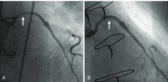
Two years later, she was admitted with unstable an- gina. Coronary angiography revealed 70% focal steno- sis of the proximal SVG to the left anterior descending artery (Fig. 1A). The SVG to the left anterior descend- ing artery was predilated with a 2.0×15 mm angiop- lasty balloon at 6 atm. After predilation, a 2.75×18 mm Cypher® SES (Cordis, Roden, The Netherlands) was deployed in the SVG and inflated at 10 atm (Fig.
1B). She tolerated the procedure well and was disch- arged without chest pain.
A follow-up coronary angiography was performed after 6 months. On admission to the hospital, the pa- tient displayed a pulse rate of 68 beats/min, blood pres- sure of 110/70 mmHg and respiratory rate of 18 br- eaths/min. Her physical examination was essentially normal. A chest X-ray demonstrated no abnormalities except sutured metallic rings around the sternum. A complete blood count, electrolytes and thyroid func- tion tests were within normal limits. Cardiac enzymes were also within normal limits. An electrocardiogram showed normal sinus rhythm without ST-segment ch- ange. Transthoracic echocardiogram revealed good re-
Received: October 14,2008 Accepted: February 25, 2009
Correspondence: Hee Jeoung Yoon, MD,Department of Internal Medicine, Daejeon St. Mary’s Hospital, College of Medicine, The Catholic University of Korea, 520-2 Daeheung-dong, Jung-gu, Daejeon 301-723, Korea Tel: 82-42-220-9114, 9504, Fax: 82-42-253-9505
E-mail: [email protected]
252·Stent Fracture in Saphenous Vein Intervention
gional wall motion with ejection fraction of 69%. The coronary angiography revealed 80% focal in-stent res- tenosis with complete stent fracture (Fig. 2A and B).
Intravascular ultrasound showed neointimal hyperpla- sia at mid-stent without presence of a stent strut (Fig. 3).
The stenosis was crossed with a guide wire and dilated with a 3.0×15 mm angioplasty balloon at 20 atm. A
final coronary angiography showed thrombolysis in myocardial infarction (TIMI) 3 distal flow of the SVG without residual stenosis (Fig. 2C).
Discussion
In-stent restenosis after implantation of SESs still
A B
Fig. 1. Angiographic views of SVG to the left anterior descending artery during the initial coronary intervention. A: angiography revealed 70% focal stenosis of the proximal SVG to the left anterior descending artery (arrow). B: after SES implantation for the SVG, a well deployed stent without residual stenosis was evident (arrow). SVG: saphenous vein graft, SES: sirolimus-eluting stent.
Fig. 2. Angiographic views of the SVG to the left anterior descending artery. A: angiography revealing 80% focal in-stent restenosis (arrow). B: complete strut fracture of mid portion of the Cypher stent was observed (arrow). C: final angiography after balloon angio- plasty revealing good distal flow of the SVG without residual stenosis. SVG: saphenous vein graft.
A B C
A B C
A
B C
Fig. 3. Angiographic and intravascular ultrasound images on fractured stent at SVG. A: proximal stent segment. B: point of stent fracture (lack of stent layer). C: distal stent segment. SVG: saphenous vein graft.
Sun Hong Yoo, et al.·253
occurs in some cases, and stent fracture was recently sug- gested as a new potential mechanism of restenosis.3)4) The “real world” clinical practice of implantation of SESs may be a safe and feasible technique for revas- cularization in patients with SVG disease with excel- lent short- and long-term clinical outcomes.6) But, the use of bare-metal stents may be associated with lower long-term mortality than the use of SES for SVG dis- ease.7) An incidence of stent fracture after SES implan- tation of 2.6% was reported.8) While the exact mecha- nism of stent fracture is unknown, stent fractures are associated with long stented segments, right coronary artery location and metal overlap.9) Multiple mechani- cal factors such as long stented segments, increased ri- gidity and pre-procedure vessel angulation might in- teract to disrupt the stent strut. The majority of stent fractures occur within 10 mm from areas of increased rigidity caused by strut overlap that may have acted as a fulcrum for metal deformation due to vessel movement.
Dynamic vessel movement and repetitive kinking of the stent during the cardiac cycle may also be implicat- ed in stent fractures.9) Many drug-eluting stent fractures occur in the proximal segments of the right coronary artery, since this artery moves more dynamically than the left coronary artery during the cardiac cycle.10)
The implantation of multiple overlapping stents sig- nificantly increases the axial stiffness of the stented seg- ment and longer stents covering longer lesions are sub- ject to higher radial forces. These may play a role in stent fracture.9) SES fracture may be due to the closed cell design of the SES compared with the paclitaxel-el- uting stent, which has an open cell design.11)
In-stent restenosis after implantation of a SES is com- monly associated with a discontinuity in stent cover- age.12) Late incomplete stent strut coverage because of stent fracture could result in incomplete inhibition of intimal hyperplasia and subsequently in restenosis at follow-up.10) Therefore, fractured stent struts cause a local mechanical irritation of the vessel and may result in inflammation and neointimal hyperplasia.11) At the fracture point, neointimal growth is putatively observ- ed because of the decrease in local drug delivery.8) Fur- thermore, exposure of a free metal strut protruding into the vessel lumen clearly could trigger platelet activa- tion and resultant stent thrombosis.11)
In our case, a short stent was deployed in the proxi- mal curvature portion of SVG to the left anterior de- scending artery. Unlike the native coronary arteries, the body of a SVG is relatively free to move in relation to the anastomotic site, depending upon the degree of peri- graft fibrosis and amount of intrathoracic space avail- able. Consequently, in vein grafts, mechanical stresses can be very high.4)13)
As yet, there is no consensus regarding the best treat- ment method of stent fracture. Re-stent therapy is con-
troversial, since there is a possibility of recurrence of stent fracture. Overlapping stents may also increase the likelihood of stent fractures from increased axial stiff- ness at the overlapping segment acting as a fulcrum for stent deformation and fracture from vessel movement.14) Furthermore, the effect of balloon angioplasty for un- covered SES restenosis is unknown.8) Instead of re-stent therapy, we treated stent fracture with balloon angio- plasty. Follow-up studies should be performed to con- firm that there is no in-stent restenosis.
Stent fracture of the SVG is relatively rare. The pre- sently-reported case is the first case of complete stent fracture of SVG in Korea.
REFERENCES
1) Moses JW, Leon MB, Popma JJ, et al. Sirolimus-eluting stents versus standard stents in patients with stenosis in a native coro- nary artery. N Engl J Med 2003;349:1315-23.
2) Kim JS, Yoon YW, Hong BK, et al. Delayed stent fracture after successful sirolimus-eluting stent (Cypher®) implantation. Kor- ean Circ J 2006;36:443-9.
3) Halkin A, Carlier S, Leon MB. Late incomplete lesion coverage following Cypher stent deployment for diffuse right coronary ar- tery stenosis. Heart 2004;90:e45.
4) Sianos G, Hofma S, Ligthart JM, et al. Stent fracture and reste- nosis in the drug-eluting stent era. Catheter Cardiovasc Interv 2004;61:111-6.
5) Lee SY, Im E, Yang WI, Kim JS, Cho YH, Shim WH. A siroli- mus-eluting stent fracture combined with a coronary artery an- eurysm. Korean Circ J 2008;38:69-71.
6) Ramana RK, Ronan A, Cohoon K, et al. Long-term clinical out- comes of real-world experience using sirolimus-eluting stents in saphenous vein graft disease. Catheter Cardiovasc Interv 2008;
71:886-93.
7) Vermeersch P, Agostoni P, Verheye S, et al. Increased late mor- tality after sirolimus-eluting stents versus bare-metal stents in diseased saphenous vein grafts: results from the randomized DE- LAYED PRISC trial. J Am Coll Cardiol 2007;50:261-7.
8) Aoki J, Nakazawa G, Tanabe K, et al. Incidence and clinical im- pact of coronary stent fracture after sirolimus-eluting stent im- plantation. Catheter Cardiovasc Interv 2007;69:380-6.
9) Yang TH, Kim DI, Park SG, et al. Clinical characteristics of stent fracture after sirolimus-eluting stent implantation Int J Car- diol 2009;131:212-6.
10) Okumura M, Ozaki Y, Ishii J, et al. Restenosis and stent fracture following sirolimus-eluting stent (SES) implantation. Circ J 2007;
71:1669-77.
11) Lee MS, Jurewitz D, Aragon J, Forrester J, Makkar RR, Kar S.
Stent fracture associated with drug-eluting stents: clinical char- acteristics and implications. Catheter Cardiovas Interv 2007;69:
387-94.
12) Lemos PA, Saia F, Ligthart JM, et al. Coronary restenosis after sirolimus-eluting stent implantation: morphological description and mechanistic analysis from a consecutive series of cases.
Circulation 2003;108:257-60.
13) Koh TW, Mathur A. Coronary stent fracture in a saphenous vein graft to right coronary artery: successful treatment by the novel use of the Jomed coronary stent graft: case report and review of the literature. Int J Cardiol 2007;119:e43-5.
14) Duda SH, Pusich B, Richter G, et al. Sirolimus-eluting stents for the treatment of obstructive superficial femoral artery disease:
six-month results. Circulation 2002;106:1505-9.