242 Copyright © 2015 The Korean Society of Cardiology Korean Circulation Journal
Introduction
Celiac Disease (CD) is a chronic small bowel immune-mediated enteropathy precipitated by exposure to dietary gluten in geneti- cally predisposed individuals.
1)CD affects approximately 1% of the European population.
2)Patients are intolerant to dietary gluten and recurrent exposure causes chronic inflammation in the small in- testines, villous atrophy and crypt hyperplasia.
3)Genetic, environ- mental and autoimmune factors play a role in its etiology and CD is associated with other autoimmune disorders such as autoim- mune thyroiditis, type 1 diabetes mellitus, collagen tissue diseases and giant cell myocarditis
4-6)Case
A 41-year-old male patient was admitted to the emergency de- partment with ongoing chest pain. The patient had no risk factors for atherosclerosis and hemodynamic parameters were stable upon admission. There was no history of trauma or collagen tissue disease and he was not currently taking any medication. He was diagnosed with CD 12 years earlier and had not been on a gluten free diet (GFD) for a year. An electrocardiogram found nonspecific ST-T changes. However, the troponin I level was 22 ng/mL, which is well above the reference range (0.02-0.06 ng/mL). The patient was therefore admitted to the coronary intensive care unit with a pri- mary diagnosis of non-ST segment elevation myocardial infarction.
An echocardiogram revealed severely hypokinetic inferior, posterior and lateral walls of the left ventricle, with an ejection fraction of 35% (Normal >52%) in the absence of a primary valve disorder.
Bloodwork hemoglobin was 9.3 g/dL, white blood cell count was 2.25×10
4/mL and platelet count was 4.41×10
5/mL. Lipid levels as well as liver and kidney function tests were normal, but erythro- cyte sedimentation rate was 19 mm/hour (0-15 mm/h) and C-re- active protein (CRP) was 12.5 mg/dL (0-6 mg/dL).
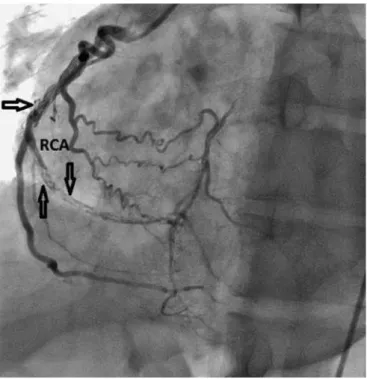
A coronary angiography (CAG) was performed and diffuse dis- sections within the left anterior descending artery (LAD) and the right coronary artery (RCA) were observed with total occlusion of second obtuse marginal artery (Figs. 1 and 2). Severely reduced left ventricular dysfunction was detected on ventriculography. Because
Case Report
http://dx.doi.org/10.4070/kcj.2015.45.3.242 Print ISSN 1738-5520 • On-line ISSN 1738-5555