Natural Product Sciences 21(4) : 293-296 (2015)
http://dx.doi.org/10.20307/nps.2015.21.4.293
293
Epi-Leptosphaerin: A New
L-Isoascorbic Acid Derivative from Marine Sponges
Roshan R. Kulkarni,A Reum Jo, Young Ho Kim, and MinKyun Na*
College of Pharmacy, Chungnam National University, Daejeon 305-764, Korea
Abstract − A new L-isoascorbic acid derivative epi-leptosphaerin (1) and two known compounds leptosphaerin (2), and verongamine (3) were isolated from sponges of the orders Verongida and Thorectidae. Compounds 1 and 2 are most likely of sponge-associated fungal origin. In the present study, isolated compounds were investigated for their inhibition of soluble epoxide hydrolase (sEH), which is considered a promising target for the management of pain, inflammation, and comorbidities associated with diabetes. Compound 3, verongamine, displayed weak inhibitory activity against sEH with an IC50 value 51.5± 1.0 µM.
Keywords −L-isoascorbic acid, soluble epoxide hydrolase, Verongida sponges
Introduction
Marine organisms, especially sponges, elaborate a wide variety of potently active metabolites.1 However, recent research has shown increasing recognition that microor- ganisms associated with sponges, tunicates, corals, etc.
are the true producers of most metabolites isolated from marine organisms.2 Indeed, microbial biomass may cons- titute up to 35% of a sponge’s volume.3 Their association with sponges attributes these microorganisms with remarkably diverse structural scaffolds; for example, out of a total of 411 compounds discovered in actinomycetes in 2013, 22% were those derived from sponge-associated species.3 This may very well result from the pressure upon microorganisms to survive conditions in which diverse species cohabitate the same sponge body. Although an extensive amount of work has been reported regarding bacterial associations with marine sponges,3 recent studies have focused on the fungal associates of marine sponges due to the potential of such fungi as sources of novel bioactive compounds.4 Deciphering the true metabolic sources of the natural products isolated from large multicellular marine organisms is important for several reasons. First, such knowledge will reduce the environ- mental burden associated with collection, as the microor- ganism of interest could, at least in principle, be cultured.
In addition, once confirmed, the producing microor- ganism can be manipulated in order to mine its hidden
chemical diversity. Another noteworthy consideration is the possibility of isolating analogous compounds, which may exhibit greater activities, from terrestrial sources, which may considerably reduce the amount of effort and cost associated with re-isolation. For example, clyclo- depsipeptides, a promising class of metabolites of fungal and plant origin, has also been isolated from fungi associated with sponges.4,5 Thus, every secondary metabolite ela- borated by a marine sponge, irrespective of its bioactivity, should be carefully studied and its origin determined.
In recent years, inhibition of soluble epoxide hydrolase (sEH), an enzyme that degrades endogenous anti-inflam- matory eicosatrienoic acids, has emerged as an effective strategy for treating cardiovascular and kidney diseases, inflammation and especially pain.6,7 Recently, a detailed understanding of the active site has enabled the design of very potent inhibitors. As exemplified by 12-(3- adamantan-1-yl-ureido) dodecanoic acid (AUDA), a transition state inhibitor belonging to the amide class, the amide carbonyl mimics an epoxy, while the carbonyl- flanking NH group interacts with Asp33 and helps stabilize the complex.6
In our ongoing mission to isolate bioactive metabolites from marine sponges of the orders Verongida and Thorectidae, our earlier studies have demonstrated the antidepressant and cytotoxic activities of numerous com- pounds and have isolated uncommon metabolites.8-11 A survey of the literature reveals that bromotyrosine-types of compounds are the mainstay of the Verongida chemical repertoire. One structural feature that is common to each of these is an amide bond that confers sEH inhibitory activity. In order to isolate compounds bearing this
*Author for correspondence
Prof. MinKyun Na, College of Pharmacy, Chungnam National Uni- versity, Daejeon 305-764, Korea
Tel: +82-42-821-5925; E-mail: [email protected]
294 Natural Product Sciences
feature, we further investigated the polar fractions of sponges, which led to the isolation of a new epi- leptosphaerin (1), the known leptosphaerin (2) and a known oxime-histamine type of bromotyrosine, veronga- mine (3) (Figure 1). Because each of these three com- pounds contained an amide bond, each was evaluated in terms of its ability to inhibit sEH. Herein, we discuss isolation, elucidate structures and evaluate the sEH- inhibitory abilities of compounds 1 - 3. The possible fungal origin of compounds 1 and 2 is also discussed.
Experimental
General Experimental Procedures− Optical rotations were measured on a JASCO DIP-1000 (Tokyo, Japan) automatic digital polarimeter. Vacuum Liquid Chromato- graphy (VLC) was performed using a Merck silica gel (70 - 230 mesh). Medium Pressure Liquid Chromatography (MPLC) was carried out using a Biotage IsoleraTM with KP-C18-HS reversed-phase C18 SNAP Cartridges (120 g and 340 g, Biotage). Preparative reversed phase HPLC (prep HPLC) separation was carried out on a YMC C18 column (250× 20.0 mm, 5 μm) using a 5 mL/min flow rate. 1H (600 MHz), 13C (150 MHz), and 2D (COSY, HSQC, HMBC) NMR spectra were recorded on a Bruker 600 MHz (Avance II) spectrometer. HRESIMS data was obtained from the Korea Basic Science Institute (Chungbuk, South Korea) on a Synapt (Waters, U.K.) high-resolution mass spectrometer. Preparatory HPLC separation was carried out using HPLC-grade solvents (Merck, Ltd.), and all solvents were distilled prior to use in VLC and MPLC separations. For the sEH bioassay, sEH and 3-phenyl- cyano(6-methoxy-2-naphthalenyl)methyl ester-2-oxiraneacetic acid (PHOME) were purchased from Cayman (A61510 and A60932, respectively), while the AUDA positive control was purchased from Sigma.
Isolation of compounds− Collection and extraction of the sponges of interest has been described in our earlier work.8,9 Dried ethanol extract (3.6 kg) was subjected to silica-gel vacuum liquid chromatography (VLC) using a
stepwise gradient of hexane/acetone/methanol/water to give 13 fractions (Fr. 1–13). Fraction 8 (2.1 g) was further divided into 13 fractions (Fr. 8-1 to 8-13) using MPLC (C18 SNAP cartridge, 120 g) and MeOH with a gradient proceeding from 80 to 100% at 40 mL/min. Fraction 8-1 (480.0 mg) was separated by prep HPLC using a methanol gradient (in water) proceeding from 0 to 50% in 60 min to yield a mixture of compounds 1 and 2 (tR 35.0 min). The mixture was subjected to further prep HPLC using a methanol gradient (in water) proceeding from 0 to 30% in 60 min to isolate pure compounds 1 (tR= 42.9 min, 1.5 mg) and 2 (tR= 44.1 min, 2.6 mg). Combined fractions 9, 10, 12, and 13 from the VLC (19.1 g) were further subdivided into 16 fractions using MPLC (C18 SNAP cartridge, 340 g) and a MeOH gradient proceeding from 10 to 100% at a flow rate of 65 mL/min. Fraction 10 from the above MPLC (90 mg) was further separated by prep HPLC using a methanol gradient (in water) proceeding from 70 to 100% over 60 min to yield pure compound 3 (52 mg, tR= 18.0 min).
epi-Leptosphaerin (1) − Colorless solid. +6.5 (c 0.07, water). Positive ion HRESIMS m/z: 224.0536 (calcd for C8H11NO5Na 224.0535). 1H NMR (Pyridine-d5,600 MHz) δ: 10.86 (1H, s, NH), 8.05 (1H, d, J = 2.0 Hz, H-3), 5.73 (1H, dd, J = 3.3, 2.0 Hz, H-4), 4.35 (1H, bs, H-5), 4.29 (2H, m, H2-6), 2.25 (3H, s, H3-8); 13C NMR (Pyridine-d5, 150 MHz) δ: 170.8 (C-1), 170.2 (C-7), 129.1 (C-3), 128.3 (C-2), 83.3 (C-4), 73.4 (C-5), 64.5 (C- 6), 23.8 (C-8).
Leptosphaerin (2)− Colorless solid. 1H NMR (Pyridine- α
[ ]D25 Fig 1. Compounds isolated from the sponges of interest.
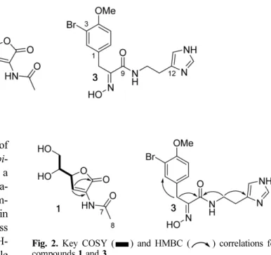
Fig. 2. Key COSY ( ) and HMBC ( ) correlations for compounds 1 and 3.
Vol. 21, No. 4, 2015 295
d5,600 MHz) δ: 8.29 (1H, d, J = 1.9 Hz, H-3), 5.68 (1H, dd, J = 5.0, 1.9 Hz, H-4), 4.43 (1H, d, J = 5.1 Hz H-5), 4.29 (2H, m, H2-6), 2.24 (3H, s, H3-8); 13C NMR (Pyridine-d5, 150 MHz) δ: 170.5 (C-1), 170.1 (C-7), 128.9 (C-3), 128.5 (C-2), 83.2 (C-4), 73.7 (C-5), 64.6 (C- 6), 23.9 (C-8).
Verongamine (3)− Brown amorphous solid. 1H NMR (MeOH-d4, 600 MHz) δ: 7.70 (1H, s, H-14), 7.43 (1H, d, J = 2.3 Hz, H-2), 7.20 (1H, dd, J = 8.5, 2.3 Hz, H-6), 6.90 (1H, d, J = 8.5 Hz, H-5), 6.86 (1H, s, H-13), 3.82 (3H, s, H3-15), 3.81 (2H, s, H2-7), 3.48 (2H, t, J = 7.1 Hz, H2-10), 2.79 (2H, t, J = 7.1 Hz, H2-11). 13C NMR (MeOH-d4,150 MHz) δ: 165.7 (C-9), 155.9 (C-4), 153.0 (C-8), 135.8 (C- 14), 135.6 (C-12), 134.7 (C-2), 131.8 (C-3), 130.4 (C-6), 118.0 (C-13), 113.1 (C-5), 112.1 (C-1), 56.7 (C-15), 40.1(C-10), 28.7 (C-7), 27.5 (C-11).
sEH assay− Fifty microliters of sEH (140 ng/mL) and 20 L of compounds 1 - 3 were dissolved in MeOH (final concentration 100 µM) and mixed in a 96-white-well plate containing 80μL of 25 mM bis-Tris-HCl buffer (pH 7.0) that contained 0.1% bovine serum albumin (BSA).
Subsequently, 50μL of 20 µM PHOME was added as a substrate to the mixture. The resulting mixture was incubated at 37oC, and, after 1 hour, the products of hydrolysis were monitored at excitation and emission wavelengths of 330 and 465 nm, respectively.
Enzyme activity (%) = [(S60− S2/ C60− C2]× 100 where C60 and S60 pertain to the fluorescence of control and inhibitor substances after 60 min, and S2 and C2 pertain to the fluorescence of control after 2 min.
Results and Discussion
The ethanol extracts of the three sponges, V. rigida, S.
aurea and S. cerebriformis, 8,9 led to the isolation of a new epi-leptosphaerin (1) as well as known leptosphaerin (2) and verongamine (3). These isolated compounds were subsequently evaluated in terms of their sEH inhibition.
Compound 1 was isolated as a colorless powder. The
13C NMR spectrum showed 8 resonances, corresponding to a methyl group, a methylene group, 2 methine carbons, an olefinic methine carbon, an olefinic quaternary carbon and 2 carbonyl carbons.
From these results, taken together with the pseu- domolecular ion peak at m/z 224.0536 (calcd for C8H11NO5Na, 224.0535), the molecular formula of 1 was determined to be C8H11NO5. The 1H and 13C spectra showed resonances for a 1,2,3-trihydroxypropyl moiety that included a methylene group at δH 4.29 (2H, m, δC
64.5) and two methine protons at δH 4.35 (1H, m, δC 73.4) and δH 5.73 (1H, dd, J = 3.3, 2.0 Hz, δC 83.3). In the 1H-
1H COrrelation SpectroscopY (COSY) spectrum, the methine proton at δH 4.35 correlated with the methylene group at δH 4.29 and the methine protonation at δH 5.73, which confirmed the 1,2,3-trihydroxypropyl linkage (Fig.
2). The methine proton at δH 5.73 also showed a COSY correlation with an olefinic proton at δH 8.05 (d, J = 2.0 Hz, δC 129.1). The proton at δH 8.05 showed a Heteronu- clear Multiple Bond Correlation (HMBC) with a carbonyl carbon at δC 170.8 and an olefinic quaternary carbon at δC
128.3 (Fig. 2). These observations led to identification of an α, β-unsaturated group with an α substitution as another fragment. The COSY correlation between δH 5.73 and δH 8.05 allowed the two fragments to be linked.
However, the chemical shifts of the olefinic methine (δH
8.05, δC 129.1) and the olefinic quaternary carbon (δC
128.3) were unusual for an α,β-unsaturated moiety and indicated an amine functionality at the α-position. When taken with the identification of an acetyl group comprised of a tertiary methyl at δH 2.25 (δC 23.8) and a quaternary carbon at δC 170.2, these results led to identification of compound 1 as a diastereomer of leptosphaerin (2), an L- ascorbic acid derivative previously isolated from the marine fungus Leptosphaeria oraemaris.12 The 1H and
13C NMR data agreed closely with that of leptosphaerin (2). In 1, the methine proton at δH 5.73 (H-4) resonated as a dd (J = 3.3, 2.0 Hz), whereas in leptosphaerin the methane proton resonated at δH 5.68 as a dd (J = 5.0, 1.9 Hz). This result, along with the positive specific rotation observed +6.5 (c 0.07, water), identified 1 as a diastereomer of leptosphaerin based on the L-isoascorbic acid scaffold. The positive specific rotation value indicated that 1 could be the C-4 epimer of leptosphaerin. To the best of our knowledge, identification of such an L- isoascorbic acid scaffold in marine sources has not yet been reported. Although synthetic production has been reported,13 the present study is the first to isolate this compound as a natural product and has assigned to it the name epi-leptosphaerin.
Although fungi of the genus Leptosphaeria have not yet been associated with Verongida and Thorectidae sponges, it is very likely that compound 1 is also produced by a species of Leptosphaeria. The area around the site of collection (the Florida Keys) is known to be rich in Sargassum algae,14 which, when collected from Japanese waters, have been revealed as a source of an epiphytic Leptosphaeria species that produces leptosins.15 Thus, Sargassum algae could form the link required for sponge and fungal association. In a related work, L-
α [ ]D25
296 Natural Product Sciences
ascorbic acid was converted into L-isoascorbic acid by a strain of Penicillium sp.16 Both genera Leptosphaeria and Penicillium belong to the ascomycetes group; thus, although ascorbic acid or its diastereomer have not been shown to be biogenetic precursors for leptosphaerins, compound 1 is, in all probability, produced by the Leptosphaeria sp. rather than by the sponge species.
Compound 2 was identified as leptosphaerin by comparison with published spectroscopic data.12
Compound 3, which was isolated as a brown amorphous powder, showed distinct resonances for a 1,2,4-trisubs- tituted phenyl ring and a histamine moiety in the 1H and
13C NMR spectra. Signals at δH 6.90 (1H, d, J = 8.5 Hz;
δC 113.1), 7.20 (1H, dd, J = 2.3, 8.5 Hz; δC 130.4), and 7.43 (1H, d, J = 2.3 Hz; δC 134.7) were assigned to the 1,2,4-trisubstituted phenyl, while resonances at δH 6.86 (1H, s; δC 118.0), δH 7.70 (1H, s; δC 135.8) and δH 2.79 (2H, t, J = 7.1 Hz; δC 27.5), δH 3.48 (2H, t, J = 7.1 Hz; δC
40.1) were assigned to the histamine moiety. In addition, a methylene group at δH 3.81 (2H, s; δC 28.7), an olefinic quaternary carbon at δC 153.0 and an amide carbonyl carbon at δC 165.7 led us to identify 3 as an oxime- histamine-type bromotyrosine derivative. In the HMBC spectrum (Fig. 2), the methylene group at δH 3.81 (H2-7) showed correlations with the C-2 (δC 134.7) and C-9 (δC
165.7) groups. The histamine was linked to the amide through the ethylene bridge, based on HMBC correlations of the methylene group at δH 3.48 (H2-10) with C-9. This analysis identified 3 as verongamine, an oxime-histamine bromotyrosine derivative.17
The isolated compounds were evaluated for their abilities to inhibit sEH activity, wherein verongamine (3) displayed weak inhibitory activity with an IC50 51.5± 1.0 µM, compared to an IC50 of 7.9± 2.7 nM for the positive control AUDA. Compounds 1 and 2 were inactive with 20.5 and 23.3% sEH at 100 µM.
In the present study, we isolated the novel compound epi-leptosphaerin, which belongs to the L-isoascorbic acid scaffold. In all probability, both compounds 1 and 2 are of fungal origin and are produced by a yet unidentified species of the fungus Leptosphaeria. Our successful isolation of 1 and 2 is significant, as association of the genus Leptosphaeria with either Verongida or Thorectidae
sponges has not, as of yet, been demonstrated. Discovery of such an association may throw new light on the true metabolic origins of other compounds isolated from these orders of sponges.
Acknowledgments
This work was supported by the research fund of Chungnam National University.
References
(1) Perdicaris, S.; Vlachogianni, T.; Valavanidis, A. Nat. Prod. Chem.
Res. 2013, 1, 114.
(2) Simmons, T. L.; Coates, R. C.; Clark, B. R.; Engene, N.; Gonzalez, D.; Esquenazi, E.; Dorrestein, P. C.; Gerwick, W. H. Proc. Natl. Acad.
Sci. 2008, 105, 4587-4594.
(3) Abdelmohsen, U. R.; Bayer, K.; Hentschel, U. Nat. Prod. Rep. 2014, 31, 381-399.
(4) Suryanarayanan, T. S. Bot. Mar. 2012, 55, 553-564.
(5) Lemmens-Gruber, R.; Kamyar, M. R.; Dornetshuber, R. Curr. Med.
Chem. 2009, 16, 1122-1137.
(6) Imig, J. D.; Hammock, B. D. Nat. Rev. Drug Discov. 2009, 8, 794- 805.
(7) Hammock, B. D.; Wagner, K.; Inceoglu, B. Pain Manag. 2011, 1, 383-386.
(8) Kochanowska, A. J.; Rao, K. V.; Childress, S.; El-Alfy, A.;
Matsumoto, R. R.; Kelly, M.; Stewart, G. S.; Sufka, K. J.; Hamann, M. T.
J. Nat. Prod. 2008, 71, 186-189.
(9) Hwang, I. H.; Oh, J.; Kochanowska-Karamyan, A.; Doerksen, R. J.;
Na, M.; Hamann, M. T. Tetrahedron Lett. 2013, 54, 3872-3876.
(10) Kulkarni, R.; Kim, J. H.; Kim, Y. H.; Oh, S.; Na, M. Nat. Prod. Sci.
2015, 21, 25-29.
(11) Hwang, I. H.; Oh, J.; Zhou, W.; Park, S.; Kim, J.-H.; Chittiboyina, A. G.; Ferreira, D.; Song, G. Y.; Oh, S.; Na, M., et al. J. Nat. Prod. 2015, 78, 453-461.
(12) White, J. D.; Badger, R. A.; Kezar III, H. S.; Pallenberg, A. J.;
Schiehser, G. A. Tetrahedron 1989, 45, 6631-6644.
(13) Miroslav, P.; Emmanuel, Z.; Fletcher Jr., H. G. Carbohydr. Res.
1975, 43, 345-354.
(14) Hanisak, M. D.; Samuel, M. A. Dev. Hydrobiol. 1987, 41, 399-404.
(15) Takahashi, C.; Numata, A.; Ito, Y.; Matsumura, E.; Araki, H.;
Iwaki, H.; Kushida, K. J. Chem. Soc. Perkin Trans. 1994, 1, 1859-1864.
(16) Kyotani, D.; Hasegawa, K.; Ohishi, H.; Wu, W.; Wang, L.;
Hasumi, K. Biosci. Biotechnol. Biochem. 2009, 73, 954-956.
(17) Mierzwa, R.; King, A.; Conover, M. A.; Tozzi, S.; Puar, M. S.;
Patel, M.; Coval, S. J.; Pomponi, S. A. J. Nat. Prod. 1994, 57, 175-177.
Received July 12, 2015 Revised September 21, 2015 Accepted September 30, 2015