Natural Product Sciences 21(4) : 289-292 (2015)
http://dx.doi.org/10.20307/nps.2015.21.4.289
289
Two New Scalaranes from a Korean Marine Sponge Spongia sp.
Inho Yang
1, Sang-Jip Nam
2,*, and Heonjoong Kang
1,3,*
1
School of Earth and Environmental Sciences, Seoul National University, NS-80, Seoul 151-747, Korea
2
Department of Chemistry and Nano Science, Global Top5 Program, Ewha Womans University, Seoul 120-750, Korea
3
Research Institute of Oceanography, Seoul National University, NS-80, Seoul 151-747, Korea
Abstract − Intensive chemical investigation of Korean marine sponge Spongia sp. has led to the isolation of two new scalaranes. The planar structures of the new compounds 1 and 2 were determined through 1D and 2D NMR spectral data analysis, while the relative stereochemistry of the compounds was determined based on the analysis of
1H-
1H coupling constants and NOESY spectroscopic data. Compounds 1 and 2 did not display any significant biological activities on farnesoid X-activated receptor (FXR) in co-transfection assay.
Keywords − Scalarane, Sesterpenoid, Spongia sp., Korean sponge, Marine natural product
Introduction
Marine sponge is well known as a rich source of natural products with diverse chemical skeletons.
1Sester- terpene is a well-known chemical skeleton with the five carbon isoprene building units and has been isolated from diverse organisms including higher plants and fungus.
2Scalarane, belonging to the sesterterpene class of natural products, is one of the marine exclusive chemical classes mainly isolated from sponges and nudibranches.
3These scalaranes have not been isolated from terrestrial organisms.
After the first report on this class of compound in 1972,
4more than 200 derivatives were reported until 2010
3and novel derivatives of scalaranes are still being reported.
5Early research revealed that these natural products possessed antifeedant and antifouling activities,
6,7as well as other variety of bioactivities had been reported such as cyto- toxicity,
8anticancer,
9antibacterial,
10and anti-inflamma- tory.
11Biological activities of scalaranes were broadened to include the inhibition of nuclear factor,
12protein tyrosine phosphatase,
13and farnesoid X-activated receptor.
14Recent efforts to discover novel marine natural products from Korean sponges resulted in the isolation of new analogs of scalarane compounds,
15also our research group has
reported several scalaranes with FXR and cytotoxic activities from two Korean sponge samples, Spongia and Psammocinia sp. repectively.
16We further investigated chemical components to discover new natural products from Spongia sp., and have isolated two new scalaranes.
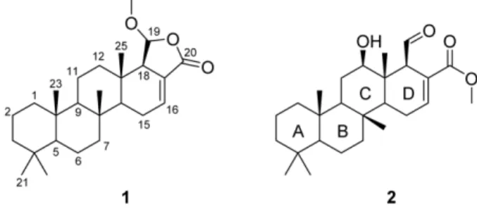
Herein, we describe the isolation and the structure eluci- dation of two new scalarenes 1 and 2 from Spongia sp.
Experimental
General experimental procedures – The optical rotation was measured using a Rudolph Research Autopol III polarimeter with a 5 cm cell. The UV spectrum was recorded in a Scinco UVS-2100 with a path length of 1 cm. Infrared spectra were recorded on a Thermo Electron Corporation spectrometer. NMR spectral spectroscopic data were obtained using Bruker Avance 600 and 500 MHz spectrometer [CDCl
3( δ
H7.26; δ
C77.0) was used as an internal standard]. HRFAB-MS data were measured on a JEOL, JMS-AX505WA mass spectrometer.
Isolation – The genus Spongia sp. sponge was collected by SCUBA at the Geoje Island, South Sea of Korea. The frozen animal (3.4 kg, wet wt.) was lyophilized and the dried specimen (600 g) was extracted with 50% MeOH in CH
2Cl
2. The extract was partitioned between CH
2Cl
2and water layers. The CH
2Cl
2-soluble layer was evaporated in vacuo and then partitioned between n-hexane and 90%
aqueous MeOH. The 90% aqueous MeOH layer was sub- sequently separated into 24 fractions with Sephadex LH- 20 column eluting with 50% MeOH in CH
2Cl
2. The fraction 20 containing the mixture of 1 and 2 was further
*Author for correspondence
Sang-Jip Nam, Department of Chemistry and Nano Science, Global Top5 Program, Ewha Womans University, Seoul 120-750, Korea Tel: +82-2-3277-6805; E-mail: [email protected]
Heonjoong Kang, School of Earth and Environmental Sciences, Seoul National University, NS-80, Seoul 151-747, Korea
Tel: +82-2-880-5730; E-mail: [email protected]
290 Natural Product Sciences
separated by reversed-phase HPLC (Phenomenex Luna C-18(2), 250 × 100 mm, 2.5 mL/min, 5 μm, 100 Å, UV = 205 nm) eluting with 70% CH
3CN in H
2O.
Compound 1: colorless oil; −1
o(0.002, CHCl
3);
UV (MeOH) λ
max(log ε) 210 (3.98) nm; IR (film) ν
max3393, 1761, 1682, 1236 cm
−1;
1H NMR data, see Table 1;
13
C NMR data, see Table 1; LRFABMS m/z 401 [M + H]
+; HRFABMS m/z 401.3052 [M + H]
+(calcd for C
26H
41O
3, 401.3056).
Compound 2: colorless oils; +8
o(0.002, CHCl
3);
UV (MeOH) λ
max(log ε) 210 (3.96) nm; IR (film) ν
max3392, 1760, 1683, 1238 cm
−1;
1H NMR data, see Table 1;
13
C NMR data, see Table 1; LRFABMS m/z 417 [M + H]
+; HRFABMS m/z 417.3017 [M + H]
+(calcd for C
26H
41O
4, 417.3005).
Results and Discussion
The molecular formula of compound 1 was established as C
26H
40O
3based on the analysis of HRFABMS data (a α
[ ]
D21α [ ]
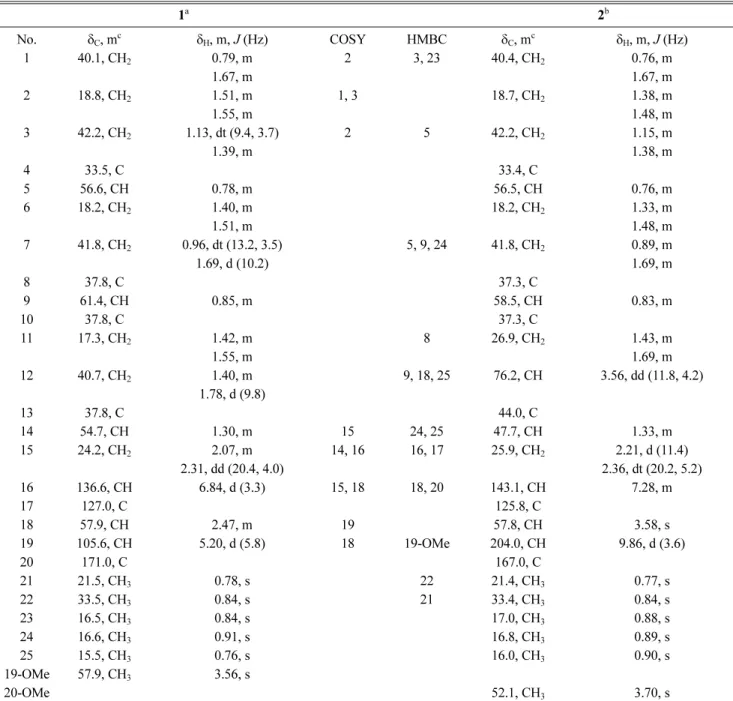
D21Table 1.
1H and
13C NMR data of 1 and 2 in CDCl
3.
1
a2
bNo. δ
C, m
cδ
H, m, J (Hz) COSY HMBC δ
C, m
cδ
H, m, J (Hz)
1 40.1, CH
20.79, m 2 3, 23 40.4, CH
20.76, m
1.67, m 1.67, m
2 18.8, CH
21.51, m 1, 3 18.7, CH
21.38, m
1.55, m 1.48, m
3 42.2, CH
21.13, dt (9.4, 3.7) 2 5 42.2, CH
21.15, m
1.39, m 1.38, m
4 33.5, C 33.4, C
5 56.6, CH 0.78, m 56.5, CH 0.76, m
6 18.2, CH
21.40, m 18.2, CH
21.33, m
1.51, m 1.48, m
7 41.8, CH
20.96, dt (13.2, 3.5) 5, 9, 24 41.8, CH
20.89, m
1.69, d (10.2) 1.69, m
8 37.8, C 37.3, C
9 61.4, CH 0.85, m 58.5, CH 0.83, m
10 37.8, C 37.3, C
11 17.3, CH
21.42, m 8 26.9, CH
21.43, m
1.55, m 1.69, m
12 40.7, CH
21.40, m 9, 18, 25 76.2, CH 3.56, dd (11.8, 4.2)
1.78, d (9.8)
13 37.8, C 44.0, C
14 54.7, CH 1.30, m 15 24, 25 47.7, CH 1.33, m
15 24.2, CH
22.07, m 14, 16 16, 17 25.9, CH
22.21, d (11.4)
2.31, dd (20.4, 4.0) 2.36, dt (20.2, 5.2)
16 136.6, CH 6.84, d (3.3) 15, 18 18, 20 143.1, CH 7.28, m
17 127.0, C 125.8, C
18 57.9, CH 2.47, m 19 57.8, CH 3.58, s
19 105.6, CH 5.20, d (5.8) 18 19-OMe 204.0, CH 9.86, d (3.6)
20 171.0, C 167.0, C
21 21.5, CH
30.78, s 22 21.4, CH
30.77, s
22 33.5, CH
30.84, s 21 33.4, CH
30.84, s
23 16.5, CH
30.84, s 17.0, CH
30.88, s
24 16.6, CH
30.91, s 16.8, CH
30.89, s
25 15.5, CH
30.76, s 16.0, CH
30.90, s
19-OMe 57.9, CH
33.56, s
20-OMe 52.1, CH
33.70, s
a