Relationship between vitamin K status, bone mineral density, and hs-CRP in young Korean women
Misung Kim
1, Heeseon Kim
2and Cheongmin Sohn
1§1
Major in Food and Nutrition, Wonkwang University, Sinyong-dong, Iksan-si, Jeonbuk 570-749, Korea
2
Department of Food Science and Nutrition, Soonchunhyang University, Chungnam 336-745, Korea
Abstract
Vitamin K intake has been reported as an essential factor for bone formation. The current study was conducted under the hypothesis that insufficient vitamin K intake would affect inflammatory markers and bone mineral density in young adult women. The study was a cross-sectional design that included 75 women in their 20s. Physical assessments, bone mineral density measurements, 24-hr dietary recalls, and biochemical assessments for high sensitivity C-reactive protein (hs-CRP) and percentages of undercarboxylated osteocalcin (%ucOC) were performed. An analysis of vitamin K nutritional status was performed comparing first, second, and third tertiles of intake based on %ucOC in plasma. Vitamin K intake levels in the first, second, and third tertiles were 94.88 ± 51.48 μg, 73.85 ± 45.15 μg, and 62.58 ± 39.92 μg, respectively (P < 0.05). The T-scores of the first and third tertiles were 1.06 and -0.03, respectively, indicating that bone mineral density was significantly lower in the group with lower vitamin K intake (P < 0.05). There was a tendency for different serum hs-CRP concentrations between the first (0.04 ± 0.02) and third tertiles (0.11 ± 0.18), however this was not statistically significant. Regression analysis was performed to identify the correlations between vitamin K nutritional status, inflammatory markers, and bone mineral density after adjusting for age and BMI. Serum hs-CRP concentrations were positively correlated with vitamin K deficiency status (P < 0.05). And bone mineral density, which was represented by speed, was negatively correlated with vitamin K deficiency status (P < 0.05). In conclusion, status of vitamin K affects inflammatory status and bone formation. Therefore, sufficient intake of vitamin K is required to secure peak bone mass in young adult women.
Key Words: Vitamin K, osteocalcin, undercarboxylated osteocalcin, bone mineral density, hs-CRP
Introduction
9)With the remarkable advancement of modern medicine as well as economic development, human longevity and the prevalence of chronic diseases due to the aging process have increased.
Osteoporosis is one representative disease that occurs in the elderly, and according to the Korean National Health and Nutrition Examination Survey in 2008, prevalence rates of osteoporosis and osteopenia were reported as 32.6% and 45.5%, respectively, in women aged 50 years or older [1]. It is therefore essential to prevent the occurrence of bone fractures and osteoporosis, among which incidence rates have abruptly increased in middle-aged and elderly individuals, by maintaining a healthy skeletal structure during the 20s, the time when maximal bone mass is achieved [2].
However, in an environment where slender women are favored from a social perspective, bone mineral density has been contradictorily reported to be lower in women in their 20s who are highly interested in body weight control as compared to those in their 30s [3]. It is therefore imperative that young women
secure maximal bone mass through healthy dietary habits and living habits [4]. Factors affecting bone mineral density include the following: age of menarche [5], body weight, amount of muscle, BMI [6], smoking [7], estrogen and progesterone status [8], and dietary intakes of vitamin D [9], isoflavones [10] and vitamin K [11]. Particularly in association with bone metabolism, vitamin K plays an essential role as a coenzyme during the carboxylation of γ-glutamic acid within the protein osteocalcin [12]. Carboxylated osteocalcin binds to hydroxyapatite and is thereby involved in bone formation. Serum osteocalcin is used as a bone metabolism marker that is associated with the homeostasis of bone formation [13].
The percentage of undercarboxylated osteocalcin (%ucOC) is referred to as the ratio of osteocalcin released to the peripheral blood in a non-carboxylated state, and it has a negative correlation with vitamin K nutritional status [14]. It is therefore used as a sensitive marker of bone metabolism in association with vitamin K deficiency [15,16].
According to studies on correlations between vitamin K and bone mineral density, there was a positive correlation between
This paper was supported by Wonkwang University in 2010.
§Corresponding Author: Cheongmin Sohn, Tel. 82-63-850-6656, Fax. 82-63-850-7301, Email. [email protected] Received: August 30, 2010, Revised: November 3, 2010, Accepted: November 6, 2010
ⓒ2010 The Korean Nutrition Society and the Korean Society of Community Nutrition
This is an Open Access article distributed under the terms of the Creative Commons Attribution Non-Commercial License (http://creativecommons.org/licenses/by-nc/3.0/) which permits unrestricted non-commercial use, distribution, and reproduction in any medium, provided the original work is properly cited.
vitamin K intake level and bone mineral density and a negative correlation with incidence of pelvic bone fracture in women [17,18]. In addition, it was also been reported that %ucOC decreased following supplementation of vitamin K
1in adult women in their 20s [19]. According to animal experiments, supplemental vitamin K suppressed the formation of osteoclasts and facilitated apoptosis in rats. This eventually led to decreased occurrence of bone loss [20]. To explain the mechanisms by which vitamin K is involved in bone metabolism, its relationship with cytokines as well as action as a coenzyme have been proposed. According to the Framingham Offspring Cohort cross- sectional study, following the measurement of vitamin K levels in subjects in a community-based setting, proinflammatory cytokines such as IL-6, intercellular adhesion molecule-1 (ICAM-1), and tumor necrosis factor receptor 2 (TNFR2) were found to be negatively correlated with vitamin K [21]. Furthermore, according to Shea et al. [22], the concentration of serum high sensitivity C-reactive protein (hs-CRP), a sensitive inflammatory marker, had a negative correlation with serum vitamin K concen- tration. Moreover, according to a cross-sectional study conducted in elderly individuals, there was a positive correlation between osteoprotegerin, an inflammatory marker suppressing bone loss reactions, and vitamin K. This implies that vitamin K is involved in inflammatory responses associated with bone metabolism. In Korea, however, there are still an insufficient number of studies on the relationship between vitamin K nutritional status, inflammation indicators, and bone mineral density.
Given the above background, we examined the relationships between %ucOC, hs-CRP (a proinflammatory marker), and bone mineral density in women in their 20s in whom maximal bone mass is formed. Thus, we attempted to provide baseline data for nutritional education on the maintenance of bone health in order to prevent osteoporosis in women.
Subjects and Methods
Subjects
The current study was approved by the Institutional Review Board (IRB) of Wonkwang University Hospital, and was conducted in women in their 20s from July to September of 2009.
Of the 86 subjects who submitted a written informed consent, 11 had study data that were incompletely collected. Seventy-five subjects were finally enrolled in the current study.
Methods
Physical examination
The height and weight of subjects were measured using an automatic measurer, a DS-102 (Jenix Co, Korea). Body mass index (BMI), body fat amount, and its percentage were measured
using an Inbody 3.0 (Biospace Co, Korea), for which the subjects wore minimal clothing by taking off their shoes and socks and removing metal items.
Measurement of bone mineral density
To measure bone mineral density, an Osteo pro smart (Bio medical technology Co, Korea) device, which is an automatic measurer of bone mineral density, was used. Bone mineral density was measured by a quantitative ultrasonographic method based on decreased velocity of ultrasound that passes bone tissue.
Data such as height, weight, date of birth, sex, and foot size were entered for the measurement of bone mineral density. In terms of body positioning, the knees were bent at a right angle in a sitting position and then the right ankle was measured in such a condition that the calf and foot formed a right angle.
Biochemical assay
Venous blood samples were taken from the subjects after more than a 12-hr fasting state. The samples were centrifuged and the serum was isolated accordingly. Blood glucose, total cholesterol, triglyceride (TG), total protein (TP), and albumin were measured using a Mindray BS-220 (Mindray, China).
Measurements of serum osteocalcin and undercarboxylated osteocalcin
The concentrations of osteocalcin and undercarboxylated osteocalcin (ucOC) in the serum were measured using a Takar kit and an Elisa Reader (Molecular Devices, USA). Percent ucOC was also calculated as shown below:
% Undercarboxylated Osteocalcin = ucOC Osteocalcin ×100
The %ucOC was analyzed based on tertiles. In each group, the first, second, and third teritiles of %ucOC were < 19.34%, 19.34-32.01%, and > 32.01%, respectively.
Measurement of serum hs-CRP
hs-CRP was measured using a Hitachi 7600-110 (Hitachi, Japan) based on optical density methods with Pureauto S CRP Latex.
Dietary assessment
Evaluations of food intake were performed via interviews by investigators who had been trained on the 24-hr recall method.
To accurately determine food intake, amounts were estimated by
eye measurements using food models. All analyses were
performed using Can-pro 3.0; a computer-aided nutritional
analysis software program developed by the Korean Nutrition
Society. For the food database of vitamin K, a pre-existing
% ucOC1)
T 1 (n = 24) T 2 (n = 26) T 3 (n = 25) Total (n = 75) F
Age (yr) 21.25 ± 1.874) 21.65 ± 2.10 22.12 ± 2.3 21.68 ± 2.13 1.030
Height (cm) 161.55 ± 5.78 160.42 ± 4.46 161.70 ± 5.32 161.21 ± 5.16 0.462
Weight (kg) 55.66 ± 6.98 55.49 ± 6.47 53.96 ± 6.16 55.04 ± 6.49 0.509
BMI (kg/m2)2) 21.38 ± 2.96 21.53 ± 2.07 20.63 ± 1.99 21.18 ± 2.37 0.359
Muscle mass (kg) 37.86 ± 4.19 36.88 ± 3.52 36.34 ± 3.60 37.01 ± 3.77 1.024
Body fat mass (kg) 15.58 ± 5.70 16.30 ± 4.18 15.31 ± 3.74 15.74 ± 4.55 0.321
% Body fat 27.48 ± 7.27 29.02 ± 5.04 28.13 ± 4.67 28.23 ± 5.70 0.450
WHR3) 0.81 ± 0.05 0.81 ± 0.04 0.80 ± 0.03 0.81 ± 0.04 0.288
1)% Undercarboxylated Osteocalcin : Tertile 1 < 19.34%, Tertile 2 : 19.34% - 32.01%, Tertile 3 > 32.01%
2)Body mass index
3)Waist-Hip Ratio
4)Mean ± SD
*P< 0.05 by F-test
Significance as determined by ANOVA-test
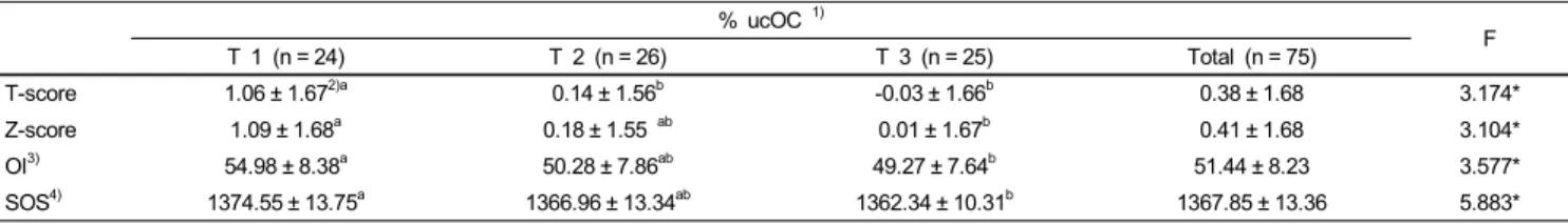
Table 1. Anthropometric measurements of the subjects by % Undercarboxylated Osteocalcin
% ucOC 1)
T 1 (n = 24) T 2 (n = 26) T 3 (n = 25) Total (n = 75) F
T-score 1.06 ± 1.672)a 0.14 ± 1.56b -0.03 ± 1.66b 0.38 ± 1.68 3.174*
Z-score 1.09 ± 1.68a 0.18 ± 1.55 ab 0.01 ± 1.67b 0.41 ± 1.68 3.104*
OI3) 54.98 ± 8.38a 50.28 ± 7.86ab 49.27 ± 7.64b 51.44 ± 8.23 3.577*
SOS4) 1374.55 ± 13.75a 1366.96 ± 13.34ab 1362.34 ± 10.31b 1367.85 ± 13.36 5.883*
1)% Undercarboxylated Osteocalcin : Tertile 1 < 19.34%, Tertile 2 : 19.34% - 32.01%, Tertile 3 > 32.01%
2)Mean ± SD
3)OI : Osteoporosis index
4)SOS : Speed of sounds
*P< 0.05
Significance as determined by ANOVA-test
a,bValues with different letters within the same line are significantly different from each other by Tukey’s test at P= 0.05.
Table 2. Bone mineral density of the subjects by % Undercarboxylated Osteocalcin
database was combined with that of the Rural Development Administration [23] and the USDA National Nutrient Database [24]. The standard guidelines for energy and nutrient intakes were based on estimated energy requirements (EER), estimated average requirements (EAR), and adequate intakes (AI). The percentages of recommended intake were analyzed to assess nutritional quality [25].
Statistical analysis
Statistical analyses were performed using SPSS (Statistical Package for Social Science) 12.0. Measurements are expressed as the mean and SD (standard deviation). ANOVA was performed to assess statistical significance depending on %ucOC between the three groups. For an inter-group analysis, a post-hoc analysis was performed with Tukey’s test. Multiple regression analysis was used to assess associations between biochemical indicators (serum %ucOC, cholesterol, and triglycerides) and the inflammatory marker hs-CRP, as well as the status of bone mineral density. The covariates that were considered in these models included age and body mass index (BMI). Statistical significance was set at P < 0.05.
Results
General characteristics and physical examination
Based on tertiles of %ucOC, the subjects’ general characteristics and physical examination findings were analyzed. The results are presented in Table 1. Between the three groups, there were no significant differences in age, height, amount of muscle, amount of body fat, waist to hip ratio, percentage of body fat, or BMI.
Mean BMI was measured as 21.18 ± 2.37 kg/m
2, which was within the normal range [26].
Analysis of bone mineral density
The results for bone mineral density measurements are presented in Table 2. The T-scores for the first and third tertiles were 1.06 and -0.03, respectively. These results indicate that T-scores were significantly lower as %ucOC values increased (P < 0.05). In the group where %ucOC values corresponded to the highest tertiles, Z-scores, OI (Osteoporosis index), and SOS (Speed of sound) were significantly lower as compared to
%ucOC values corresponding to the lowest tertiles (P < 0.05).
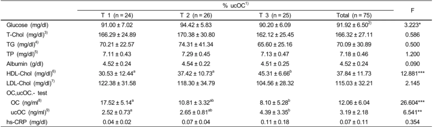
% ucOC1)
T 1 (n = 24) T 2 (n = 26) T 3 (n = 25) Total (n = 75) F
Glucose (mg/dl) 91.00 ± 7.02 94.42 ± 5.83 90.20 ± 6.09 91.92 ± 6.502) 3.223*
T-Chol (mg/dl)3) 166.29 ± 24.89 170.38 ± 30.80 162.12 ± 25.45 166.32 ± 27.11 0.586
TG (mg/dl)4) 70.21 ± 22.57 74.31 ± 41.34 65.60 ± 25.16 70.09 ± 30.89 0.500
TP (mg/dl)5) 7.11 ± 0.43 7.29 ± 0.45 7.13 ± 0.47 7.18 ± 0.46 1.200
Albumin (g/dl) 4.52 ± 0.24 4.54 ± 0.22 4.51 ± 0.25 4.52 ± 0.24 0.090
HDL-Chol (mg/dl)6) 30.53 ± 12.44a 37.42 ± 10.73a 45.31 ± 6.66b 37.84 ± 11.73 12.881***
LDL-Chol (mg/dl)7) 122.38 ± 31.58 118.30 ± 34.79 104.56 ± 28.32 115.03 ± 32.21 2.145
OC,ucOC.- test
OC (ng/ml8) 17.52 ± 5.14a 10.81 ± 3.32ab 8.10 ± 5.28b 12.06 ± 6.04 26.604***
ucOC (ng/ml)9) 2.52 ± 0.73a 2.65 ± 0.81ab 4.39 ± 3.35b 3.19 ± 2.18 6.541**
hs-CRP (mg/dl) 0.04 ± 0.02 0.07 ± 0.04 0.11 ± 0.18 0.07 ± 0.11 0.354
1)% Undercarboxylated Osteocalcin : Tertile 1 < 19.34%, Tertile 2 : 19.34% - 32.01%, Tertile 3 > 32.01%
2)Mean ± SD
3)T-Chol : Total Cholesterol
4)TG : Triglyceride
5)TP : Total-Protein
6)HDL-Chol : High Density Lipoprotein-Cholesterol
7)LDL-Chol : Low Density Lipoprotein-Cholesterol
8)Osteocalcin
9)Undercarboxylated Osteocalcin
*P< 0.05, **P< 0.01, ***P< 0.001 Significance as determined by ANOVA - test
a, b, cvalues with different letters within the same line are significantly different from each other by Tukey’s test at P= 0.05.
Table 3. Biochemical assessments of subjects by % Undercarboxylated Osteocalcin
DRI for reference
% ucOC1) T 1 F
(n = 24) % of
Mean/DRI T 2
(n = 26) % of
Mean/DRI T 3
(n = 25) % of
Mean/DRI Total
(n = 75)
Energy (kcal) EER2) 1616.02 ± 398.07a 77 1589.27 ± 525.94b 76 1245.76 ± 453.06b 59 1483.33 ± 487.855) 4.941*
Protein (g) EAR3) 59.58 ± 17.48a 170 59.27 ± 20.33b 169 44.41 ± 16.56b 126 54.42 ± 19.35 5.644*
Fat (g) 52.49 ± 17.62a 48.44 ± 21.07ab 35.88 ± 17.93b 45.55 ± 20.02 5.143**
Carbohydrate (g) 221.81 ± 68.70 228.69 ± 81.87 186.53 ± 76.88 212.43 ± 77.43 2.218
Fiber (g) AI4) 16.08 ± 6.49 64 15.32 ± 9.47 61 12.05 ± 6.15 48 14.48 ± 7.66 1.984
Calcium (mg) EAR 407.01 ± 148.11 70 453.26 ± 272.21 78 318.17 ± 140.47 55 393.43 ± 203.67 3.041 Phosphorus (mg) EAR 857.50 ± 221.64a 148 857.47 ± 332.07a 148 635.58 ± 243.55b 110 783.52 ± 287.84 5.564**
Iron (mg) EAR 10.71 ± 3.63ab 97 12.71 ± 7.89a 116 8.31 ± 2.70b 76 10.60 ± 5.55 4.382*
Sodium (mg) AI 3620.30 ± 1220.05 241 3324.43 ± 2479.47 222 2807.04 ± 1299.30 187 3246.64 ± 1788.80 1.314 Potassium (mg) AI 2075.37 ± 699.56 44 2130.76 ± 1142.40 45 1819.63 ± 987.10 39 2009.33 ± 963.21 0.743
Zinc (mg) EAR 7.12 ± 2.53 102 7.06 ± 2.50 101 5.63 ± 1.88 80 6.60 ± 2.39 3.298*
Vitamin A (μgRE) EAR 775.85 ± 432.98 169 632.91 ± 571.64 138 512.48 ± 277.46 111 638.51 ± 452.91 2.137
Vitamin B1 (mg) EAR 1.04 ± 0.38 116 1.09 ± 0.58 121 0.80 ± 0.38 89 0.89 ± 1 2.841
Vitamin B2 (mg) EAR 1.00 ± 0.89 98 1.26 ± 1.38 126 0.75 ± 0.40 75 01.00 ± 0.89 2.198
Vitamin B6 (mg) EAR 1.68 ± 0.70ab 140 1.81 ± 0.81a 151 1.28 ± 0.45b 107 1.59 ± 0.70 4.319*
Niacin (mg) EAR 12.09 ± 4.07 110 14.08 ± 6.60 128 11.56 ± 7.01 105 12.60 ± 6.09 1.226
Vitamin C (mg) EAR 60.85 ± 23.79 81 69.56 ± 66.80 93 50.86 ± 28.10 68 60.54 ± 44.72 1.119 Folate (μg) EAR 221.22 ± 111.42 69 201.70 ± 124.22 63 154.54 ± 69.22 48 192.23 ± 106.82 2.656
Vitamin E (mg) AI 15.23 ± 8.49 15 12.20 ± 6.78 12 11.10 ± 7.64 11 12.80 ± 7.59 2.004
Vitamin K (μg) AI 94.88 ± 51.48a 146 73.85 ± 45.15ab 114 62.58 ± 39.92b 96 76.82 ± 46.97 3.146*
Cholesterol (mg) 328.05 ± 119.31 290.37 ± 161.48 226.21 ± 164.87 281.04 ± 154.31 2.879
C: P: F (%)6) 54 : 15 : 30 58 : 15 : 27 58 : 14 : 27 57 : 15 : 28
1)% Undercarboxylated Osteocalcin : Tertile 1 < 19.34%, Tertile 2 : 19.34% - 32.01%, Tertile 3 > 32.01%
2)EER : Estimated Energy Requirements
3)EAR Estimated Average Requirements
4)AI : Adequate Intake
5)Mean ± SD
6)C: P: F (%) : Carbohydrate : Protein : Fat
*P< 0.05, **P< 0.01
Significance as determined by ANOVA-test
Table 4. Nutrient intakes of subjects by % Undercarboxylated osteocalcin
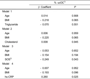
% ucOC1)
β Coeffient P
Model 1
Age 0.014 0.908
BMI - 0.218 0.065
Triglyceride - 0.070 0.551
Model 2
Age 0.006 0.959
BMI - 0.220 0.065
Cholesterol 0.008 0.945
Model 3
Age - 0.053 0.652
BMI - 0.154 0.194
SOS2) - 0.249 0.043
Model 4
Age - 0.007 0.952
BMI - 0.193 0.096
hs-CRP 0.260 0.025
1)% ucOC : % Undercarboxylated Osteocalcin
2)SOS : Speed of sound
Table 5. Associations between serum %ucOC and biochemical markers and bone mineral density
Biochemical assessment
Blood glucose, total cholesterol, triglycerides, total protein, and albumin were measured in the study subjects and the results are presented in Table 3. The first, second, and third tertiles of blood glucose were 91.00 ± 7.02 mg/dl, 94.42 ± 5.83 mg/dl, and 90.20
± 6.09 mg/dl (P < 0.05), respectively, and were in normal range.
There were no significant differences in total cholesterol, triglyceride, total protein, and albumin between the three groups.
Concentrations of osteocalcin and non-carboxylated osteocalcin were 17.52 ± 5.14 ng/ml and 2.52 ± 0.73 ng/ml in the highest tertile group, and 8.10 ± 5.28 ng/ml and 4.39 ± 3.35 ng/ml in the lowest tertile group, respectively, and there were significant difference between the groups (P < 0.001, P < 0.01). Tertile values of serum hs-CRP showed an increasing tendency as values of %ucOC increased; the first, second, and third tertiles of serum hs-CRP were 0.04 ± 0.02 mg/dl, 0.07 ± 0.04 mg/dl, and 0.11 ±
0.18 mg/dl, respectively, but these were not statistically significant.
Assessment of daily intake
Inter-group comparisons of nutrient intake amounts depending on values of %ucOC are presented in Table 4. Calorie intake and protein intake were found to be highest in the group where values of %ucOC were lowest. The first, second, and third tertile values of daily vitamin K intake were measured as 94.88 ± 51.48 μ g, 73.85 ± 45.15 μg, and 62.58 ± 39.92 μg, respectively, and were significantly different (P < 0.05). There were also significant differences in intake amounts of nutrients such as iron, phosphorus, zinc, and vitamin B
6between the classified groups based on values of %ucOC (P < 0.05). Following the calculations
of percentages of recommended nutrient intakes, all study subjects were found to have intakes of protein, phosphorus, vitamin A, vitamin B
2, niacin, vitamin B
6, and vitamin B
1that were greater than the recommendations. As compared to the recommendations, however, all the study subjects consumed insufficient amounts of calcium, folic acid, and vitamin C.
Correlations between serum %ucOC, inflammatory markers, and bone mineral density
We examined the relationships between %ucOC, biochemical markers, and bone mineral density, and the results are presented in Table 5. There was no association between serum %ucOC and serum triglycerides or between serum %ucOC and cholesterol, after adjusting for covariates. There was a negative correlation between serum %ucOC and hs-CRP. However, there was a positive correlation between serum %ucOC and bone mineral density, which was measured by an ultrasonographic method.
Discussion
We examined concentrations of serum hs-CRP, an inflam- matory marker, along with bone mineral density and vitamin K nutritional status in young women in their 20s. Then, we also examined the relationships between these parameters. The 20s is the period of age in which bone mass reaches a maximum.
During this period, bone health and nutritional status are important to prevent osteoporosis during the elderly years [27].
In the current study, the mean T-score of the study subjects was 0.38. In particular, in the group with the highest %ucOC and where the degree of vitamin K nutritional status was relatively lower, the T-score was measured as -0.03. This value corres- ponded to the normal range according to WHO criteria. It is presumed, however, that further risks of developing osteopenia would increase considering that the enrolled subjects were women in their 20s who maintained a maximal degree of bone mass.
As a marker indicating bone formation, osteocalcin is a
bone-specific protein composed of 49 amino acids. It is also one
of the non-collagen proteins forming bone matrix, which is used
as a marker to predict degree of bone formation [28-30]. In regard
to the concentration of serum osteocalcin, following the synthesis
of osteocalcin in osteoblasts, osteocalcin accumulates in the bone
matrix and its residual amount is measured following its release
into the blood. Accordingly, an increased concentration of serum
osteocalcin is indicative of increased bone formation [31]. In our
study, the mean concentration of serum osteocalcin was 12.06
ng/ml. This value was relatively higher than 11.8 ng/ml, as
measured by Hong [32] in postmenopausal women, and 8.30
ng/ml as measured by Sung et al. [33]. These results indicate
that degrees of bone formation are relatively higher in younger
women.
In association with the maintenance of a healthy skeletal structure, protein plays a key role in forming and then maintaining maximal bone mass. Yet, excessive intake of animal protein decreases calcium absorption from the kidneys and this has a detrimental effect on the formation of skeletal structure [34]. In this study, when compared with the EAR for protein (35 g), the subjects were found to have a protein intake ratio of 155.48%. Intake amounts of essential nutrients for the formation of skeletal structure, including calcium, zinc, folic acid, and vitamin C, were all found to be insufficient as compared to their EAR values. In particular, in the group where %ucOC was highest, degrees of nutrient insufficiency were also found to be relatively higher. Accordingly, in women in their 20s, in whom maximal bone mass is reached, an appropriate intake of protein is mandatory. Therefore, these subjects should be instructed to consume vitamins and minerals that might be easily deficient.
In our study, vitamin K was found to be appropriately consumed at an amount of 78.82 μg, as compared to the mean daily AI of 65 μg proposed by the KDRIs. To date, not many studies have been conducted to analyze the status of vitamin K intake in Korea. According to Hong [32] who examined postmenopausal women, vitamin K was found to be consumed at an amount of 379 μg. According to a study conducted to analyze vitamin K intake in young Japanese women, the average intake was 155.9 μg [35], and it was approximately 80 μg in American women under age 45 [36]. In our study, the amount of vitamin K intake was relatively lower than the results of previous studies. This might be due to factors associated with differences in dietary habits between countries, a deficiency of green leafy vegetables containing higher amounts of vitamin K, and lower food intake because of excessive interest in body weight control in younger women.
In such a condition where osteocalcin is not carboxylated due to the depletion of vitamin K, osteocalcin is not effective for bone formation and is released to the blood as a form of ucOC.
ucOC has therefore been used as a marker indicating vitamin K nutritional status [37]. In recent years, however, the proportion of uncarboxylated osteocalcin has been measured in proportion to the amount of osteocalcin, and this is referred to as %ucOC.
Therefore, %ucOC has been proposed as a more sensitive indicator of vitamin K nutritional status [37]. In the current study, the degree of vitamin K intake was relatively lower in the group where %ucOC was relatively higher as compared to other groups.
These results are in agreement with previous reports demons- trating a negative correlation between vitamin K intake and
%ucOC [38,39]. However, the young women in the present study had a relatively high degree of undercarboxylated serum osteocalcin (mean 32.5%) than subjects in other studies (21.9%
for healthy girls in Denmark [38], 7.5% for healthy adults [19], 13.0% for postmenopausal women in the USA [39]).
According to a cohort study that was conducted to examine
the correlation between proximal femur fracture and level of vitamin K intake in the elderly, incidence of fracture was relatively lower in subjects with higher intakes of vitamin K [16,40]. It was also reported that risks of developing hip joint fractures were decreased following the intake of vitamin K [41].
Furthermore, it is recommended that vitamin K should be ingested at an amount of > 500 μg/day for the prevention of bone fractures [42]. In the current study, which was conducted in women in their 20s, there was a negative correlation between
%ucOC and bone mineral density, which is in agreement with previous reports that studied elderly patients and patients with bone fractures. Therefore, in women in their 20s during which maximal bone mineral density is reached, special attention should be given to vitamin K intake to ensure adequate stores of the nutrient.
Osteoporosis is a disease occurring as a result of imbalances in bone remodeling due to the predominance of bone resorption over bone formation, during which various key factors are involved in bone physiology and regeneration [43]. Pro-inflammatory cytokines involved in inflammatory responses, such as interleukin (IL)-6 and CRP, have also been revealed to regulate bone metabolism even in healthy individuals without immunological diseases [44]. IL-6 is synthesized from osteoblasts, monocytes, and T-cells and it facilitates the differentiation and activation of osteoclasts [45]. C-reactive protein (CRP) belongs to the pentaxin family of immune-recognition proteins, and it is a sensitive marker for systemic inflammatory responses. Further- more, it was recognized that IL-1, IL-6, and TNF-α are regulatory factors for the synthesis of CRP [46,47]. According to Bae et al. [48] who examined the correlation between bone mineral density and hs-CRP, as concentrations of serum hs-CRP increased, bone mineral density decreased and bone turnover rate was increased [49]. These authors therefore proposed that serum hs-CRP is closely associated with bone metabolism and inflammatory responses. According to a study on correlations between vitamin K and inflammatory responses, there were positive correlations between %ucOC and two inflammatory markers (IL-6 and hs-CRP). Moreover, in the current study following regression analysis, as values of %ucOC indicating vitamin K deficiency increased, concentrations of serum hs-CRP also significantly increased. Because there remains an insufficient amount of definite data on inflammatory markers and mechanisms associated with vitamin K, further studies are needed.
The limitations of the current study are as follows: The study was a cross-sectional design and it did not clarify whether there are causal relationships between vitamin K nutritional status, bone mineral density, and inflammatory markers. Further studies are therefore warranted to clarify any causal relationships.
Nevertheless, the current study is of great significance in that
it includes an analysis of correlations between vitamin K
nutritional status and inflammatory responses in healthy adult
women who had not yet developed specific types of bone disease.
References
1. Ministry of Health and Welfare. Korea National Health and Nutrition Examination Survey Report (KNHANES Ⅳ); 2008.
2. Kim MS, Koo JO. Comparative analysis of food habits and bone density risk factors between normal and risk women living in the Seoul area. Korean Journal of Community Nutrition 2008;13:
125-33.
3. Koo JO, Ahn HS, Yoo SY. Study of bone mineral density, body composition and dietary habits of 20-30 years women. Korean Journal of Community Nutrition 2008;13:489-98.
4. Mayoux-Benhamou MA, Leyge JF, Roux C, Revel M. Cross- sectional study of weight-bearing activity on proximal femur bone mineral density. Calcif Tissue Int 1999;67:179-83.
5. Yu SH, Lee YS, Lee JS. Some factors affecting bone density of Korean college women. The Korean Journal of Nutrition 1998;31:36-45.
6. Cho DS, Lee JY. Bone mineral density and factors affecting in female college students. Korean Journal of Women Health Nursing 2008;14:297-305.
7. Joo NS, Kong MH, Kim BT, Park SB, Lee TY, Kim KM. Impact of smoking and alcohol intake on bone mineral density in men.
Journal of the Korean Academy of Family Medicine 2006;27:
911-6.
8. Choi SK, Yoon JH, Kim ES, Oh JK. The effect of replacement therapy on bone mineral density of the lumbar spine and hip in postmenopausal women. Journal of the Korean Academy of Family Medicine 1998;19:86-94.
9. Cooper L, Clifton-Bligh PB, Nery ML, Figtree G, Twigg S, Hibbert E, Robinson BG. Vitamin D supplementation and bone mineral density in early postmenopausal women. Am J Clin Nutr 2003;77:1324-9.
10. Sung CJ, Kim SY, Kim MH, Kim EY. The effect of isoflavone supplementation by soymilk on bone mineral density in under- weight college women. The Korean Journal of Nutrition 2003;36:
470-5.
11. Hong YJ, Choue RW. Correlation of dietary vitamin K intakes and bone mineral density in postmenopausal women. The Korean Journal of Nutrition 1997;30:299-306.
12. Korean Society of Bone Metabolism. Physician’s guide for diagnosis & treatment of osteoporosis; 2008. p.87.
13. Han JK. The effect of progressive resistance exercise on osteocalcin or bone density in postmenopausal women. Korean Journal of Sport Science 2008;17:571-8.
14. Yamauchia M, Yamaguchi T, Nawata K, Takaoka S, Sugimoto T. Relationships between undercarboxylated osteocalcin and vitamin K intakes, bone turnover, and bone mineral density in healthy women. Clin Nutr 2010;29:761-5.
15. Binkley NC, Suttie JW. Vitamin K nutrition and osteoporosis.
J Nutr 1995;125:1812-21.
16. Booth SL, Tucker KL, Chen H, Hannan MT, Gagnon DR, Cupples LA, Wilson PWF, Ordovas J, Schaefer EJ, Dawson- Hughes B, Kiel DP. Dietary vitamin K intakes are associated with hip fracture but not with bone mineral density in elderly men and women. Am J Clin Nutr 2000;71:1201-8.
17. Kaneki M, Hosoi T, Ouchi Y, Orimo H. Pleiotropic actions of vitamin K: protector of bone health and beyond? Nutrition 2006;22:845-52.
18. Booth SL, Broe KE, Gagnon DR, Tucker KL, Hannan MT, McLean RR, Dawson-Hughes B, Wilson PWF, Cupples LA, Kiel
DP. Vitamin K intake and bone mineral density in women and men. Am J Clin Nutr 2003;77:512-6.
19. Binkley NC, Krueger DC, Kawahara TN, Engelke JA, Chappell RJ, Suttie JW. A high phylloquinone intake is required to achieve maximal osteocalcin gamma-carboxylation. Am J Clin Nutr 2002;76:1055-60.
20. Hwang CS, Chung Hy, Kang YS, Moon IG, Yim CH, Han KO, Jang HC, Yoon HK, Han IK, Choi TB. The antiresorptive effects of vitamin K2 on osteoblasts and osteoclasts. Korean Journal of Bone Metabolism 2001;8:115-21.
21. Shea MK, Booth SL, Massaro JM, Jacques PF, D’Agostino RB Sr, Dawson-Hughes B, Ordovas JM, O’Donnell CJ, Kathiresan S, Keaney JF Jr, Vasan RS, Benjamin EJ. Vitamin K and vitamin D status: associations with inflammatory markers in the Framingham Offspring study. Am J Epidemiol 2008;167:313-20.
22. Shea MK, Dallal GE, Dawson-Hughes B, Ordovas JM, O’Donnell CJ, Gundberg CM, Peterson JW, Booth SL. Vitamain K, circulating cytokines, and bone mineral density in older men and women. Am J Clin Nutr 2008;88:356-63.
23. National Rural Resources Development Institute. Seventh revision food composition table; 2006. p.16-95.
24. United States Department of Agriculture [Internet]. Search the USDA national nutrient database for standard reference [Cited 2009 September 01]. Available from: http://www.nal.usda.gov/
fnic/foodcomp/search.
25. Korean Nutrition Society. Dietary reference intakes for Koreans;
2005.
26. WHO Western Pacific Region. The Asia-pacific perspective:
Redefining obesity and its Treatment; 2000.
27. Jea EJ, Byoun KE, Youn JE, Lee BK, Kim HS. Effects of body composition and nutrients intake on the calcaneal broadband ultrasound attenuation in college students. Korean Journal of Community Nutrition 2009;14:590-9.
28. Huh M, Mun YJ, Lim HS, Jo HH, Kim MR, Kim EJ, Kim JH, Kim JH. The effects of vitamin K2 (glakay) and vitamin K2 plus hormone replacement therapy on bone mineral density and bone metabolism in postmenopausal woman. The Journal of Korean Society of Menopause 2005;11:206-12.
29. Price PA, Williamson MK, Lothringer JW. Origin of the vitamin K- dependent bone protein found in plasma and its clearance by kidney and bone. J Biol Chem 1981;256:12760-6.
30. Delmas PD. Biochemical markers of bone turnover I: Theoretical considerations and clinical use in osteoporosis. Am J Med 1993;
95:11S-6S.
31. Liu G, Peacock M. Age-related changes in serum undercarbox- ylated osteocalcin and its relationships with bone density, bone quality, and hip fracture. Calcif Tissue Int 1998;62:286-9.
32. Hong JY. The effests of vitmain K supplements on serum osteocalcin caraboxylation in postmenopausal women. The Korean Journal of Nutrition 1999;32:726-31.
33. Sung CJ, Choi YH, Kim MH, Choi SH, Cho KO. A study of nutrient intake and serum levels of osteocalcin, Ca, P, and Mg and their correlation to bone mineral density in Korean postmenopausal women residing in rural areas. Korean Journal of Community Nutrition 2002;7:111-20.
34. Chung HY. Osteoporosis diagnosis and treatment 2007. Journal of Korean Endocrine Society 2008;23:76-108.
35. Kamao M, Suhara Y, Tsugawa N, Uwano M, Yamaguchi N, Uenishi K, Ishida H, Sasaki S, Okano T. Vitamin K content of foods and dietary vitamin K intake in Japanese young women.
J Nutr Sci Vitaminol 2007;53:464-70.
36. Booth SL, Suttie JW. Dietary intake and adequacy of vitamin K.
J Nutr 1998;128:785-8.
37. van Summeren MJ, van Coeverden SC, Schurgers LJ, Braam LA, Noirt F, Uiterwaal CS, Kuis W, Vermeer C. Vitamin K status is associated with childhood bone mineral content. Br J Nutr 2008;100:852-8.
38. O’Connor E, Molgaard C, Michaelsen K, Jakobsen J, Lamberg- Allardt CJ, Cashman KD. Serum percentage undercarboxylated osteocalcin, a sensitive measure of vitamin K status, and its relationship to bone health indices in Danish girls. Br J Nutr 2007;97:661-6.
39. Binkley N, Harke J, Krueger D, Engelke J, Vallarta-Ast N, Gemar D, Checovich M, Chappell R, Suttie J. Vitamin K treatment reduces undercarboxylated osteocalcin but does not alter bone turnover, density, or geometry in healthy postmenopausal north American women. J Bone Miner Res 2009;24:983-91.
40. Feskanich D, Weber P, Willett WC, Rockett H, Booth SL, Colditz GA. Vitamin K intake and hip fractures in women: a prospective study. Am J Clin Nutr 1999;69:74-9.
41. Shiraki M, Shiraki Y, Aoki C, Miura M. Vitamin K2 (menate- trenone) effectively prevents fractures and sustains lumbar bone mineral density in osteoporosis. J Bone Miner Res 2000;15:
515-21.
42. Bügel S, Søensen AD, Hels O, Kristensen M, Vermeer C,
Jakobsen J, Flynn A, Mølgaard C, Cashman KD. Effect of phylloquinone supplementation on biochemical markers of vitamin K status and bone turnover in postmenopausal women.
Br J Nutr 2007;97: 373-80.
43. Arron JR, Choi Y. Bone versus immune system. Nature 2000;
408:535-6.
44. Muller B. Cytokine imbalance in non-immunological chronic disease. Cytokine 2002;18:334-9.
45. Manolagas SC. Birth and death of bone cells: basic regulatory mechanisms and implications for the pathogenesis and treatment of osteoporosis. Endocr Rev 2000;21:115-37.
46. Gabay C, Kushner I. Acute-phase proteins and other systemic responses to inflammation. N Engl J Med 1999;340:448-54.
47. Weinhold B, Ruther U. Interleukin-6-dependent and independent regulation of the human C-reactive protein gene. Biochem J 1997;327:425-9.
48. Bae SJ, Son HY, Pyun DK, Nah SS, Koh JM, Kim GS. Higher Circulating hs-CRP levels are associated with lower bone mineral density in healthy pre-and postmenopausal women: evidence for a link between systemic inflammation and osteoporosis. Osteoporos Int 2005;16:1263-71.
49. Shea MK, Dallal GE, Dawson-Hughes B, Ordovas JM, O’Donnell CJ, Gundberg CM, Peterson JW, Booth SL. Vitamin K, circulating cytokines, and bone mineral density in older men and women. Am J Clin Nutr 2008;88:356-63.