서론
골유착(osseointegration) 개념이 도입된 이후,1,2 치과 용 임플란트는 상실된 치아의 재건을 위해 널리 이용되었 으며, 이와 관련하여 장기간에 걸친 높은 성공률이 보고 되고 있다.3,4 최근 Niedermaier 등은 7년간의 후향적 연구 에서 97.0%의 임플란트 생존율을,4 Hasegawa 등은 10년 간의 후향적 연구에서 96.7%의 임플란트 생존율을 보고 하였다.5
임플란트와 관련하여 보고된 성공 기준(Success crite- ria) 가운데, Roos 등이 제안한 다음의 기준이 가장 널리
받아들여지고 있다; 임상적으로 동요도가 없을 것, 방사 선 사진 상 임플란트 주위로 방사선 투과상이 없을 것, 임 플란트 식립 1년 후 매년 0.2 mm 미만의 수직적 골 결손 을 보일 것, 통증, 감염, 신경병증, 감각이상 또는 하악관 의 침범이 없을 것, 5년 실패율이 10%를 미만일 것.6 성공 률(Success rate)은 위의 조건을 모두 만족시키며 이상적 으로 기능중인 임플란트에 적용되는 개념이기 때문에 약 간의 골소실을 동반하였지만 구내에서 안정적인 상태를 가지는 일반적인 임플란트는 이에 포함되지 않는다. 따 라서 임상 보고에서 가장 널리 통용되는 성공 기준은 임 플란트가 구내에서 생리적으로 기능하는지 혹은 제거되
*Correspondence to: Hyun-Joo Kim
Clinical Assistant Professor, Department of Periodontology, School of Dentistry, Pusan National University, Dental Research Institute, 20 Geumo-ro, Beomeo-ri, Mulgeum-eup, Yangsan, 50612, Republic of Korea
Tel: +82-55-360-5190, Fax: +82-55-360-5194, E-mail: [email protected] Received: March 9, 2018/Last Revision: March 27, 2018/Accepted: April 2, 2018
Factors associated with the survival rate and the marginal bone loss of dental implant over 7-years loading
Jung-Hyeok Choi, Jae-kwon Koh, Eun-Young Kwon, Ji-Young Joo, Ju-Youn Lee, Hyun-Joo Kim*
Department of Periodontology, School of Dentistry, Pusan National University, Dental Research Institute, Yangsan, Republic of Korea
Purpose: The purpose of this study was to analyze the factors affecting the survival rate and the marginal bone level of dental
implants that have functioned over 7-years. Materials and Methods: In 92 patients, 178 dental implants were included. Implant- related factors (diameter, length, prosthetic splint), patient-related factors (gender, smoking, plaque index, compliance to supportive periodontal therapy) and surgery-related factors (proficiency of surgeon, bone graft) were evaluated via clinical and radiographic examination. The marginal bone level was determined by intraoral standard radiography at the mesial and distal aspects of each implant using an image analysis software program. Results: The survival rate of all the implants was 94.94% and the marginal bone level was 0.89 ± 1.05 mm, these results are consistent with other studies that present long-term good clinical results. Implant length and plaque index among several factors were statistically significant for implant survival rate (P < 0.05). Smoking and the presence of regeneration surgery were statistically significant for the marginal bone level (P < 0.05). Conclusion: Dental implant that have functioned over 7-years showed favorable long-term survival rates and marginal bone level. Implant length and plaque control should be considered for improving the long-term clinical results. It is needed that careful application of bone regeneration technique and smoking control for maintaining of marginal bone level. (J Dent Rehabil Appl Sci 2018;34(2):116-26)Key words: dental implants; survival rate
Copyright© 2018 The Korean Academy of Stomatognathic Function and Occlusion.
It is identical to Creative Commons Non-Commercial License.
cc
었는지를 의미하는 생존율(Survival rate)이다.7 성공률은 최상의 상태를 나타내지만, 생존율이란 여전히 기능은 하지만 이상적인 상태는 아닌 경우까지 포함함을 의미한 다. Misch 등은 방사선상 골흡수가 임플란트 길이의 1/2 이상이거나, 동요도가 있으면 실패라고 간주하였고, 본 연구에서도 이 기준을 적용하였다.8
임플란트 예후에 영향을 주는 요인에는 임플란트 관련 요소, 환자 관련 요소, 술자 관련 요소 등 여러 가지가 보 고되고 있다. 그 중 임플란트 관련 요소로는 임플란트 길 이,9-11 직경,12 식립 위치, 표면처리 방식, 단일 임플란트 또 는 인접 임플란트간 고정 여부13 등이 있으며, 임플란트 길이가 짧을수록, 직경은 증가할수록 실패율이 높았다.
환자 관련 요소로는 성별,14 흡연,15 전신질환, 치주질환 이력, 구강위생 상태를 반영하는 치태지수16 등이 있으며, 그 중 흡연과 치태지수가 실패율과 연관이 있다고 보고 되고 있다. 술자 관련 요소로는 수술방법, 술자 숙련도,17 골재생술 시행여부18 등이 보고되고 있으나 연관성에 대 한 견해는 아직 논의 중에 있다. 임플란트의 장기적 예후 에 영향을 주는 변연골 수준도 다양한 요인들과 연관성 이 있다고 보고되고 있다.19,20 수술 시 외상, 교합적 과부 하, 감염 등도 임플란트 변연골 수준에 영향을 주는 것으 로 알려져 있으나 이에 관하여 이견이 존재한다.
이에 본 연구는 부산대학교치과병원 치주과에서 임플 란트를 식립하고, 7년 이상 기능한 임플란트를 대상으로 하여 임플란트 요인, 환자 요인, 수술 요인 등 다양한 요 인들과 임플란트 주위 변연골 수준과 생존율의 상관관계 를 분석하고자 하였다.
연구 대상 및 방법
1. 연구대상
부산대학교치과병원 치주과에서 임플란트 매식수술을 시행한 후, 기능한지 7년 이상 경과된 임플란트를 대상으 로 하였다. 후향적 연구에 대한 설명 및 서면 동의서 작성 후, 연구를 진행하였다. 본 연구는 부산대학교치과병원 생명윤리심의위원회의 승인(PNUDH-2017-005)하에 진 행되었다.
2. 연구방법
본 연구에서 임플란트 평가는 구내에서 생리적으로 기
능하는 임플란트는 생존으로 간주하였으며, 방사선상 골흡수가 임플란트 길이의 1/2 이상이거나, 동요도가 있 으면 실패로 간주하였다.8
1) 임상검사
치태지수는 다음의 기준으로 측정하였다.
- 치태지수(modified Plaque Index; mPI)21 0: 치태 없음
1: 탐침으로만 식별 가능한 치태 2: 눈으로 식별 가능한 치태 3: 많은 치태
2) 진료기록부 검사
다음의 항목들이 기록되었다.
① 임플란트 길이
② 임플란트 직경
③ 임플란트 식립 위치
④ 임플란트 상부 보철물 연결 유무
⑤ 성별
⑥ 흡연 유무
⑦ 유지치주치료 순응도
⑧ 골재생술 시행 유무
⑨ 술자 숙련도
3) 방사선학적 검사
각 임플란트 마다 표준 치근단방사선 사진을 평행촬 영법으로 촬영하였다. 모든 방사선 사진은 컴퓨터상에서 디지털화하여 Image analysis software프로그램(AxioVi- sion, Carl Zeiss, Oberkochen, Germany)을 이용하여 측 정하였다. 각 표준 치근단방사선 사진의 흑화도 및 대조 도는 프로그램상의 최적화 값으로 교정하였고, 측정값 의 표준화는 임플란트 고정체 길이를 기준으로 하였다.
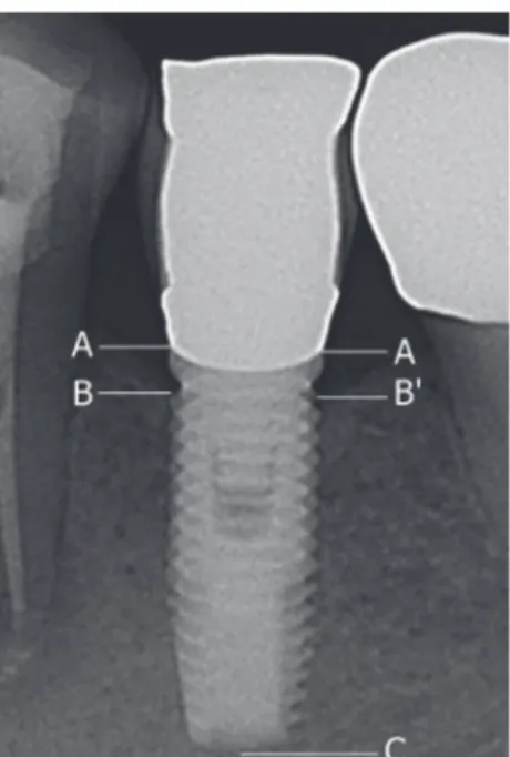
임플란트 고정체-지대주 연결부에서 첫 번째로 나타나는 골과 고정체 접촉점 간의 거리를 근심 및 원심측에서 각 각 측정하였다(Fig. 1). 변연골 수준은 1인의 검사자에 의 해 1회 측정하였고, 평균 변연골 수준은 근심 및 원심측 변연골 수준의 평균으로 계산하였다.
4) 통계분석
임플란트 길이, 직경, 식립 위치, 보철물 연결고정 유 무, 성별, 흡연 유무, 치태지수, 유지치주치료 순응도, 골 재생술 시행 유무, 술자 숙련도가 임플란트 생존율에 미
치는 영향을 알아보기 위하여 이분형 로지스틱 회귀분 석(binary logistic regression analysis)을 시행하였다. 이 후 변수 간 비교를 위하여 다중회귀분석(multiple logistic regression analysis)을 시행하였다. 그리고 임플란트 길 이, 임플란트 직경, 임플란트 식립 위치, 임플란트 보철 물 연결고정 유무, 성별, 흡연 유무, 유지치주치료 순응 도, 골재생술 시행 유무가 임플란트 변연골 수준에 미 치는 영향을 알아보기 위하여 독립표본 T-검정법을 사 용하였으며, 치태지수 및 술자 숙련도에 따른 평균 변연 골 수준의 관계를 알아보기 위하여 일원배치 분산분석법 (one way ANOVA)을 시행하였다. 사후검정법(post hoc analysis)으로는 Bonferroni 검정을 채택하였다. 모든 통 계는 SPSS 21.0 버전(SPSS ver. 21, IBM SPSS Inc., Chi- cago, USA) 프로그램을 이용하여 시행하였으며, 유의수 준은 0.05 이하인 경우 유의하다고 평가하였다.
결과
연구에 참여한 환자 수는 총 92명(남성 31명, 여성 61 명, 평균 연령 58.73 ± 10.55세), 식립된 임플란트는 178
개이었다. 178개 중 9개가 실패하여 전체 임플란트 생존 율은 94.94%로 나타났다. 임플란트의 평균 변연골 흡수 는 0.89 ± 1.05 mm로 나타났다.
1. 임플란트의 요인별 분포 현황
연구에 포함된 모든 임플란트 고정체는 RBM (resorb- able blasting media)으로 표면 처리된 외부 연결형이었으 며, 임플란트의 요인별 분포는 다음과 같다(Table1).
Table 1. Distribution of implants according to variable
factorsSurvived implant N
(%)
Failed implant N
(%)
Total implant N
(%) Age
≤ 49 27 (15.2) 0 (0) 27 (15.2) 50 - 59 60 (33.7) 6 (3.3) 66 (37.0) 60 - 69 58 (32.6) 3 (1.7) 61 (34.3)
≥ 70 24 (13.5) 0 (0) 24 (13.5) Loading period
11 Y 11 (6.2) 0 (0) 11 (6.2) 10 Y 30 (16.9) 5 (2.8) 35 (19.7)
9 Y 36 (20.2) 2 (1.0) 38 (21.2) 8 Y 29 (16.3) 1 (0.6) 30 (16.9) 7 Y 63 (35.4) 1 (0.6) 64 (36.0) Length
8.5 15 (8.4) 2 (1.1) 17 (9.5) 10 50 (28.1) 5 (2.8) 55 (33.9) 11.5 60 (33.7) 1 (0.6) 61 (34.3) 13 44 (24.7) 1 (0.6) 45 (25.3) Diameter
3.3 3 (1.7) 0 (0) 3 (1.7) 3.75 6 (3.4) 0 (0) 6 (3.4) 4 51 (28.6) 3 (1.7) 54 (30.3) 5 109 (61.2) 6 (3.4) 115 (64.6) Position
Maxillary
anterior 5 (2.8) 0 (0) 5 (2.8) Maxillary
canine 5 (2.8) 0 (0) 5 (2.8) Maxillary
posterior 65 (36.5) 4 (2.3) 69 (38.8) Mandibular
anterior 2 (1.1) 0 (0) 2 (1.1) Mandibular
canine 0 (0) 0 (0) 0 (0) Mandibular
posterior 92 (51.7) 5 (2.8) 97 (54.5)
Fig. 1. Distances from bone to implant (mesial, distal)
were determined.
A - B: Distance of marginal bone level (mesial) A - B’: Distance of marginal bone level (distal)
A - C: Length of fixture (implant-abutment junction-apex) χ = (A - B) * Fixture length / (A - C)
χ’ = (A - B’) * Fixture length / (A - C) Mean marginal bone level = (χ + χ’) / 2
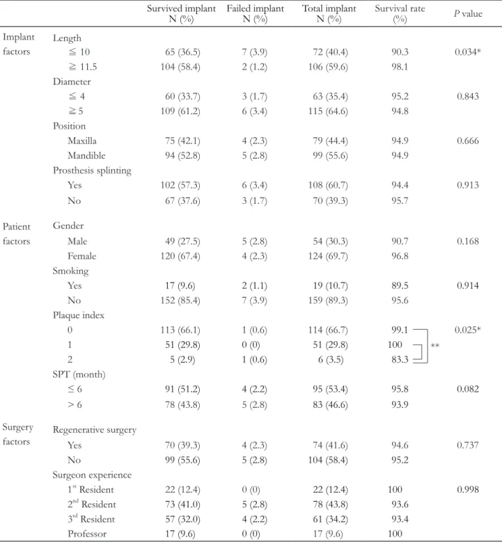
2. 임플란트 생존율에 영향을 주는 요인
임플란트 요인, 환자 요인, 수술 요인과 임플란트 생존 율과의 상관관계를 평가하였다(Table 2). 임플란트 요인에 서 임플란트 길이는 임플란트 생존율과 통계적인 유의성
이 관찰되었고(P < 0.05), 직경, 식립 위치, 보철물 연결고 정 유무는 임플란트 생존율과 통계적인 유의성이 관찰되 지 않았다. 환자 요인에서 치태지수는 임플란트 생존율과 통계적인 유의성이 관찰되었고(P < 0.05), 성별, 흡연 유 무, 유지치주치료 순응도는 임플란트 생존율과 통계적인
Table 2. Association of implant survival rate with variable factors by binary logistic regression analysis Survived implant
N (%) Failed implant
N (%) Total implant
N (%)
Survival rate(%) P value
Implant
factors Length
≦ 10 65 (36.5) 7 (3.9) 72 (40.4) 90.3 0.034*
≧ 11.5 104 (58.4) 2 (1.2) 106 (59.6) 98.1 Diameter
≦ 4 60 (33.7) 3 (1.7) 63 (35.4) 95.2 0.843
≧ 5 109 (61.2) 6 (3.4) 115 (64.6) 94.8 Position
Maxilla 75 (42.1) 4 (2.3) 79 (44.4) 94.9 0.666
Mandible 94 (52.8) 5 (2.8) 99 (55.6) 94.9 Prosthesis splinting
Yes 102 (57.3) 6 (3.4) 108 (60.7) 94.4 0.913
No 67 (37.6) 3 (1.7) 70 (39.3) 95.7
Patient factors
Gender
Male 49 (27.5) 5 (2.8) 54 (30.3) 90.7 0.168
Female 120 (67.4) 4 (2.3) 124 (69.7) 96.8 Smoking
Yes
17 (9.6) 2 (1.1) 19 (10.7) 89.5 0.914
No 152 (85.4) 7 (3.9) 159 (89.3) 95.6
Plaque index
0 113 (66.1) 1 (0.6) 114 (66.7)
99.1
0.025*1
51 (29.8) 0 (0) 51 (29.8) 100
2
5 (2.9) 1 (0.6) 6 (3.5) 83.3
SPT (month)
≤ 6
91 (51.2) 4 (2.2) 95 (53.4) 95.8 0.082
> 6 78 (43.8) 5 (2.8)
83 (46.6) 93.9
Surgeryfactors Regenerative surgery
Yes 70 (39.3) 4 (2.3) 74 (41.6) 94.6 0.737
No
99 (55.6) 5 (2.8) 104 (58.4) 95.2
Surgeon experience
1st Resident 22 (12.4) 0 (0)
22 (12.4) 100 0.998
2nd Resident73 (41.0) 5 (2.8) 78 (43.8) 93.6
3rd Resident
57 (32.0) 4 (2.2) 61 (34.2) 93.4
Professor
17 (9.6) 0 (0)
17 (9.6)100
* P < 0.05, ** P < 0.01.
SPT: supportive periodontal therapy.
Plaque index was analyzed by one way ANOVA for comparison between variables.
**
유의성이 관찰되지 않았다. 수술 요인에서 골재생술 시 행 유무, 술자 숙련도는 임플란트 생존율과 통계적인 유 의성이 관찰되지 않았다.
각 요인별로 이분형 로지스틱 회귀 분석을 시행한 후, 통 계적으로 유의성이 있는 임플란트 길이와 치태지수를 다 중 회귀 분석으로 분석하였다. 그 결과, 임플란트 길이는 10 mm 이하인 그룹에 비하여 11.5 mm 이상인 그룹에서 통계적으로 높은 임플란트 생존율을 보여주었다(Table 3).
치태지수는 0과 1인 그룹이 2인 그룹에 비하여 통계적으 로 유의하게 임플란트 생존율이 높았으며(P < 0.01), 치 태지수가 0과 1인 그룹 간에는 통계적으로 유의한 차이 가 없었다(Table 2).
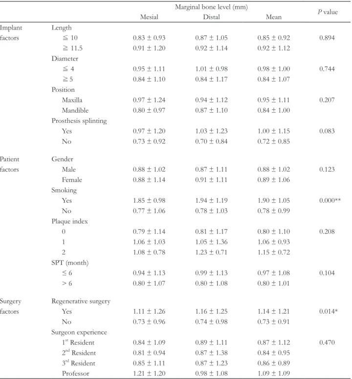
3. 평균 변연골 수준에 영향을 주는 요인
본 연구에서 임플란트 요인, 환자 요인, 수술 요인과 평 균 변연골 수준과의 상관관계를 평가하였다. 임플란트 요 인에서 임플란트 길이, 직경, 식립 위치, 보철물 연결고정 유무는 평균 변연골 수준과 통계적인 유의성이 관찰되지 않았다(Table 4). 환자 요인에서 흡연은 평균 변연골 수준 과 통계적인 유의성이 관찰되었고(P < 0.05), 성별, 치태 지수, 유지치주치료 순응도는 평균 변연골 수준과 통계 적인 유의성이 관찰되지 않았다(Table 4). 수술 요인에서 골재생술 시행 유무는 평균 변연골 수준과 통계적인 유 의성이 관찰되었고(P < 0.05), 술자 숙련도는 통계적인 유의성이 관찰되지 않았다(Table 4).
각 요인별로 독립표본 T-검정법 및 일원배치 분산분석 법을 시행한 후, 통계적으로 유의성이 관찰되었던 흡연 및 골재생술 시행 유무를 다중 회귀 분석으로 분석하였다. 그 결과, 비흡연 그룹에 비하여 흡연 그룹에서 평균 변연골 흡수량이 유의하게 증가하였다(P < 0.05, Table 5). 그리고 재생골 그룹은 건전골 그룹에 비하여 평균 변연골 흡수량 이 유의하게 증가하였다(P < 0.05, Table 5).
고찰
총 178개의 임플란트 중 9개가 실패하여 전체 임플란트 생존율은 94.94%였으며, 평균 변연골 수준은 0.89 ± 1.05 mm였다. Jung 등은 5년 후 임플란트 생존율은 97.2%, 10 년 후 임플란트 생존율은 95.2%로 보고하였으며,22 Simo- nis 등은 10년 생존율은 89.23%, 16년 생존율은 82.94%
로 보고 한 바 있다.23 본 연구에서도 기능 7년 후의 생존 율은 98.4%, 10년 후의 생존율은 89.1%로 관찰되어 앞 선 연구들과 유사하게 시간이 경과함에 따라 임플란트의 평균 생존율이 감소하는 것으로 나타났다.
임플란트 관련 요소 중 임플란트 길이가 임플란트 생 존율과 통계적 유의성이 있는 것으로 나타났다. 임플란트 길이는 11.5 mm 이상인 그룹에서 높은 생존율을 보여주었 는데, 이는 임플란트 길이가 길수록, 임플란트 생존율이 높아진다고 보고한 여러 연구와 일치하였다.9-11 선택된 임 플란트의 길이는 사용 가능한 골의 양과 질 혹은 하악골 의 모양과 관련이 있다.24 앞 선 연구에 의하면, 가용할 수 있는 골의 질이 나쁠수록, 골의 양이 적을수록 짧은 임플 란트를 식립 하는 경향이 있다. 따라서 임플란트 길이가 짧을수록 식립 시 수술 부위 골 환경이 긴 임플란트에 비 해 좋지 못했던 것으로 미루어 짐작해 볼 수 있으며 골질 및 골의 양 등이 임플란트의 길이와 생존율의 관계에 영 향을 주었을 것이다.
이 외에 임플란트 생존율에 영향을 줄 수 있을 것이라 고 고려하였던 임플란트 관련 요소 중 통계적 유의성이 관찰된 것은 없었다. 그 중 임플란트 직경에 관하여 살펴 보면, 이전 연구에서 직경이 증가함에 따라 임플란트 실 패율이 증가한다는 보고가 있었지만,12 본 연구에서는 임 플란트 직경이 증가함에 따라 임플란트 실패율이 증가하 는 경향성을 볼 수 있었지만 통계적인 유의성은 없었다.
하지만 좀 더 최근의 연구에 의하면 임플란트 직경과 생 존율 사이에는 관계가 없다는 보고가 있다.25,26 이는 아마
Table 3. Association of implant survival rate with variable factors by multiple regression analysis
B ± SE β t
P valueLength 9.364 ± 3.482 0.210 2.689 0.008*
Plaque index -4.759 ± 1.465 -0.190 -2.200 0.042*
* P < 0.05.
SE: standard errors.
Data are shown as mean ± standard deviation.
Table 4. Association of marginal bone level with variable factors by student’s T-test and One-way ANOVA
Marginal bone level (mm)P value
Mesial Distal Mean
Implant Length
factors ≦ 10 0.83 ± 0.93 0.87 ± 1.05 0.85 ± 0.92 0.894
≧ 11.5 0.91 ± 1.20 0.92 ± 1.14 0.92 ± 1.12 Diameter
≦ 4 0.95 ± 1.11 1.01 ± 0.98 0.98 ± 1.00 0.744
≧ 5 0.84 ± 1.10 0.84 ± 1.17 0.84 ± 1.07 Position
Maxilla 0.97 ± 1.24 0.94 ± 1.12 0.95 ± 1.11 0.207 Mandible 0.80 ± 0.97 0.87 ± 1.10 0.84 ± 1.00
Prosthesis splinting
Yes 0.97 ± 1.20 1.03 ± 1.23 1.00 ± 1.15 0.083 No 0.73 ± 0.92 0.70 ± 0.84 0.72 ± 0.85
Patient Gender
factors Male 0.88 ± 1.02 0.87 ± 1.11 0.88 ± 1.02 0.123
Female 0.88 ± 1.14 0.91 ± 1.11 0.89 ± 1.06 Smoking
Yes 1.85 ± 0.98 1.94 ± 1.19 1.90 ± 1.05 0.000**
No 0.77 ± 1.06 0.78 ± 1.03 0.78 ± 0.99 Plaque index
0 0.79 ± 1.14 0.81 ± 1.17 0.80 ± 1.10 0.208
1 1.06 ± 1.03 1.05 ± 1.36 1.06 ± 0.93
2 1.08 ± 0.78 1.23 ± 0.71 1.15 ± 0.72
SPT (month)
≤ 6 0.94 ± 1.13 0.99 ± 1.13 0.97 ± 1.08 0.104
> 6 0.80 ± 1.07 0.80 ± 1.08 0.80 ± 1.01 Surgery Regenerative surgery
factors Yes 1.11 ± 1.26 1.16 ± 1.25 1.14 ± 1.21 0.014*
No 0.73 ± 0.96 0.74 ± 0.98 0.73 ± 0.91 Surgeon experience
1st Resident 0.84 ± 1.09 0.89 ± 1.11 0.87 ± 1.12 0.470 2nd Resident 0.81 ± 0.94 0.87 ± 1.38 0.84 ± 0.95
3rd Resident 0.85 ± 1.11 0.87 ± 1.23 0.86 ± 0.89 Professor 1.21 ± 1.20 0.98 ± 1.08 1.09 ± 1.09
* P < 0.05, ** P < 0.01.
Data are shown as mean ± standard deviation.
Plaque index and surgeon experience were analyzed by one-way ANOVA for comparison between variables. Another factors were analyzed by Student’s t-test.
Table 5. Association of marginal bone level of implants with variable factors by multiple regression analysis
B ± SE β t
P valueSmoking 1.323 ± 0.277 0.381 4.783 0.000**
Regenerative surgery 0.348 ± 0.159 0.162 2.184 0.030*
* P < 0.05, ** P < 0.01.
SE: standard errors.
Data are shown as mean ± standard deviation.
도 초기 골융합 개념의 도입 이후 시간이 흐름에 따라 임 플란트에 대한 지식이 축적되면서 적절한 케이스 선택이 가능해지고 임플란트 수술법의 알맞은 사용과 임플란트 형태 및 디자인의 발전에 따른 것으로 생각된다. 이와 같 은 맥락으로 Javed와 Romanos는 위축된 상악 구치부에 서 임플란트 직경과 생존율에 대한 상관관계를 조사한 systematic review에서 임플란트 직경은 부가적인 요소 라고 하였다.27 그들의 결론에 따르면 장기간에 걸쳐 임플 란트의 높은 생존율을 얻기 위해서는 임플란트 수술법을 잘 계획하여 식립 당시 높은 고정력을 얻어야 하며 이와 함께 주기적인 유지관리를 시행하는 것이 필요하다.
본 연구에서 조사하였던 환자 관련 요소 중 치태지수 가 임플란트 생존율과 밀접한 관련이 있었다. 치태지수 는 2 이상일 때 임플란트 생존율을 낮추는 요인으로 작용 하였다. 이는 Higuchi 등의 연구와 상응하는 결과를 보여 주었으며, 구강위생관리가 임플란트 생존율에 영향을 주 는 요인이 된다는 것을 의미한다.16 이와 유사하게 Serino 와 Ström은 임플란트 주위염이 부적절한 치태조절과 관 련이 있다고 보고 하였다.28 이들의 연구에 따르면 임플 란트의 74%에서 부적절한 치태조절이 관찰되었다. 특 히 잔존 자연치의 치주질환 유무와 관계없이 임플란트 주위염이 관찰되었으며 이는 임플란트의 구조적 특성으 로 인해 치태조절을 위한 효과적인 접근이 어렵기 때문으 로 해석하였다. 따라서 다수의 연구들이 임플란트의 생 존율을 높이기 위해 효과적인 치태조절의 중요성을 강 조하였으며 이를 위해 유지치주치료가 필요하다고 하였
다.29-31 유지관리 순응도와 임플란트 생존율의 상관관계
를 살펴보았을 때, 본 연구에서 유지치료 간격이 6개월 이상인 그룹에서 임플란트 실패율이 증가하였지만 통계 적으로 유의하지는 않았다. 정확한 유지관리 순응도 평 가를 위해서는 정기적 내원 시기를 좀 더 세분화해서 상 관관계를 평가할 필요가 있다. 과거 치주질환 기왕력이 임플란트 생존율에 미치는 영향에 관하여서는 이견이 존 재한다. 많은 연구가 치주질환의 기왕력이 있는 환자군 에서 건강한 환자군과 비교해 보았을 때 더 낮은 생존율 과 더 많은 임플란트 주위골 소실을 보인다고 보고하였
다.15,32,33 반면 Wahlströme 등은 치주염의 기왕력 보다 과
부하나 이갈이 습관 등이 임플란트에게 더 해로운 요소 라고 보고 하였으며,34 Gianserra 등은 5년 기능한 임플란 트에서 치주염의 기왕력이 임플란트 실패율을 높이지 않 는다고 보고 하였다.35 그러나 이 두 연구는 모두 5년 이 하의 짧은 기간동안 평가하였기 때문에 다양한 환자 그
룹의 임상 결과를 비교 평가하기에는 부족하다.36 치주염 의 기왕력과 임플란트의 실패의 상관관계에 대한 이견과 는 별개로 양쪽 모두 계속유지관리의 중요성은 강조하 고 있다. 특히 최근에 acid-etched와 sandblasted 임플란 트와 같이 표면처리하여 미세 요철 구조를 가진 임플란 트가 널리 사용됨에 따라 계속유지관리의 중요성은 더욱 증가하고 있다.37
평균 변연골 수준에 영향을 주는 요인으로는 흡연 및 골재생술 시행 유무가 통계적으로 유의한 상관관계를 가졌다. Galindo-Moreno 등 역시 임플란트 주위 변연골 흡수에 영향을 줄 것으로 예상되는 인자(성별, 나이, 흡 연, 음주 그리고 치주질환의 기왕력)를 살펴보았으며 이 중 흡연이 가장 결정적인 영향을 미친다고 보고 하였다.38 흡연 그룹은 평균 변연골의 흡수가 증가하였는데, 이는 흡연이 초기 치유 기간 동안 임플란트 주위 변연골 흡수 량을 증가시키기 때문으로 보인다.39 최근 Duan 등은 흡 연과 구강 미생물총의 관계에 주목하여 연구하였으며, 비흡연가에 비해 흡연가에서 미생물총이 해로운, 즉 위험 성이 높은 방향으로 변하였음을 보고하였다.40 특히 Por- phylomonas gingivalis가 흡연가에서 유의하게 높았으며 이는 식립 이후 골 치유 과정 동안 진행되는 변연골 흡수 의 심도에 관련된 것으로 밝혀졌다. 다만 이 연구는 임플 란트 식립 후 초기 3개월간 임플란트 주위 변연골 흡수를 살펴본 것이므로 장기간에 걸친 연구가 이후 필요할 것 으로 보인다.
또한 평균 변연골 흡수량은 재생골에서 통계적으로 유 의하게 증가하였는데, 골재생술의 방법 및 술 전 골질에 대한 자료가 부족하였으므로 이러한 부분을 고려하여 해 석해야 할 것이다. Massimo 등은 수직으로 증대 시킨 이 식골에 식립한 임플란트의 변연골 흡수에 관하여 이식을 시행하지 않은 자연골에서와 유사하다고 보고 하였다.18 이보다 훨씬 이전에 다양한 연구들이 수평적으로 증대 시킨 이식골에 식립된 임플란트에서 자연골과 유사한 변 연골 흡수를 보고 하였다.41,42 그러나 상악동 내로 증대된 이식골에 식립된 임플란트에서는 상반된 연구결과가 보 고된 바 있다.38 상악동 내에 형성된 신생골의 stiffness가 자연골보다 작을 때 임플란트에 가해지는 힘이 변연골에 집중되는 경향이 있으며 이로 인해 임플란트 변연골의 흡 수가 더 진행되게 된다.19 따라서 임플란트 식립 후 초기 치유과정과 기능 부하 후 초기 시기 동안, 상악동 이식술 에 사용된 이식재의 성질에 따라 stiffness가 변하기 때문 에 이식재 종류가 초기 변연골 흡수에 영향을 미칠 것으
로 짐작해 볼 수 있다. 변연골 흡수에 대한 골재생술의 영 향을 좀 더 정확히 평가하기 위해서는 골재생 술식의 방 법 및 재료 그리고 시간에 따른 변연골 흡수량을 연구할 필요가 있다.
본 연구에서 흡연과 골 이식 유무는 모두 임플란트 생 존율과는 유의성이 없었으나 임플란트 변연골 흡수와는 유의성이 있는 것으로 밝혀졌다. 이는 우리가 임플란트 의 장기적 예후를 평가하는 요소로 성공률이 아닌 생존 율을 선택하였기 때문인 것으로 생각해볼 수 있다. 이상 적인 상태가 아닌 구강 내에서 여전히 기능중인 상태를 평가요소로 선택하였기 때문에 흡연과 골 이식이 임플란 트의 변연골 흡수에는 영향을 미치는 요소이지만 임플 란트 생존율에는 그러한 경향이 있기는 하지만 유의성이 있는 차이를 만들지는 못하였다. 본 연구에서 고려해본 여러 인자들은 사실 독립적 이라기 보다는 둘 혹은 그 이 상으로 서로 연결되어 있다고 생각해볼 수 있다. 예를 들 어, 여러 연구들이 흡연과 치주염의 기왕력이 합쳐질 때 임플란트 변연골 흡수가 더 많이 일어난다고 보고하였
다.15,20 따라서 하나의 요소를 완전히 독립적으로 평가하
기란 어려운 문제이며 향후 서로 관련이 있는 인자들 간 의 배제 혹은 병합을 통해 co-effect에 대한 평가가 함께 고려되어야 할 것으로 보인다.
결론
본 연구에서 부산대학교치과병원 치주과에서 식립하 고 7년 이상 기능한 임플란트의 생존율은 94.94%, 변연 골 수준은 0.89 ± 1.05 mm로 우수한 결과를 보여주었 다. 임플란트 길이가 길수록, 치태지수가 0 또는 1인 경우 즉 구강위생관리 수준이 양호한 경우 임플란트 생존에 유리하였다. 임플란트 주변 변연골 수준은 흡연가와 골 재생술 시행군에서 더 낮게 관찰되었다.
Acknowledgements
이 성과는 2017년도 정부(과학기술정보통신부)의 재원 으로 한국연구재단의 지원을 받아 수행된 연구임(NRF- 2017M3A9B6062026).
ORCID
Jung-Hyeok Choi http://orcid.org/0000-0001-6065-9219
Jae-kwon Koh http://orcid.org/0000-0002-2771-7218 Eun-Young Kwon http://orcid.org/0000-0001-9555-0360 Ji-Young Joo http://orcid.org/0000-0002-4050-5797 Ju-Youn Lee http://orcid.org/0000-0002-0772-033X Hyun-Joo Kim http://orcid.org/0000-0001-7553-6289
References
1. Brånemark PI, Adell R, Breine U, Hansson BO, Lindström J, Ohlsson A. Intra-osseous anchorage of dental prostheses: I. experimental studies. Scand J Plast Reconstr Surg 1969;3:81-100.
2. Brånemark R, Skalak R. An in-vivo method for biomechanical characterization of bone-anchored implants. Med Eng Phys 1998;20:216-9.
3. Flores-Guillen J, Álvarez-Novoa C, Barbieri G, Martín C, Sanz M. Five-year outcomes of a ran- domized clinical trial comparing bone level im- plants with either submerged or transmucosal heal- ing. J Clin Periodontol 2018;45:125-35.
4. Niedermaier R, Stelzle F, Riemann M, Bolz W, Schuh P, Wachtel H. Implant-supported immedi- ately loaded fixed full-arch dentures: evaluation of implant survival rates in a case cohort of up to 7 years. Clin Implant Dent Relat Res 2017;19:4-19.
5. Hasegawa T, Kawabata S, Takeda D, Iwata E, Saito I, Arimoto S, Kimoto A, Akashi M, Suzuki H, Komori T. Survival of Brånemark System MK III implants and analysis of risk factors associated with implant failure. Int J Oral Maxillofac Surg 2017;46:
267-73.
6. Roos J, Sennerby L, Lekholm U, Jemt T, Gröndahl K, Albrektsson T. A qualitative and quantitative method for evaluating implant success: a 5-year ret- rospective analysis of the Brånemark implant. Int J Oral Maxillofac Implants 1997;12:504-14.
7. ten Bruggenkate CM, van der Kwast WA, Ooster- beek HS. Success criteria in oral implantology. A re- view of the literature. Int J Oral Implantol 1990;7:
45-51.
8. Misch CE, Perel ML, Wang HL, Sammartino G, Galindo-Moreno P, Trisi P, Steigmann M, Rebaudi A, Palti A, Pikos MA, Schwartz-Arad D, Chouk- roun J, Gutierrez-Perez JL, Marenzi G, Valavanis
DK. Implant success, survival, and failure: the International Congress of Oral Implantologists (ICOI) Pisa Consensus Conference. Implant Dent 2008;17:5-15.
9. van Steenberghe D, Lekholm U, Bolender C, Fol- mer T, Henry P, Herrmann I, Higuchi K, Laney W, Linden U, Astrand P. Applicability of osseointe- grated oral implants in the rehabilitation of partial edentulism: a prospective multicenter study on 558 fixtures. Int J Oral Maxillofac Implants 1990;5:272- 81.
10. Herrmann I, Lekholm U, Holm S, Kultje C. Evalu- ation of patient and implant characteristics as po- tential prognostic factors for oral implant failures.
Int J Oral Maxillofac Implants 2005;20:220-30.
11. Renouard F, Nisand D. Impact of implant length and diameter on survival rates. Clin Oral Implants Res 2006;17:35-51.
12. Ivanoff CJ, Gröndahl K, Sennerby L, Bergström C, Lekholm U. Influence of variations in implant di- ameters: a 3-to 5-year retrospective clinical report.
Int J Oral Maxillofac Implants 1999;14:173-80.
13. Mendonça JA, Francischone CE, Senna PM, Matos de Oliveira AE, Sotto-Maior BS. A retrospective evaluation of the survival rates of splinted and non-splinted short dental implants in posterior par- tially edentulous jaws. J Periodontol 2014;85:787- 94.
14. Moy PK, Medina D, Shetty V, Aghaloo TL. Int J Oral Maxillofac Implants 2005;20:569-77.
15. Aglietta M, Siciliano VI, Rasperini G, Cafiero C, Lang NP, Salvi GE. A 10-year retrospective analysis of marginal bone-level changes around implants in periodontally healthy and periodontally compro- mised tobacco smokers. Clin Oral Implants Res 2011;22:47-53.
16. Higuchi KW, Folmer T, Kultje C. Implant survival rates in partially edentulous patients: a 3-year pro- spective multicenter study. J Oral Maxillofac Surg 1995;53:264-8.
17. Melo MD, Shafie H, Obeid G. Implant survival rates for oral and maxillofacial surgery residents: a retrospective clinical review with analysis of resi- dent level of training on implant survival. J Oral Maxillofac Surg 2006;64:1185-9.
18. Simion M, Jovanovic SA, Tinti C, Benfenati SP.
Long-term evaluation of osseointegrated implants inserted at the time or after vertical ridge augmen- tation. A retrospective study on 123 implants with 1-5 year follow-up. Clin Oral Implants Res 2001;12:
35-45.
19. Inglam S, Suebnukarn S, Tharanon W, Apatananon T, Sitthiseripratip K. Influence of graft quality and marginal bone loss on implants placed in maxillary grafted sinus: a finite element study. Med Biol Eng Comput 2010;48:681-9.
20. Wennström J, Zurdo J, Karlsson S, Ekestubbe A, Gröndahl K, Lindhe J. Bone level change at implant-supported fixed partial dentures with and without cantilever extension after 5 years in func- tion. J Clin Periodontol 2004;31:1077-83.
21. Mombelli A, van Oosten MA, Schurch E Jr, Lang NP. The microbiota associated with successful or failing osseointegrated titanium implants. Oral Mi- crobiolo Immunol 1987;2:145-51.
22. Jung RE, Zembic A, Pjetursson BE, Zwahlen M, Thoma DS. Systematic review of the survival rate and the incidence of biological, technical, and aes- thetic complications of single crowns on implants reported in longitudinal studies with a mean follow- up of 5 years. Clin Oral Implants Res 2012;23:2-21.
23. Simonis P, Dufour T, Tenenbaum H. Long-term implant survival and success: A 10-16-year follow- up of non-submerged dental implants. Clin Oral Implants Res 2010;21:772-7.
24. Herrmann I, Lekholm U, Holm S, Kultje C. Evalu- ation of patient and implant characteristics as po- tential prognostic factors for oral implant failures.
Int J Oral Maxillofac Implants 2005;20:220-30.
25. Romeo E, Lops D, Margutti E, Ghisolfi M, Chi- apasco M, Vogel G. Long-term survival and success of oral implants in the treatment of full and partial arches: a 7-year prospective study with the ITI den- tal implant system. Int J Oral Maxillofac Implants 2004;19:247-59.
26. Lemmerman KJ, Lemmerman NE. Osseointegrat- ed dental implants in private practice: a long-term case series study. J Periodontol 2005;76:310-9.
27. Javed F, Romanos GE. Role of implant diameter on long-term survival of dental implants placed in
posterior maxilla: a systematic review. Clin Oral In- vestig 2015;19:1-10.
28. Serino G, Ström C. Peri-implantitis in partially edentulous patients: association with inadequate plaque control. Clin Oral Implants Res 2009;20:
169-74.
29. Berglundh T, Persson L, Klinge B. A systematic review of the incidence of biological and techni- cal complications in implant dentistry reported in prospective longitudinal studies of at least 5 years. J Clin Periodontol 2002;29 Suppl 3:197-212.
30. Lang NP, Tonetti MS. Periodontal risk assessment (PRA) for patients in supportive periodontal thera- py (SPT). Oral Health Prev Dent 2003;1:7-16.
31. Renvert S, Persson GR. Supportive periodontal therapy. Periodontol 2000 2004;36:179-95.
32. Matarasso S, Rasperini G, Iorio Siciliano V, Salvi GE, Lang NP, Aglietta M. A 10-year retrospective analysis of radiographic bone-level changes of implants supporting single-unit crowns in peri- odontally compromised vs. periodontally healthy patients. Clin Oral Implants Res 2010;21:898-903.
33. Simonis P, Dufour T, Tenenbaum H. Long-term implant survival and success: a 10-16-year follow- up of non-submerged dental implants. Clin Oral Implants Res 2010;21:772-7.
34. Wahlström M, Sagulin GB, Jansson LE. Clinical follow-up of unilateral, fixed dental prosthesis on maxillary implants. Clin Oral Implants Res 2010;21:
1294-300.
35. Gianserra R, Cavalcanti R, Oreglia F, Manfredonia MF, Esposito M. Outcome of dental implants in patients with and without a history of periodontitis:
a 5-year pragmatic multicentre retrospective cohort study of 1727 patients. Eur J Oral Implantol 2010;
3:307-14.
36. Karoussis IK, Salvi GE, Heitz-Mayfield LJ, Brägger
U, Hämmerle CH, Lang NP. Long-term implant prognosis in patients with and without a history of chronic periodontitis: a 10-year prospective cohort study of the ITI Dental Implant System. Clin Oral Implants Res 2003;14:329-39.
37. Quirynen M, Abarca M, Van Assche N, Nevins M, van Steenberghe D. Impact of supportive peri- odontal therapy and implant surface roughness on implant outcome in patients with a history of peri- odontitis. J Clin Periodontol 2007;34:805-15.
38. Galindo-Moreno P, Fernández-Jiménez A, Avila- Ortiz G, Silvestre FJ, Hernández-Cortés P, Wang HL. Marginal bone loss around implants placed in maxillary native bone or grafted sinuses: a retro- spective cohort study. Clin Oral Implants Res 2014;
25:378-84.
39. Shibli JA, Piattelli A, Iezzi G, Cardoso LA, Onuma T, de Carvalho PS, Susana d, Ferrari DS, Mangano C, Zenóbio EG. Effect of smoking on early bone healing around oxidized surfaces: a prospective, controlled study in human jaws. J Periodontol 2010;
81:575-83.
40. Duan X, Wu T, Xu X, Chen D, Mo A, Lei Y, Cheng L, Man Y, Zhou X, Wang Y, Yuan Q. Smoking may lead to marginal bone loss around non-submerged implants during bone healing by altering salivary microbiome: a prospective study. J Periodontol 2017;88:1297-308.
41. Nevins M, Mellonig JT, Clem DS 3rd, Reiser GM, Buser DA. Implants in regenerated bone: long- term survival. Int J Periodontics Restorative Dent 1998;18:34-45.
42. Palmer RM, Smith BJ, Palmer PJ, Floyd PD, Jo- hannson CB, Albrektsson T. Effect of loading on bone regenerated at implant dehiscence sites in hu- mans. Clin Oral Implants Res 1998;9:283-91.
*교신저자: 김현주
(50612)경남 양산시 물금읍 범어리 금오로 20 부산대학교 치의학전문대학원 치주과 Tel: 055-360-5193|Fax: 055-360-5194|E-mail: [email protected] 접수일: 2018년 3월 9일|수정일: 2018년 3월 27일|채택일: 2018년 4월 2일
7년 이상 기능한 임플란트의 변연골 흡수와 생존율에 영향을 주는 요인
최정혁, 고재권, 권은영, 주지영, 이주연, 김현주*
부산대학교 치의학전문대학원 치주과
목적: 본 연구는 7년 이상 기능한 임플란트의 생존율과 평균 변연골 수준에 영향을 미치는 요인을 분석하고자 하였다.
연구 재료 및 방법: 92명의 환자에서 178개의 임플란트를 대상으로 하였다. 임상적 및 방사선학적 검사를 통해 임플란트 관련 요인(임플란트 직경, 임플란트 길이, 상부 보철물 고정 유무), 환자 관련 요인(성별, 흡연, 치태지수, 유지 치주치료 순응도) 및 수술 관련 요인(술자 숙련도, 골재생술 시행 유무)을 조사하였다. 구내 표준 방사선 촬영 이 후 각 임플란트의 근심 및 원심 변연골 수준은 이미지 분석 소프트웨어 프로그램을 사용하여 측정하였다.
결과: 임플란트의 생존율은 94.94%였고, 평균 변연골 흡수는 0.89 ± 1.05 mm였다. 임플란트 길이와 치태지수는 임플란 트 생존율과 통계적으로 유의하였다(P < 0.05). 흡연과 골재생술 시행 유무는 변연골 흡수와 통계적으로 유의하였다(P
< 0.05).
결론: 본 연구에서 7년 이상 기능한 임플란트는 양호한 생존율과 변연골 수준을 보였다. 임플란트의 장기적인 유지를 위 해서는 임플란트의 길이, 치태조절에 유의하며 변연골 수준의 유지를 위해서는 골재생술의 신중한 적용, 흡연의 조절이 필요하다.
(구강회복응용과학지 2018;34(2):116-26)
주요어: 치과용 임플란트; 생존율