Helicobacter pylori 감염 위염에서의 비만세포 증가와 Stem Cell Factor Receptor (c-kit)의 발현
원광보건대학 임상병리과
제갈 승주
Mast Cell Increase and Stem Cell Factor Receptor (c-kit) Expression in Helicobacter pylori-infected Gastritis
Seung-Joo Jekal
Department of Clinical Laboratory Science, Wonkwang Health Science College, Iksan 570-750, Korea
It is known that mast cells (MCs) are increased in H. pylori-infected gastritis and its increase is mediated by stem cell factor (c-kit ligand). To determine the mechanism of mast cell recruitment and activation by stem cell factor, weinvestigated the expression of stem cell factor receptor (c-kit) in H. pylori-positive and -negative gastric mucosa. Biopsy specimens from 16 H. pylori-negative and 20 positive subjects were examined. H. pylori infection in gastric mucosa was examined by the Warthin-Starry method. MC and c-kit were identified by immunohistochemisty, using a monoclonal antihuman MC tryptase antibody and a polyclonal anti-human c-kit antibody. Densities of MC and c-kit positive cell were measured by a computerized image analysis system. MCs were detected in the lamina propria of both H. pylori-positive and -negative gastric mucosa. Densities of MC and c-kit positive cell were significantly greater in H.
pylori-positive than -negative subjects. c-kit was located on the surface of MCs. These results indicate that stem cell factors may be one of the factors involved in mast cell increase and that they activate mast cells by binding with c-kit.
Key Words : Helicobacter pylori, Mast cell, c-kit 대한임상검사학회지 : 37권 제1호, 41-46, 2005
1)
I. 서 론
Helicobacter pylori (H. pylori)는 활동성 및 만성 위염 에서 다양한 병리학적 반응을 일으켜 몇 종류의 위십이 지장 질환을 유도한다 (Wallace 등, 1991; Nagajima 등, 1997). 이 같은 다양한 병리학적 진행 과정에 있어 H.
교신저자 : 제갈승주, (우)570-750 전북 익산시 신룡동 344-2, 원광보건대학 임상병리과
Tel : 063-840-1215 E-mail : [email protected]
pylori 역할을 이해하기 위해서는 감염에 대한 가능한 다
양한 반응을 조사하여야 한다. 우리는 이전 논문에서 위 점막 내의 비만세포가 H. pylori 감염 위염조직에서 증가 하고 활성화된다는 것을 증명하여 H. pylori 유도성 염증 의 진행과정에 비만세포가 관여한다는 가능성을 제시하 였다 (제갈, 2000).
비만세포는 알러지 및 비알러지성 염증에서 일차 행동 세포 (effector cells)의 하나로 알려져 있다 (Schleimer 등, 1985). 비만세포의 탈과립은 이미 형성되어 있거나 새로 이 합성된 히스타민, 헤파린, 단백질분해효소, 프로스타
그란딘 D2, 류코트리엔 C4와 같은 전염증성 매개물질이 나, 염증성 진행반응을 개시 또는 활성화하는 사이토카인 을 세포 밖으로 방출하는 유도 역할을 한다 (Schleimer 등, 1985). 이와 반대로 비만세포는 다른 염증세포들에 의 해 동원되거나 활성화되기도 한다 (Galli, 1992; Marshall 와 Bienenstock, 1994).
H. pylori 감염 위점막에서 비만세포의 동원과 활성화 를 위해서는 감염 자체가 이 기전에 방아쇠 역할을 해야 만 한다. 비만세포의 증가는 비만세포 화학주성인자 (mast cell chemotactic factors)와 비만세포 성장인자 (mast cell growth factors)의 두 가지 요인에 의한 것으로 알려 져 있다. 사람 비만세포 화학주성인자는 stem cell factor, IL-3, laminin 및 transforming growth factor-β (Gruber 등, 1994; Nisson 등, 1994)가 있으며 이들은 섬유모세포, 내 피세포, 림프구 및 대식세포에서 생성되는 것으로 보고 되어 있다. 이중 특히 stem cell factor는 개의 심장 치유 과정 (Nikolaos 등, 1998), 간섬유증 (Gaca 등, 1999) 및 사 람 H. pylori 감염 위염 (Bamba 등, 2002)에서 비만세포의 동원을 촉진하는 것으로 최근 보고되었다. 그러나 H.
pylori 감염 위염에서 stem cell factor (c-kit ligand)에 대한 수용체 (receptor)가 비만세포에서 발현되는가에 관한 보 고는 아직 이루어져 있지 않다.
따라서 본 연구는 H. pylori-음성 및 -양성 위염조직에 서 비만세포의 수적 차이와 비만세포에서의 stem cell factor receptor (c-kit)의 발현율의 차이를 조사하여 stem cell factor가 비만세포의 동원을 직접 촉진하는가를 확인 하기 위하여 시도하였다.
II. 재료 및 방법
1. 재료
원광대학병원 내과에서 위내시경 검사소견이 위염으로 의심되는 환자에서 채취한 36개의 위생검 조직 검체를 조사하였다. 생검은 모두 유문동에서 채취되었다. 본 연 구에는 위암이나 위선종은 포함시키지 않았다. 환자의 연 령은 12세에서 76세 사이였으며 평균 연령은 47세이었다.
2. 염색방법
실험에 사용된 조직은 모두 10 % 중성완충 포르말린에 고정한 후, 탈수 및 투명과정을 거쳐 파라핀에 포매하였
다. 블록은 회전형 박절기 (Microm, Germany)로 4 um 두 께의 절편을 제작하여 통상적인 방법에 따라 hemato- xylin-eosin (HE) 염색표본을 작성하였다. 또한 H. pylori 감염을 확인하기 위해 Warthin-Starry 염색과 비만세포와 c-kit 양성세포를 동정하기 위해 면역조직화학적 염색을 다음과 같이 시행하였다.
1) Warthin-Starry염색
파라핀 절편을 자일렌에 탈파라핀하여 함수한 후 Kerr (1938)의 Warthin과 Starry의 변법에 따라 43 ℃의 1 % silver nitrate 용액에 옮겨 30~60분간 은을 침투시킨 다음 56℃의 현상액 (2 % silver nitrate 25 ml, 5 % gelatin 37.5
ml, 0.15 % hydroquinone 20 ml)에 2-5분간 환원시킨 후 증류수로 수세하고 탈수, 투명화하여 canada balsam으로 봉입하였다. Silver nitrate, gelatin, hydroquinine은 모두 Sigma 제품 (Sigma, Japan)을 사용하였다.
2) 면역 조직화학적 염색
Mast cell tryptase-양성 세포 및 c-kit-양성 세포의 관찰 을 위해 4 um 두께의 파라핀 절편을 제작하여 절편 탈락 방지를 목적으로 Probe on plus slide (Fisher scientific, Pittsburgh, USA)에 붙이고 55 ℃의 슬라이드 건조기에서 40분간 건조시킨 후 microprobe system (Fisher scientific, Pittsburgh, USA)을 이용하여 자일렌에 탈파라핀, 하강계 열 알코올에 함수한 다음 면역조직화학 염색을 시행하였 다. 그 과정을 간단히 요약하면 절편을 수세 후 조직내 내 인성 과산화효소의 활성을 차단하기 위해 auto blocker (Research genetics, Huntsville, USA)로 45 ℃에서 5분간 작용시킨 후, immuno/DNA buffer (Research genetics, Huntsville, USA)로 세척한 다음 mast cell tryptase 염색 절편은 pepsin용액 (Research genetics, Huntsville, USA)에 넣어 항원성을 부활시켰고, c-kit 염색 절편의 경우는 citrate buffer (pH 6.0)에 넣어 microwave oven에서 10분간 가열하여 항원성을 부활시켰다. 그 다음 두 절편 모두 비 특이적 반응을 차단하기 위해 non-immune serum (Zymed, San Francisco, USA)에 넣어 45 ℃에서 7분간 작용시켰다.
이어 mast cell tryptase 염색을 위해 일차항체로 monoclonal mouse anti-human mast cell tryptase (Dako A/S; Glostrup, Denmark)를 1:100으로 희석하여 45 ℃에 서 30분간, c-kit 염색의 경우는 polyclonal rabbit anti-human c-kit (Dako A/S; Glostrup, Denmark)를 각각 작용시킨 다음 두 절편 모두 Immno/DNA buffer로 적당
Fig. 1. Microscopic pictures of the mucosa with H.pylori-positive gastritis stained with anti-human mast cell tryptase (A) and anti-human c-kit (B). Many mast cells and c-kit positive cells are demonstrated in the lamina propria. Original magnification: x400
히 세척하고 이차 항체로 universal secondary antibody (Zymed, San Francisco, USA)로 45 ℃에서 15분간 작용시 킨 후 enzyme conjugate (HRP-conjugate) (Zymed, San Francisco, USA)로 45 ℃에서 10분간 작용시켰다. 그 후 DAB (Sigma, St Louis, MO)로 45 ℃에서 10분간 발색시 킨 후 Gill's hematoxylin으로 대조염색 한 다음 탈수, 투 명한 후 canada balsam을 사용하여 봉입하였다. 또한 c-kit 양성세포가 비만세포인가를 확인하기 위해 c-kit 염색 후 에 Enerbeck (1966)의 방법에 따라 1 % alcian blue 8GX (0.7N HCl에 용해한 후 농염산으로 pH 0.3으로 조정)으 로 이중 염색하였다.
3. 화상 분석
H. pylori-양성과 -음성 위염 조직으로부터 mast cell tryptase와 c-kit 양성세포 평균 수를 구하기 위해 CCD 카 메라 (Thosiba U-CMAD-2, Japan)가 부착된 현미경 (Olymphus BX 50, Olymphus Optical Lts., Japan)과 분석 소프트웨어인 Image-Pro Plus ver 3.01 (Media Cyberbetics Inc., USA)를 사용하여, 전 시야로부터 200배에서 화상을 획득하여 1 mm2 당 세포 수를 산출하였다.
4. 통계 분석
H. pylori-양성과 -음성 위염조직에서 mast cell tryptase-양성 세포와 c-kit 양성 세포 수의 차이 비교를 위해 t-검정 (t-test)를 하여 결과가 p<0.05이면 통계학적으
로 유의하다고 판정하였다. 통계 프로그램은 SPSS ver 10.0을 사용하였다.
III. 결 과
위내시경 생검 조직을 Warthin-Starry 염색한 결과 총 36개의 검체에서 H. pylori-음성 위염조직이 16개, H.
pylori-양성 위염조직이 20개였다.
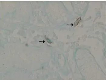
위염조직의 점막을 mast cell tryptase 면역 염색한 결과 비만세포는 주로 위점막의 고유층에 주로 분포하였고, 핵 은 한 개로 세포질 중앙부에 위치하였으며 원형 또는 타 원형의 짙은 자색으로 염색되었으며 세포질 과립은 짙은 갈색을 띤 난원형, 장타원형, 방추형의 건재한 세포 또는 탈과립 상태로 관찰되었다 (Fig. 1A). 또한 c-kit 면역염색 결과 c-kit 양성 세포 역시 주로 위 점막의 고유층에 분포 하였고, 세포 형태도 비만세포와 매우 유사한 형태로 관 찰되었으며 DAB 발색 반응부위가 세포막 주변에 출현하 였다 (Fig. 1B). c-kit 양성세포가 비만세포인가를 확인하 기 위해 대조염색으로 alcian blue 염색을 한 결과 c-kit 양성세포의 세포질 안쪽이 alcian blue 양성을 나타내어 비만세포임을 확인하였다 (Fig. 2).
화상 분석 프로그램을 사용하여 비만세포 밀도를 구한 결과 H. pylori-음성 위염조직 (47.7 ± 9.6 cells/mm2)에 비 해 H-pylori-양성 위염조직 (103.9 ± 35.5 cells/1mm2)에서 통계학적으로 유의한 증가를 나타내었다 (p<0.001) (Fig.
3). 또한 c-kit 양성세포의 밀도도 H-pylori-음성 위염조직
Fig. 2. A microscopic picture of the mucosa with H.
pylori-positive gastritis stained with alcian blue (pH 0.3) and immunohistochemical double-staining for c-kit positive cells.
Mast cells are stained blue with alcian blue in the cytoplasm, and c-kit positive cells (arrow) are stained brown along by the cell membrane. Original magnification: x400.
Fig. 3. Comparison of mast cell density in the gastric mucosa of the antrum between H. pylori-negative and H. pylori-positive gastritis. Error bars indicate SD. H. pylori-negative gastritis, N=16; H. pylori-positive gastritis, N=20. **P<0.01 compared with H. pylori-negative gastritis.
(33.9±12.6 cells/mm2)에 비해 H-pylori-양성조직 (81.8±26 cells/mm2)에서 통계학적으로 매우 유의한 증가를 보였다
(p<0.001) (Fig. 3).
IV. 고 찰
Stem cell factors (SCF)는 사람 피부 비만세포의 성장 과 분화뿐만 아니라 화학 주성 인자로서 병변 부위에 비 만세포를 동원하며 (Nilsson 등, 1994), 사람과 쥐의 간섬
Fig. 4. Comparison of stem cell factor receptor (c-kit)-positive cell number in the gastric mucosa of the antrum between H.
pylori-negative and H. pylori-positive gastritis. Error bars indicate SD. H. pylori-negative gastritis, N=16; H. pylori- positive gastritis, N=20. **P<0.01 compared with H. pylori- negative gastritis.
유증 발생과정 (Gaca 등, 1999)이나 H. pylori 감염 위염 조직에서 비만세포의 증가와 밀접히 관련되어 있는 것으 로 보고 되어 있다 (Bamba 등, 2002). 앞서 연구한 우리의 H. pylori 감염 위염조직에서의 toluidine blue염색을 사용 한 결과에서도 비만세포가 H. pylori-음성 위염조직에 비 해 H. pylori-양성 위염조직에서 유의한 증가를 보였으며, 이 결과는 염증 강도의 증가와도 상관성이 있는 것으로 조사되었다 (제갈, 2000).
본 실험에서는 염증 강도에 기인한 요인을 제거하기 위하여 단핵세포 침윤 정도를 표준화한 상태에서 H.
pylori -양성과 H. pylori-음성 위염조직 사이의 비만세포 밀도를 비교하였다. Anti-human mast cell trypatse를 사용 한 면역 염색을 사용하여 비만세포의 밀도를 조사한 결 과 H. pylori-음성 위염 환자에 비해 H.pylori-양성 환자에 서 유의하게 증가하였다. 이 결과는 H. pylori 감염에서의 비만세포의 밀도는 단핵세포 침윤 강도뿐만 아니라 다른 요인이 관계한다는 것을 의미한다.
Bamba 등 (2002)은 비만세포 증가 기전을 규명하기 위 해 PCNA 항체를 사용하여 비만세포 증식을 조사한 결과 H. pylori-양성 위염조직이 H. pylori-음성 조직에 비해 유 의한 증가를 보였고, 단핵세포 침윤 강도가 동일한 예의 비교에서도 PCNA-양성 비만세포 비율이 높았으며, H.
pylori 감염 치료 후에는 유의한 감소를 나타내었다. 이러 한 결과는 비만세포가 사람 위점막 조직에서 국소적으로 증식할 뿐만 아니라 H, pylori-양성 위염 조직에 비만세포 증식인자가 존재한다는 것을 의미한다. 또한 이들은 H.
pylori-양성 위염 조직에서 SCF-양성 세포가 존재한다는 것을 확인하였고, SCF-양성 세포의 밀도가 H. pylori-양 성 위염 조직에서 유의하게 증가하며, 이는 SCF-양성 세 포의 증가가 비만세포의 동원과 증식 및 화학 주성에 관 계하는 것으로 추정하였다. 이는 SCF가 비만세포 증식인 자로서 섬유모세포를 포함한 여러 종류의 세포로부터 생성되고 (Galli 등, 1994), SCF는 비만세포 화학 유인인 자 (chemoattractant)로서 비만세포와 섬유모세포의 결합 을 매개하며 (Adachi 등, 1992), 비만세포 중개제 (mediators)의 방출을 유도한다는 것과 일치한다 (Bischoff 와 Dahinden, 1992). 그리고 섬유모세포와 비만세포의 혼 합 세포배양 실험에서 이 SCF의 효과는 두 세포의 결합 과 그 후에 일어나는 비만세포 표면에 있는 tyrosine kinase receptor인 c-kit의 이합체 형성 (dimerisation)의 결 과이며 (Galli 등, 1993), 이 경로에는 세포의 증식, 분화 및 운동성을 조절하는 protein kinase C (serine/threonine kinase)가 관련된 것으로 알려져 있다 (Nishizuka, 1988).
우리의 실험에서도 H. pylori-양성 위염 조직이 H.
pylori-음성 위염 조직에 비해 c-kit-양성 세포가 매우 유 의하게 증가하였으며, 이 c-kit-양성세포는 alcian blue, pH 0.3 대조염색에 의해 대부분 비만세포인 것으로 확인 되었다. 또한 c-kit-양성 세포에서 염색 반응부위가 세포 막 가장자리를 따라 나타난 것은 수용체가 염색된 것임 을 알 수 있었다. 이 결과는 비만세포가 성장인자에 대한 수용체를 세포 표면에 가지며 (Britto와 Borojevic, 1997), 이 세포 표면 수용체는 조직 비만세포에 유일한 것 (Farini 등, 1993)이라는 주장과 일치한다. 또한 섬유모세 포 외에 비만세포도 SCF를 분비하며 (Bamba 등, 2002), 쥐 복강 비만세포가 H. pylori 수용성 추출물에서 탈과립 을 일으키는 것으로 알려져 있다 (Tufano 등, 1994). 따라 서 만일 비만세포가 스스로 SCF를 생성 또는 분비하고, 이 SCF가 H. pylori 감염에서 어떤 모르는 기전에 의해 방출된 수용체와 결합한다면, 비만세포는 그들 스스로가 위 점막에서 autocrine 또는 paracrine 경로에 의해 조절되 고 있다고 가정할 수 있다.
V. 결 론
우리는 H. pylori 감염 위점막 조직에서 비만세포가 증 가하며, 비만세포의 세포막에는 c-kit이 존재한다는 것을 확인하였다. 따라서 우리는 비만세포가 H. pylori 감염 위
점막 조직의 염증 및 그 후 궤양 형성 과정에서 중요한 역할을 한다고 가정한다. 그러나 이 가정에 대한 확인을 위해서는 앞으로도 SCF와 c-kit의 결합 및 H. pylori 감염 위염 조직의 진행과정에 있어서의 비만세포의 역할에 대 한 좀더 정밀한 연구가 미래의 과제로 남아 있다.
참 고 문 헌
1. Adachi BS, Ebi Y, Nishikawa S, Hayashi S, Yamazaki M, Kasugai T. Necessity of extracellular domain of W (c-kit) receptors for attachement of murine cultured mast cells to fibroblasts. Blood 79:650-656, 1992
2. Bamba N, Nakajima S, Andoh A, Bamba M, Sugihara H, Bamba T, Hattori T. Stem cell factor expressed in human gastric mucosa in relation to mast cell increases in Helicobacter pylori-infected gastritis. Dig Dis Sci 47:274-282, 2002
3. Bischoff SC, Dahinden CA. c-kit ligand : A unique potentiator of mediator release by human lung mast cells. J Exp Med 175:237-244, 1992
4. Britto JM, Borojevic R. Liver granuloma in schistosomiasis: Mast cell dependant induction of SCF expression in hepatic stellate cells is mediated by TNF-α. J Leukoc Biol 62:389-395, 1997
5. Enerback L. Mast cells in rat gastrointestinal mucosa.
Acta Pathol Microbiol Immunol Scand 66:303-312, 1966
6. Falini B, Flenghi L, Pileri S. PG-M1, a new monoclonal antibody directed against a fixative- resistant epitope on the macrophage restricted form on the CD68 molucule. Am J Pathol 142:1359-1372, 1993
7. Frangogiannis NG, Perrard JL, Mendoza LH, Burns AR, Lindsey ML, Ballantyne CM, Michael LH, Smith CW, Entman ML. Stem cell factor induction is associated with mast cell accumulation after canine myocardial ischemia and reperfusion. Circulation 98:687-698, 1998
8. Gaca MDA, Pickering JA, Arthur MJP, Benyon RC.
Human and rat hepatic stellate cells produce stem cell
factor: a possible mechanism for mast cell recruitment in liver cirrhosis. J Hepatol 30:850-858, 1999 9. Galli SJ. New concepts about the mast cell. N Engl J
Med 317:30-34, 1987
10. Galli SJ, Tsai M, Wershil BK. The c-kit receptor, stem cell factor and mast cells. What each is teaching us about the other. Am J Pathol 142:965-974, 1993 11. Galli SJ, Zsebo KM, Geissler EN. The c-kit ligand,
stem cell factor. Adv Immunol 55:1-96, 1994
12. Gruber BL, Marchese MJ, Kew RR. Transforming growth factor β-1 mediate mast cell chemotaxis. J Immunol 152:5860-5867, 1994
13. Kerr DA. Improved Warthin-Starry method of staining spirochetes in tissue section. Am J Clin Pathol 8:63-67, 1938
14. Marshall JS, Bienenstock J. The role of mast cells in inflammatory reactions of the airways, skin and intestine. Curr Opin Immunol 6:853-859, 1994
15. Nakajima S, Krishnan B, Ota H, Segura AM, Hattori T, Graham DY, Genta RM. Mast cell involvement in gastritis with or without Helicobacter pylori infection.
Gastroenterology 114;746-754, 1997
16. Nilsson G, Butterfield JH, Nisson K, Siebahn A.
Stem cell factor is a chemotactic factor for human mast cells. J Immunol 153:3717-3123, 1994
17. Nishizuka Y. The molecular heterogeneity of protein kinase C and its implication for cellular regulation.
Nature 334:661-665, 1998
18. Schleimer RP, Fox CC, Neclerio RM. Role of human basophils and mast cells in the pathogenesis of aller- gic disease. J Allergy Clin Immunol 76:369-374, 1985 19. Tufano MA, Rossano F, Catalanotti P, Liguori G,
Capasso C, Ceccarelli MT, Marinelli P. Immunobiolo- gical activities of Helicobacter pylori porins. Infect Immun 62:1392-1399, 1944
20. Wallace JL, Keenan CM, Mugridge KG. Possible mechanisms and mediators of gastritis associated with Helicobacter pylori infection. Scand J Gastroenterol 26 (suppl 187):65-70, 1991
21. 제갈 승주. Helicobacter pylori 위염에서의 비만세포 와 염증 반응과의 관련성. 임상병리검사과학회지 32:260-268, 2000