Ⅰ. Introduction
Acupuncture is an oriental ancient therapeutic method, and has been used for the treatment of pa- tients with various diseases from old times.
The theory of meridians and acupuncture points is the basic theory of acupuncture therapy.
The science of meridians expounds the routes that connect different parts of the body based on the clinical application of acupuncture points.
Meridians not only connect all parts of the body, but also coordinate their functions, they also influ- ence various complicated pathological changes and adjust the states of the viscera.
Galactosamine (GalN) is a hepatotoxin that induces liver injury through the formation of the
太衝 및 陽池의 電針이 實驗的으로 誘發된 白鼠의 肝保護效果에 미치는 影響
윤 대 환1⋅나 창 수1
1동신대학교 한의과대학 경혈학교실
Hepatoprotective effects of Electro-Acupuncture at Taechung (LR3) and Yangji (TE4) on experimental liver injury in rats
Dae-Hwan Youn1, Chang-su Na1
1Dept. of Meridian and Acupoint, College of Oriental Medicine, Dongshin University
Abstract
목 적 : 본 연구는 太衝 및 陽池穴에 대한 電鍼이 galactosamine을 이용한 백서의 肝毒性을 실험적으로 유발시킨 모델에서 豫防效果를 알아보고자 혈청중의 γ-GTP, GOT, GPT, LDH, total bilirubin, total cholesterol, triglyceride의 변화를 관찰하였다.
방 법 : 간독성은 각 군들은 간독성을 유발하지 않고 무처치한 정상군, 간독성을 유발하고 난 후 무 처치한 대조군, 각각 10 Hz, 50 Hz, 100 Hz 전침을 20일간 10회 시행한 후 간독성을 유발한 Pre 10, Pre 50, Pre 100군 등으로 분류하였다.
결 과 : 太衝․陽池穴에 대한 電鍼 治療의 豫防效果에서는 Pre 10군에서는 γ-GTP, GOT, GPT, total cholesterol, triglyceride이 Pre 50군에서는 γ-GTP, GOT, GPT, LDH, total bilirubin, total cholesterol 이, Pre 100군에서는 γ-GTP, GOT, LDH, total bilirubin, total cholesterol이 대조군에 비해 감소하였 다.
결 론 : 위의 결과를 종합해보면 간독성에 대하여 太衝․陽池穴의 電鍼을 시행한 모든 군에서 간기능 과 지질대사에서 간손상에 대한 유의한 예방효과를 나타내었다.
Key words : 간 독성, 전침, 예방효과, 태충, 양지
▪교신저자 : 윤대환, 전남 나주시 대호동 252 동신대학교 한의과대학 경혈학교실, Tel. 061-330-3527, Fax. 061-330-2900, E-mail : [email protected]
‧투고 : 2006/05/26 ‧심사 : 2006/06/07 ‧채택 : 2006/06/19
T h e K orea n Jou rn al of M erid ia n & A cu p oin t
highly reactive hydroxyl radical lipid peroxidation and damage to the cell membrane1). It has been used as an animal model of fulminant hepatic fail- ure to develop artificial liver support, and it can be commonly seen at human viral hepatitis2,3).
Recent reports on the study of acupuncture are as follows.
Liu et al4) reported effects on the reduction of biochemical and pathological parameters of liver injury after acupuncture, and Lin et al5) reported the prevention of ANIT-induced hyperbilirubine- mia and cholangitis. Huang et al6) and Chakrabarti et al7) reported the improvement of the hepatic me- tabolism and promoting the membrane transport action in endotoxic shock and the hepatic functions on the liver in rats. Shimoju-Kobayashi et al8) re- ported increases of hepatic glucose output in re- sponses to electro-acupuncture (EA) stimulation.
In this study we investigated hepatoprotective effects of EA at Taechung (LR3)․Yangji (TE4) on the GalN-induced liver injury in rats.
Ⅱ. Materials & Methods 1. Animals
Male Sprague Dawley rats (Damul Science Co., Korea) weighing 160±10 g were used for the experiment. Each animal was housed at a con- trolled temperature (22±3 ℃) and was maintained under light-dark cycles, each cycle consisting of 12 hr of light and 12 hr of darkness (lights on from 09:00 to 19:00 hr) with food (Pellet, Samyang, Korea) and water made available ad libitum. The experimental procedures were performed in ac- cordance with the animal care guidelines of the
NIH and the Korean Academy of Medical Sciences.
2. Liver injury and electro-acupuncture
To induce liver injury in the experimental ani- mals, a single intraperitoneal injection of GalN (400 mg/kg in saline : Sigma Chemical Co., St. Louis, MO, USA) was given to each animal, while animals of the normal group received equivalent amounts of normal saline.
In the EA groups, EA treatment was given to each animal every other day 10 times for 20 days at 1 time per 2 days on bilateral acupuncture points.
The needles were then connected to the Dual Impedance Research stimulator (Harvard, USA) and were electrically stimulated at 1 mV intensity, 1 msec duration during 5 minutes.
Before 20 days in liver injury, as treatment to study hepatoprotective effects, four stainless steel 0.5 mm diameter needles (3-0.5, Haenglim, Korea) were acupunctured at a point corresponding to LR3 and TE4 in rats.
LR3 is located on the dorsum of foot, in the depression distal to the junction of the first and second metatarsal bones, when the palm is turned upward, TE4 is in the depression at the point on the transverse crease of the wrist dorsum crossed with the vertical interstice between the third and fourth metacarpal bones. When the knee is flexed, LR 8 is in the depression above the medial end of the transverse popliteal crease, posterior to the me- dial epicondyle of the femur.
3. Groups
Animals are divided into 8 groups (n=8 in each
group). Normal group is not liver injury-induced and not treated, Control group is the liver in- jury-induced and not treated.
The Pre 10, Pre 50, Pre 100 groups executed by the hepatoprotective treatment are carried out electro-acupuncture at LR3․TE4 with each 10 Hz, 50 Hz, 100 Hz electrical stimulation before the liver is injury-induced.
4. Assays of serums
Rats are sacrificed by the decapitation after final treatment, and then blood samples collected from the stump are put at a room temperature to
allow coagulation. Then serums are immediately centrifuged at 5,000 rpm for 10 minutes, and they are collected and stored at -70 ℃ for γ-GTP, GOT, GPT, LDH, total bilirubin, total cholesterol, and triglyceride.
The activity of γ-GTP is measured using di- agnostic kits (AM158-K, Asan, Korea) at 635 nm by the spectrophotometer (Unikon922, Kontron, Italy). That of other parameters in serum without γ-GTP is determined using diagnostic kits by the photometer (5010, Robert Riele GmbH &Co.
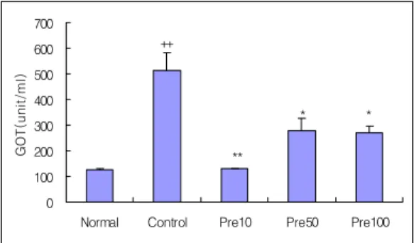
Germany): GOT (GOT-SL Kit at 340 nm, ELITech, France); GPT (GPT-SL Kit at 340 nm, ELITech, France); total bilirubin (T. bilirubin-SL Fig. 2. Hepatoprotective effect of electro-acupu- ncture at Taechung (LR3) and Yangji (TE4) on serum GOT in liver injury rats induced by D-galactosamine. Normal, not liver in- jury-induced and not treated group. Con- trol, the liver injury-induced and not treated group. Pre 10. before the liver injury- in- duced electro-acupuncture with 10 Hz elec- trical stimulation at bilateral acupuncture point of LR3․TE4. Pre 50, before the liver injury-induced electro-acupuncture with 50 Hz electrical stimulation at bilateral acu- puncture point of LR3․TE4. Pre 100, before the liver injury-induced electro-acupunc- ture with 100 Hz electrical stimulation at bilateral acupuncture point of LR3․TE4.
Results are shown as mean±S.E. ++, p<0.01 as compared with the corresponding data of normal group. *, p<0.05 , **, p<0.01 as compared with the corresponding data of control group.
0 100 200 300 400 500 600 700
Normal Control Pre10 Pre50 Pre100
GOT(unit/ml)
**
*
* ++
Fig. 1. Hepatoprotective effect of electro-acupun- cture at Taechung (LR3) and Yangji (TE4) on serum γ-GTP in liver injury rats in- duced by D-galactosamine. Normal, not liver injury-induced and not treated group.
Control, the liver injury-induced and not treated group. Pre10. before the liver in- jury-induced electro-acupuncture with 10Hz electrical stimulation at bilateral acu- puncture point of LR3․TE4. Pre50, before the liver injury-induced electro-acupunc- ture with 50Hz electrical stimulation at bi- lateral acupuncture point of LR3․TE4.
Pre100, before the liver injury-induced el- ectro-acupuncture with 100Hz electrical stimulation at bilateral acupuncture point of LR3․TE4. Results are shown as mean
±S.E. +, p<0.05 as compared with the corre- sponding data of normal group. *, p<0.05 as compared with the corresponding data of control group.
0 20 40 60 80 100
Normal Con
trol Pre
10 Pre50
Pre100
r-GTP(mUnit/ml)
* * *
+
Kit at 546 nm, ELITech, France); LDH (LDH-SL Kit at 340 nm, ELITech, France); total cholesterol (T. cholesterol-SL Kit at 546 nm, ELITech, France); triglyceride (Triglyceride -SL Kit at 505 nm, ELITech, France).
5. Data Analysis
Data are analyzed using SPSS 10.0.5 for Windows (SPSS Inc. USA) by student t-test.
Results are expressed as mean ± standard error (S.E.). Differences are considered significant for p<0.05.
Ⅲ. Results
1. Effects of EA on γ-GTP in serums
The γ-GTP was 75.8±0.27 mUnit/ml in the normal group, 91.1±3.88 mUnit/ml in the control group, 78.5±1.52 mUnit/ml in the Pre10 group, 79.2±1.72 mUnit/ml in the Pre 50 group and 76.8±1.65 mUnit/ml in the Pre 100 group.
These observations indicate that the control group is significantly increased as compared with normal group (p<0.05), and that Pre 10 , Pre 50 and Pre 100 groups are significantly decreased on the γ-GTP in serums to be compared with the Fig. 3. Hepatoprotective effect of electro-acupu-
ncture at Taechung (LR3) and Yangji (TE4) on serum GPT in liver injury rats induced by D-galactosamine. Normal, not liver injury-induced and not treated group.
Control, the liver injury-induced and not treated group. Pre 10. before the liver in- jury-induced electro-acupuncture with 10 Hz electrical stimulation at bilateral acu- puncture point of LR3․TE4. Pre 50, before the liver injury-induced elec- tro-acupuncture with 50 Hz electrical stim- ulation at bilateral acupuncture point of LR3․
TE4. Pre 100, before the liver in- jury-induced electro-acupuncture with 100 Hz electrical stimulation at bilateral acu- puncture point of LR3․TE4. Results are shown as mean±S.E. ++, p<0.01 as com- pared with the corresponding data of normal group. *, p<0.05 , **, p<0.01 as compared with the corresponding data of control group.
0 50 100 150 200 250 300 350 400 450
Normal Control Pre10 Pre50 Pre100
GPT(unit/ml)
**
* ++
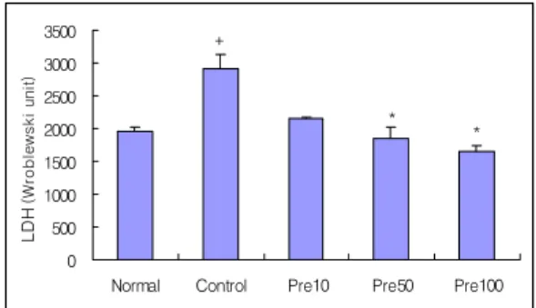
Fig. 4. Hepatoprotective effect of electro-acupu- ncture at Taechung (LR3) and Yangji (TE4) on serum LDH in liver injury rats induced by D-galactosamine. Normal, not liver injury-induced and not treated group.
Control, the liver injury-induced and not treated group. Pre 10. before the liver in- jury-induced electro-acupuncture with 10 Hz electrical stimulation at bilateral acu- puncture point of LR3․TE4. Pre 50, before the liver injury-induced elec- tro-acupuncture with 50 Hz electrical stim- ulation at bilateral acupuncture point of LR3․
TE4. Pre 100, before the liver in- jury-induced electro-acupuncture with 100 Hz electrical stimulation at bilateral acu- puncture point of LR3․TE4. Results are shown as mean±S.E. +, p<0.05 as compared with the corresponding data of normal group. *, p<0.05 as compared with the cor- responding data of control group.
0 500 1000 1500 2000 2500 3000 3500
Normal Control Pre10 Pre50 Pre100
LDH(Wroblewski unit)
* *
+
control group (p<0.05)(Fig. 1).
2. Effects of EA on GOT in serum
The GOT was 128.2±3.50 unit/ml in the nor- mal group, 512.9±68.91 unit/ml in the control group, 132.0±0.31 unit/ml in the Pre 10 group, 276.5±48.26 unit/ml in the Pre 50 group and 269.5±25.37 unit/ml in the Pre 100 group.
These observations indicate that the control group is significantly increased as compared with normal group (p<0.01), and that Pre 10 (p<0.01) , Pre 50 (p<0.05) and Pre 100 (p<0.05) groups are significantly decreased on the GOT in serums to
be compared with the control group (Fig. 2).
3. Effects of EA on GPT in serum
The GPT was 72.1±2.10 unit/ml in the normal group, 326.0±65.61 unit/ml in the control group, 72.5±9.24 unit/ml in the Pre 10 group, 171.4±15.46 unit/ml in the Pre 50 group and 239.5±30.31 unit/ml in the Pre 100 group.
These observations indicate that the control group is significantly increased as compared with normal group (p<0.01), and that Pre 10 (p<0.01) and Pre 50 (p<0.05) groups are significantly de- creased on the GPT in serums to be compared with Fig. 5. Hepatoprotective effect of electro-acupu-
ncture at Taechung (LR3) and Yangji (TE4) on serum total bilirubin in liver injury rats induced by D-galactosamine. Normal, not liver injury-induced and not treated group. Control, the liver injury-induced and not treated group. Pre 10. before the liver injury-induced electro-acupuncture with 10 Hz electrical stimulation at bilateral acu- puncture point of LR3․TE4. Pre 50, before the liver injury-induced electro-acupu- ncture with 50 Hz electrical stimulation at bilateral acupuncture point of LR3․TE4. Pre 100, before the liver injury-induced elec- tro-acupuncture with 100 Hz electrical stimulation at bilateral acupuncture point of LR3․TE4. Results are shown as mean±S.E. ++, p<0.01 as compared with the corresponding data of normal group.
*, p<0.05 as compared with the correspond- ing data of control group.
0.0 0.5 1.0 1.5 2.0 2.5 3.0 3.5
Normal Control Pre10 Pre50 Pre100
Total billilubine (mg/dl)
* *
++
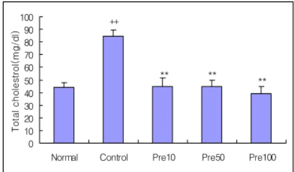
Fig. 6. Hepatoprotective effect of electro-acupu- ncture at Taechung (LR3) and Yangji (TE4) on serum total cholesterol in liver injury rats induced by D-galactosamine. Normal, not liver injury-induced and not treated group. Control, the liver injury-induced and not treated group. Pre 10. before the liver injury-induced electro-acupuncture with 10 Hz electrical stimulation at bilateral acu- puncture point of LR3․TE4. Pre 50, before the liver injury-induced electro-acupunc- ture with 50 Hz electrical stimulation at bi- lateral acupuncture point of LR3․TE4. Pre 100, before the liver injury-induced elec- tro-acupuncture with 100 Hz electrical stimulation at bilateral acupuncture point of LR3 ․TE4. Results are shown as mean±S.E. ++, p<0.01 as compared with the corresponding data of normal group.
**, p<0.01 as compared with the corre- sponding data of control group.
0 10 20 30 40 50 60 70 80 90 100
Normal Control Pre10 Pre50 Pre100
Total cholestrol(mg/dl)
** **
**
++
the control group (Fig. 3).
4. Effects of EA on LDH in serum
The LDH was 1962.8±64.22 Wroblewski unit in the normal group, 2903.0±230.87 Wroblewski unit in the control group, 2153.3±16.29 Wroblewski unit in the Pre 10 group, 1854.6±156.68 Wroblewski unit in the Pre 50 group and 1660.8±77.90 Wroblewski unit in the Pre 100 group.
These observations indicate that the control group is significantly increased as compared with normal group (p<0.05), and that Pre 50 and Pre 100
groups are significantly decreased on the LDH in serums to be compared with the control group (p<0.05)(Fig. 7).
5. Effects of EA on Total bilirubin in serum
The total bilirubin in serum was 0.3±0.02 mg/dl in the normal group, 2.6±0.41 mg/dl in the control group, 2.6±0.12 mg/dl in the Pre 10 group, 1.0±0.23 mg/dl in the Pre 50 group and 1.1±0.25 mg/dl in the Pre 100 group.
These observations indicate that the control group is significantly increased as compared with normal group (p<0.01), and that Pre 50 and Pre 100 groups are significantly decreased on the total bi- lirubin in serums to be compared with the control group (p<0.05)(Fig. 5).
6. Effects of EA on Total cholesterol in serum
The total cholesterol was 44.0±3.60 mg/dl in the normal group, 84.6±4.95 mg/dl in the control group, 44.8±6.96 mg/dl in the Pre 10 group, 44.5±5.44 mg/dl in the Pre 50 group and 39.3±5.16 mg/dl in the Pre 100 group.
These results suggest that the control group is significantly increased as compared with normal group (p<0.01), and that Pre 10, Pre 50 and Pre 100 groups are significantly decreased on the total cholesterol in serums to be compared with the con- trol group (p<0.01)(Fig. 6).
7. Effects of EA on Triglyceride in serum
The triglyceride was 32.0±5.44 mg/dl in the normal group, 58.6±6.78 mg/dl in the control group, Fig. 7. Hepatoprotective effect of electro-acupun-
cture at Taechung (LR3) and Yangji (TE4) on serum triglyceride in liver injury rats induced by D-galactosamine. Normal, not liver injury-induced and not treated group.
Control, the liver injury-induced and not treated group. Pre 10. before the liver in- jury-induced electro-acupuncture with 10 Hz electrical stimulation at bilateral acu- puncture point of LR3․TE4. Pre 50, before the liver injury-induced electro-acupun- cture with 50 Hz electrical stimulation at bilateral acupuncture point of LR3․TE4. Pre 100, before the liver injury-induced elec- tro-acupuncture with 100 Hz electrical stimulation at bilateral acupuncture point of LR3 ․TE4. Results are shown as mean±S.E. ++, p<0.01 as compared with the corresponding data of normal group.
**, p<0.01 as compared with the corre- sponding data of control group.
0 10 20 30 40 50 60 70
Normal Control Pre10 Pre50 Pre100
Triglyceride(mg/dl)
** **
++
32.7±2.94 mg/dl in the Pre 10 group, 36.7±1.82 mg/dl in the Pre 50 group and 49.6±3.88 mg/dl in the Pre 100 group.
These observations indicate that the control group is significantly increased as compared with normal group (p<0.01), and that Pre 10 and Pre 50 groups are significantly decreased on the trigly- ceride in serums to be compared with the control group (p<0.01)(Fig. 7).
Ⅳ. Discussion
In recent years, carbon tetrachloride(CCl4) and GalN are well known for highly toxic chemical materials inducing the liver injury and the toxic ef- fects of them on the liver have been steadily re- ported for years.
CCl4 treatment inducing fatty liver, is man- ifested histologically as hepatic steatosis, cen- trilobular necrosis, and cirrhosis. Hepatic steatosis of the liver is a multifactorial phenomenon which is thought to be caused by a blockage of lipoprotein secretion9), impaired sysnthesis or peroxidation of phospholipids10,11), and the toxic effects of free alkyl radicals on cell membranes12,13), and disturbances in methylation reactions14).
GalN treatment induce the features of acute hepatitis in rats. The toxic effect of GalN is con- nected with an insufficiency of UDP-glucose and UDP-galactose and the loss of intracellular calcium homeostasis. These changes affect cell membranes and organelles and the synthesis of proteins and nucleic acids15). After GalN application, the location of proteoglycans is changed in the rat liver16). GalN also inhibits the energy metabolism of hep- atocytes17).
The special acupuncture points (Yuan-Source, Five acupuncture points) retain re- flecting the pathological changes of the viscera and they are clinically used to diagnose and treat the disorders of the related viscera and organs.
LR3 is used in the clinical treatment of liver disease, headache, vertigo, redness, and swelling pain of eyes, glaucoma, nearsightedness, apoplexy, epilepsy, infantile convulsion, irregular menstru- ation, vomiting, hiccup, stomachache, and flaccid- ity18).
TE4 is commonly used in the clinical treat- ment of tinnitus, deafness, sore throat, flaccidity and bi-syndrome of the upper limbs and diabetes18). So this study is designed to investigate hep- atoprotective effects of EA at LR3․TE4 on GalN-induced liver injury in rats.
When the liver cell plasma membrane is dam- aged, varieties of enzyme normally located on the cytosol are released into the bloodstream19). The serum levels of the γ-GTP, GPT, GOT, and total bilirubin, the markers of liver enzymes, are em- ployed as important indicators of liver dam- age4,5,19-21). Lactate dehydrogenase (LDH) is one of the important enzymes in diagnosis of specific or- gan diseases. Vertebrate tissues and serums con- tain the characteristic distributions of the LDH iso- zymes22,23).
In this study, the control group significantly shows a decrease of markers of liver enzymes in serum.
On the hepatoprotective effect of EA at LR3․
TE4, Pre 10 group is significantly decreased on the γ-GTP, GOT and GPT in serums to be compared with the control group, and Pre 50 group is de-
creased on the γ-GTP, GOT, GPT, LDH, and total bilirubin, and Pre 100 group is decreased on the γ -GTP, GOT, LDH, and total bilirubin.
These results support that all of EA groups are effective on markers of liver enzymes, which concern liver functions in serums, in GalN-induced liver injury in rats. Hence, these data suggest that electro-acupuncture treats and prevents the en- zyme disorder of liver injury.
The evaluation of the concentration of serum lipids was reported by many investigations after toxic liver damage in rats24,25). In this study, the control group significantly shows an increase of concentration of serum lipids.
On the hepatoprotective effect of EA at LR3․
TE4, Pre 10 group is significantly decreased on to- tal cholesterol, and triglyceride in serums to be compared with the control group, and the Pre 50 group is decreased on total cholesterol, and trigly- ceride, and the Pre 100 group is decreased on total cholesterol. These results support that the EA has especially preventive effect on lipid metabolism progression in GalN-induced liver injury in rats by the decrease of concentration of serum lipid agents such as cholesterol and triglyceride. Theses results support that the EA has effect of treatment on lipid metabolism progression in GalN-induced liver in- jury in rats by the increase of concentration of total cholesterol.
In sum, the data suggest that the hepatother- apeutic effect on liver injury is intensive to treat only liver meridian channel within the EA at LR3․
TE4.
Ⅴ. Conclusion
The hepatoprotective effects of EA at LR3․
TE4 on liver injury induced by GalN in rats are observed as follows.
1. On the hepatoprotective effect of EA at LR3․TE4, the γ-GTP, GOT, GPT, total cholesterol and triglyceride are sig- nificantly decreased in the Pre 10 group.
2. On the hepatoprotective effect of EA at LR3․TE4, γ-GTP, GOT, GPT, LDH, total bilirubin, total cholesterol and triglyceride are significantly decreased in the Pre 50 group,
3. On the hepatoprotective effect of EA at LR3․TE4, γ-GTP, GOT, LDH, total bilir- ubin and total cholesterol are significantly decreased in the Pre 100 group.
Based on the results, we have indicated that the electro-acupuncture may contribute to its pre- ventive and hepatotherapeutic effect on the pro- gression of GalN-induced liver injury in rats.
References
1. Barry H, Gutteridge JMC. Free Radicals in Biology and Medicine 2nd edition. Oxford : Claredon Press. 1989 ; 254–5.
2. Decker K, Keppler D. Galactosamine hepatitis:
key role of the nucleotide deficiency period in the pathogenesis of cell injury and cell death.
Rev Physiol Biochem Pharmacol. 1974 ; (71) : 77-106.
3. Gu CH, Cao R, Wang GX. Protective effect of prostaglandin E on hepatocytes and its value
of early treatment of severe viral hepatitis.
Zhonghua Nei Ke Za Zhi. 1991 ; 30(1) : 17-20.
4. Liu HJ, Hsu SF, Hsieh CC, Ho TY, Hsieh CL, Tsai CC, Lin JG. The effectiveness of Tsu-San-Li (St-36) and Tai-Chung (Li-3) acupoints for treatment of acute liver damage in rats. Am J Chin Med. 2001 ; 29(2) : 221-6.
5. Lin JG, Yang SH, Tsai CH. Acupuncture pro- tection against experimental hyper- bilirubinemia and cholangitis in rats. Am J Chin Med. 1995 ; 23(2) : 131-7.
6. Huang W, Huang K, Xu Q, Wang Z, Sun Y, Cai H, Zhang X, Lin J. Histochemical ob- servation of the effect of electroacupuncture on the livers of rats with endotoxic shock. Zhen Ci Yan Jiu. 1995 ; 20(3) : 36-9.
7. Chakrabarti AK, Chatterjee K, Ghosh JJ, Ganguly A. Electroacupuncture and its effect on rat hepatic functions. Acupunct Electrother Res. 1983 ; 8(2) : 111-26.
8. Shimoju-Kobayashi R, Maruyama H, Yoneda M, Kurosawa M. Responses of hepatic glucose output to electro-acupuncture stimulation of the hindlimb in anaesthetized rats. Auton Neurosci. 2004 ; 115(1-2) : 7-14.
9. Recknagel RO, Malamed S. The osmotic nature of mitochondrial swelling produced by carbon tetrachloride and inorganic phosphate. J Biol Chem. 1958 ; 232(2) : 705-13.
10. Shimizu Y. Effect of carbon tetrachloride ad- ministration on the synthesis of triglycerides and phospholipids in rat liver. J Lipid Res. 1969
; 10(5) : 479-86.
11. Terao J, Asano I, Matsushita S. High-perform- ance liquid chromatographic determination of
phospholipid peroxidation products of rat liver after carbon tetrachloride administration. Arch Biochem Biophys. 1984 ; 235(2) : 326-33.
12. James JL, Friend DS, MacDonald JR, Smuckler EA. Alterations in hepatocyte plasma mem- brane in carbon tetrachloride poisoning.
Freeze-fracture analysis of gap junction and electron spin resonance analysis of lipid fluidity. Lab Invest. 1986 ; 54(3) : 268-74.
13. Tomasi A, Albano E, Lott KA, Slater TF. Spin trapping of free radical products of CC14 acti- vation using pulse radiolysis and high energy radiation procedures. FEBS Lett. 1980 ; 122(2) : 303-6.
14. Varela-Moreiras G, Alonso-Aperte E, Rubio M, Gasso M, Deulofeu R, Alvarez L et al., Carbon tetrachloride-induced hepatic injury is associated with global DNA hypomethylation and homocysteinemia: effect of S-ad- enosylmethionine treatment. Hepatology. 1995
; 22(4 Pt 1) : 1310-5.
15. Keppler D, Decker K. Studies on the mecha- nism of galactosamine-1-phosphate and its in- hibition of UDP-glucose pyrophosphorylase.
Eur J Biochem. 1969 ; 10(2) : 219-25.
16. Sasaki S, Koide N, Shinji T, Tsuji T.
Immunohistochemical study of proteoglycans in D-galactosamine-induced acute liver injury in rats. J Gastroenterol. 1996 ; 31(1) : 46-54.
17. Mangeney-Andreani M, Sire O, Montagne- Clavel J, Nordmann R, Nordmann J. Inhibitory effect of D-galactosamine administration on fatty acid oxidation in rat hepatocytes. FEBS Lett. 1982 ; 145(2) : 267-70.
18. Zhu A, Huang Y, Tao J, Li Z. Shanghai: house
of shanghai university of traditional chin medicine. Chinese Acup moxibus. 2002 : 64, 82, 84, 86, 222-3.
19. Inuma Y, Kubota M, Yagi M, Kanada S, Yamazaki S, Kinoshita Y. Effects of the herbal medicine Inchinko-to on liver function in post- operative patients with biliary atresia-a pilot study. J Pediatr Surg. 2003 ; 38(11) : 1607-11 20. Ferencikova R, Cervinkova Z, Drahota Z.
Hepatotoxic effect of D-galactosa mine and protective role of lipid emulsion. Physiol Res.
2003 ; 52(1) : 73-8.
21. Ozardali I, Bitiren M, Karakilcik AZ, Zerin M, Aksoy N, Musa D. Effects of selenium on his- topathological and enzymatic changes in ex- perimental liver injury of rats. Exp Toxicol Pathol. 2004 ; 56(1-2) : 59-64.
22. Chung HW, Lee CK. Detoxification Effect of Red Ginseng Extract on Toxicity of Methy- lmercury Chloride to LDH in the Liver, Kidney and Serum of Mouse. Korean J of Zoology.
1987 ; 30(3) : 231-8.
23. Richterich R, Schafroth P, Aebi H. A study of lactic dehydrogenase isoenzyme pattern of hu- man tissues by adsorption-elution on Sepha- dex-DEAE. Clin Chim Acta. 1963 ; 8 : 178-92.
24. Cartwright CK, Ragland JB, Weidman SW, Sabesin SM. Alterations in lipoprotein compo- sition associated with galactosamine-induced rat liver injury. J Lipid Res. 1982 ; 23(5) : 667-79.
25. Venukumar MR, Latha MS. Effect of Cosci- nium fenestratum on hepatotoxicity in rats.
Indian J Exp Biol. 2004 ; 42(8) : 792-7.