D I A B E T E S & M E T A B O L I S M J O U R N A L
This is an Open Access article distributed under the terms of the Creative Commons Attribution Non-Commercial License (http://creativecommons.org/licenses/by-nc/4.0/) which permits unrestricted non-commercial use, distribution, and reproduction in any medium, provided the original work is properly cited.
Nonalcoholic Fatty Liver Disease in Diabetes. Part I:
Epidemiology and Diagnosis
Yong-ho Lee1,2, Yongin Cho1,2, Byung-Wan Lee1,2, Cheol-Young Park3, Dae Ho Lee4, Bong-Soo Cha1,2, Eun-Jung Rhee3
1Department of Internal Medicine, 2Institute of Endocrine Research, Yonsei University College of Medicine, Seoul,
3 Division of Endocrinology and Metabolism, Department of Internal Medicine, Kangbuk Samsung Hospital, Sungkyunkwan University School of Medicine, Seoul,
4Department of Internal Medicine, Wonkwang University Hospital, Wonkwang University School of Medicine, Iksan, Korea
Nonalcoholic fatty liver disease (NAFLD) and diabetes are common metabolic disorders whose prevalence rates are expected to rise worldwide, corresponding to aging and increasingly obese populations. Compared to the general population (around 25%), 50% to 70% of people with diabetes have NAFLD, and NAFLD severity (including fibrosis) tends to be worsened by the presence of diabetes. NAFLD is considered an emerging risk factor for type 2 diabetes mellitus and a contributor to the development of chronic diabetes-related complications. This reciprocal relationship demonstrates the importance of confirming suspected NAFLD in pa- tients with diabetes. Due to the invasive nature of liver biopsy to assess NAFLD status, various alternative non-invasive modalities have been developed and validated. Here, we summarized the epidemiology of NAFLD in patients with diabetes and reviewed cur- rently available imaging modalities and biomarker-based prediction models for their ability to detect liver steatosis and/or fibrosis.
Keywords: Diabetes mellitus; Diagnosis; Epidemiology; Non-alcoholic fatty liver disease
Corresponding author: Eun-Jung Rhee https://orcid.org/0000-0002-6108-7758 Division of Endocrinology and Metabolism, Department of Internal Medicine, Kangbuk Samsung Hospital, Sungkyunkwan University School of Medicine, 29 Saemunan-ro, Jongno-gu, Seoul 03181, Korea
E-mail: [email protected]
INTRODUCTION
The prevalence of diabetes is rising. According to Diabetes At- las 2017, 425 million people suffer from diabetes worldwide;
the prevalence rate in Korea is 14.4% according to the Diabetes Fact Sheet 2018 [1,2]. Type 2 diabetes mellitus (T2DM) is caused by derangements in multiple organs and by various pathophysiological abnormalities, such that it is implicated in many other diseases related to insulin resistance and obesity, such as nonalcoholic fatty liver disease (NAFLD), sleep apnea, arthritis, cardiovascular disease (CVD), and metabolic syn- drome (MetS) [3,4].
NAFLD is considered the hepatic manifestation of ectopic fat accumulation caused by abdominal obesity and insulin re- sistance; its presence also reflects visceral, intramuscular, epi- cardial, and perivascular fat accumulation. Recent evidence
suggests that NAFLD should be considered a cardiovascular risk factor [4]. NAFLD is a strong clinical signal for insulin re- sistance and MetS, and is considered a confirmative risk factor for T2DM [2]. Prevalence of NAFLD in patients with T2DM is much higher than in the general population. This review sum- marizes the epidemiology of NAFLD relative to T2DM, and compares various approaches to diagnose and assess NAFLD characteristics, including steatohepatitis and fibrosis.
PREVALENCE OF NAFLD IN THE GENERAL POPULATION
The current prevalence of NAFLD is proportional to increased rates of obesity. NAFLD incidence in the general population is about 25%, but it rises to more than 90% for very obese indi- viduals undergoing weight reduction procedures/surgeries [5].
https://doi.org/10.4093/dmj.2019.0011 pISSN 2233-6079 · eISSN 2233-6087
In addition, like obesity, NAFLD displays different characteris- tics among various ethnic groups and across countries [6].
In the general Korean population, some discrepancies exist in NAFLD prevalence. In a study by Park et al. [7] in which 4,967 young, healthy Koreans participated in a health screen- ing program, the age-adjusted NAFLD prevalence rate was 21.6% (men), 11.2% (women), and 16.1% in the whole popula- tion. According to Jeong et al. [8], in a cross-sectional study of 140,000 participants in a health screening program, the NAFLD prevalence rate was 25.2%, with some differences based on where participants lived.
PREVALENCE OF NAFLD IN PATIENTS WITH T2DM
NAFLD prevalence in patients with T2DM is much higher than in the general population. In a systemic review by Lonar- do et al. [9], the NAFLD incidence rate in patients with T2DM is 50% to 75%, according to ethnicity, across studies. In reverse, the prevalence of diabetes in patients with NAFLD is higher than in the general population. In a study by Browning et al.
[10] of 2,287 urban people participating in the Dallas Heart Study, hepatic triglyceride content was measured by 1H-nucle- ar magnetic resonance spectroscopy (MRS), with prevalence of T2DM or impaired fasting glucose (IFG) reported as 18% to 33% in patients with NAFLD.
There is discrepancy between the studies regarding preva- lence of NAFLD in patients with T2DM, which may be ac- counted for by the diagnostic tools used [11]. The prevalence of NAFLD was 20.3% when plasma alanine aminotransferase was used to define NAFLD, whereas the prevalence reached 72.8% when MRS was used to define NAFLD. In addition, NAFLD prevalence in patients with T2DM significantly in- creased with increasing obesity [11].
NAFLD prevalence in Asians is never lower than in Cauca- sians. In 929 Korean patients with T2DM who visited a univer- sity-based diabetes clinic, NAFLD was assessed by ultrasono- gram [12], and 63.3% of patients had NAFLD. In 8,352 Japa- nese patients who received health check-ups from 2009 to 2010, NAFLD incidence was 25.6% in those with normal fast- ing glucose, 56.2% in those with IFG, and 68% in those with T2DM defined as fasting blood glucose ≥126 mg/dL, suggest- ing NAFLD prevalence could differ by glycemic status [13].
NAFLD AS A RISK FACTOR FOR T2DM
There is convincing evidence that NAFLD often precedes the development of T2DM [14]. Some suggest that the convention- al paradigm of NAFLD to represent ‘hepatic manifestation’ of MetS is outdated, as recently NALFD has been shown to be an early predictor and determinant for development of diabetes.
By examining the pathophysiological aspects of insulin resis- tance in NAFLD development, we can understand the intimate relationship between these conditions. NAFLD is associated with hepatic and peripheral insulin resistance, causing insuffi- cient suppression of hepatic gluconeogenesis, decreased glyco- gen synthesis, and increased lipid accumulation [3]. The influx of high amounts of free fatty acid (FFA) from white adipose and dietary lipids into the liver promotes further glucose synthesis.
De novo synthesis of FFA is driven by sterol regulatory element binding-protein 1c (Srebp-1c) and carbohydrate response ele- ment binding-protein (ChREBP), catalyzed by hyperinsu- linemia and hyperglycemia. Further factors such as decreased lipid export through very-low density lipoprotein, increased he- patic insulin resistance via diacylglycerol, and increased oxida- tive stress mediated due to exhaustion of antioxidant capacities of superoxide dismutase and glutamate peroxidase, ultimately result in development of NAFLD and progression to nonalco- holic steatohepatitis (NASH) in insulin resistant patients.
Meta-analyses have shown a 1.6- to 6.8-fold increased risk for diabetes in subjects with NAFLD according to established diag- nostic methods, definitions, and the risk factors included for adjustment in the model [4,15]. In 5,372 non-diabetic Koreans in whom health screening was performed repeatedly in 5 years;
interval, after adjustment for confounding factors, relative risk for T2DM was 1.97 in all participants and 2.29 after excluding frequent drinkers [16]. In another study analyzed in nondiabet- ic 11,091 Koreans in whom health screening was performed re- peatedly in 5 years; interval, the odds ratio for T2DM was 2.05 after adjustment for confounding factors, and this risk was highest in those with the highest baseline insulin levels [17].
Although NAFLD is a definite risk factor for T2DM, risk could be attenuated if NAFLD is resolved. In 13,218 nondiabet- ic Korean participants in a health promotion center, incident diabetes was assessed at 5 years [18] by dividing them into six groups according to hepatic steatosis status at baseline and 5 years later. Using “no steatosis at baseline or follow-up” as the reference group, steatosis at baseline and follow-up showed a 2.78-fold increased risk for T2DM, and worsening severity of
steatosis at follow-up showed a 7.38-fold increased risk. How- ever, steatosis at baseline but not at follow-up showed no in- creased risk for T2DM compared to the reference group, sug- gesting that resolution of steatosis attenuated T2DM risk. Simi- lar results were observed in another study of 4,604 Japanese participants at 10 years of follow-up [19]. In a study by Bae et al.
[20], NAFLD status was assessed in 7,849 non-diabetic subjects over 5 years. Those with NAFLD for 5 consecutive years showed a 1.55-fold increased risk for T2DM compared with those who never had NAFLD, and participants with intermittent NAFLD showed no increased risk for T2DM, suggesting that persistent NAFLD presence affects risk for T2DM more strongly.
While it is evident that NAFLD is a significant risk factor for T2DM, certain conditions promote that risk. In a retrospective study of 12,853 non-diabetic Korean participants, those with any of insulin resistance, overweight/obesity, and steatosis in- dependently doubled the risk of T2DM [21]. However, when these three conditions co-occurred, risk increased 14-fold, suggesting their combined influence on T2DM risk. In a study by Bae et al. [22], participants with IFG and NAFLD showed a 9-fold increased risk for T2DM at 5 years compared with the group without IFG and NAFLD. In addition, there was an ad- ditive effect of NAFLD on development of T2DM in individu- als with MetS, and the risk for T2DM increased when elevated liver enzymes or increased fibrosis scores were accompanied by NAFLD [23-25].
It is apparent that NAFLD is an established risk factor for T2DM. Further, NAFLD resolution attenuates T2DM risk, but when obesity, insulin resistance, or hyperglycemia is combined with NAFLD, T2DM risk significantly increases.
T2DM AS A RISK FACTOR FOR NASH AND FIBROSIS
While presence of NAFLD may not predict any clinical abnor- malities other than T2DM, progression of NAFLD to NASH or fibrosis, and further development of cirrhosis, is a serious problem. A 10% to 35% of those with normal liver histology progresses to steatosis and 12% to 40% of those with hepatic steatosis progress to steatohepatitis, and up to 15% of patients with NASH are known to progress to cirrhosis [26].
The factors that affect progression of NAFLD to NASH are unknown. One study reports on 129 biopsy-proven subjects with NAFLD that were followed for 13.7 years [27]. Although baseline glycemic status was unknown, 78% of participants at
follow-up had diabetes, and insulin resistance was more prom- inent in those with progressive versus nonprogressive fibrosis, suggesting dysglycemia as a factor for progression to fibrosis.
In 1,918 Hong Kong patients with diabetes in whom fibroscan was performed, 17.7% showed significant liver stiffness [28].
In a Rotterdam study of 3,041 participants, those with baseline diabetes and NAFLD showed a significantly higher probability of liver stiffness compared as those without diabetes or NAFLD, and age, diabetes and NAFLD were the factors that aggravated liver stiffness [28,29]. In 108 patients who had two liver biopsies in a median interval of 6.6 years, there was a sig- nificantly higher proportion of patients with T2DM among those who progressed to fibrosis compared with those who did not [30]. In one study of biopsy-proven NAFLD patients, the proportion of those with diabetes significantly increased as fi- brosis stage worsened from 0 to 4 [31]; and in another study, diabetes was the significant predictor for fibrosis in patients with biopsy-proven NAFLD [32].
Based on these results, Tilg et al. [3] recently suggested a clinical algorithm to screen and manage NAFLD by which pa- tients with NAFLD should be screened for MetS and T2DM and, as patients with T2DM are at very high risk for NAFLD, screening for NAFLD should be performed irrespective of se- rum liver enzyme levels and elastography should be consid- ered in these patients to detect liver stiffness.
NAFLD AS AN AGGRAVATING FACTOR FOR DIABETIC COMPLICATIONS
There is evidence that presence of NAFLD in patients with T2DM aggravates diabetic complications [33]. In 2,839 T2DM patients, those with NAFLD showed a significantly higher prevalence of CVD [34]. The risk for macrovascular complica- tions remained significantly increased after adjustment for multiple risk factors. In 2,103 patients with T2DM and NAFLD ascertained by history and ultrasonogram, the risk for proliferative retinopathy and chronic kidney disease, respec- tively, were 1.75- and 1.87-fold increased compared with those who did not have NAFLD, suggesting risk for microvascular as well as macrovascular complications is significantly associated with NAFLD in T2DM [35].
Other study results suggest increased mortality in patients with T2DM and NAFLD. In 337 patients with diabetes fol- lowed for 10.9 years, more liver-related deaths occurred in those with versus without NAFLD [36]. In addition, in a mul-
tivariate model, NAFLD significantly predicted death in pa- tients with T2DM after adjustment for confounding factors.
Increased risk for diabetic complications in the presence of NAFLD was similarly observed in patients with type 1 diabetes mellitus (T1DM). In 250 T1DM patients, NAFLD prevalence was 44.4%, and the prevalence of CVD and microvascular dis- ease was significantly higher in those with versus without NAFLD [37,38]. These results suggest a deleterious effect of NAFLD on vasculature, which is commonly seen in patients with T1DM and T2DM.
DIAGNOSIS OF NAFLD
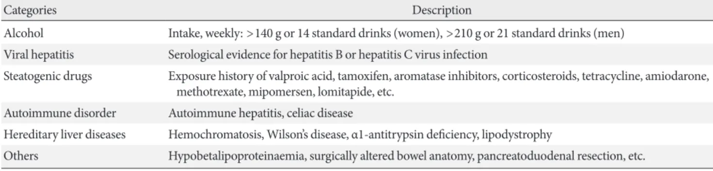
In individuals with and without diabetes, a two-step process should be used to diagnose NAFLD: (1) existence of hepatic steatosis, either by imaging or histology; and (2) exclusion of secondary causes of liver steatosis as described in Table 1 [39,40]. Other endocrinologic disorders such as hypothyroid- ism, hypopituitarism, and hypogonadism are also associated with NAFLD; therefore, related laboratory assessments should be considered for patients with signs or symptoms of these en- docrinopathies.
NAFLD spans the spectrum of fatty liver conditions, from simple steatosis (nonalcoholic fatty liver [NAFL]) and steato- hepatitis (NASH) to advanced fibrosis or cirrhosis. By histo- logic liver biopsy, NAFL is defined as ≥5% hepatic steatosis with no evidence of hepatocellular injury (hepatocyte balloon- ing). NASH is a combination of NAFL and inflammation with hepatocyte injury (e.g., ballooning), with or without fibrosis.
IMAGING MODALITIES TO ASSESS HEPATIC STEATOSIS
Despite its invasive nature and sampling variability [41], liver
biopsy remains the gold standard to assess hepatic steatosis, inflammation, and fibrosis in patients with NAFLD. It should be considered for patients suspected to have advanced fibrosis or when exclusion of other chronic liver diseases is needed. To overcome its high costs and risks, alternative noninvasive im- aging, clinical, and laboratory modalities have been developed and validated to diagnose NAFLD.
Currently, ultrasonography (US), computed tomography (CT), magnetic resonance imaging (MRI), MRS, and transient elastography (TE) are widely available tools to measure hepatic steatosis (Table 2) [42-51].
Ultrasonography
US is the recommended first line screening method for pa- tients with T2DM by the European NAFLD guidelines [42].
Although US has interobserver variability and limited sensitiv- ity to detect mild (<20%) steatosis [52] or morbid obesity (body mass index [BMI] >40 kg/m2) [53], it can provide addi- tional information about liver structure. In a meta-analysis, US achieved pooled sensitivity of 84.8% and pooled specificity of 93.6% to detect ≥20% to 30% steatosis, compared with histol- ogy [52]. However, US is limited in accuracy and reproducibil- ity as echogenicity, an essential sonographic feature to assess degree of fat content, can be affected by various patient (obesi- ty, coexistent kidney disease) and acquisition (device, operator, instrument settings) factors.
Computed tomography
CT scanners are standardized to obtain pixel value measure- ments relative to water using a dimensionless unit known as the Hounsfield unit (HU). Radiodensity of water is 0 HU by definition and air is defined as −1,000 HU [54]. In non-con- trast CT, normal liver parenchyma and fat are about 50 to 60 and −20 to −100 HU, respectively. Due to inconsistency in HU
Table 1. Secondary causes of hepatic fat accumulation
Categories Description
Alcohol Intake, weekly: >140 g or 14 standard drinks (women), >210 g or 21 standard drinks (men) Viral hepatitis Serological evidence for hepatitis B or hepatitis C virus infection
Steatogenic drugs Exposure history of valproic acid, tamoxifen, aromatase inhibitors, corticosteroids, tetracycline, amiodarone, methotrexate, mipomersen, lomitapide, etc.
Autoimmune disorder Autoimmune hepatitis, celiac disease
Hereditary liver diseases Hemochromatosis, Wilson’s disease, α1-antitrypsin deficiency, lipodystrophy
Others Hypobetalipoproteinaemia, surgically altered bowel anatomy, pancreatoduodenal resection, etc.
calibration by external factors, ‘fat-free spleen’ can be used as an internal reference [55]. Although various criteria to define steatosis have been proposed, hepatic HU less than 40 or (liver HU−spleen HU) less than −10 can detect steatosis [43] with sensitivity and specificity ranging from 46% to 72% and 88%
to 95%, respectively [56]. Similar to US, CT has limited sensi- tivity to detect mild steatosis (<30% liver fat). Radiation expo- sure is an additional drawback. Although CT can quantitative- ly measure hepatic steatosis, it is an unfavorable modality to diagnose fatty liver in the clinical setting. A few clinical trials of patients with T2DM applied CT scan to monitor changes in hepatic steatosis [57,58].
Magnetic resonance imaging
To date, MRI showed the most powerful performance for as-
sessing steatosis. MRI-based methods are generally divided into two classes: MRS and MRI-proton density fat fraction (MRI-PDFF). MRS is considered the gold standard for quanti- fication of hepatic triglyceride (fat) content [42,44], as it is sen- sitive enough to detect trace amounts of liver fat. MRS is more a biochemical-based technique than imaging. It quantifies the signal intensity of proton at frequencies corresponding to wa- ter or fat within the voxel-of-interest, and the fat signal fraction is calculated. In a recent meta-analysis with histology as the reference, MRS showed high diagnostic accuracy to detect mild steatosis (histological grade ≥5%) with sensitivity and specificity of 89% and 84%, respectively [59]. However, major limitations of MRS are: high cost; time consumption; need for specialized expertise and devices; and small volume of mea- surements (usually <3×3×3 cm in size), which may cause Table 2. Summary of currently used imaging devices for quantification of hepatic steatosis and fibrosis
Device Detection criteria Accuracy reproducibility
quantification
Hepatic volume of assessment
Time
accessibility Cost Specific comments Hepatic steatosis
US Specific sonographic
findings [51] + +++ +
(bedside) + Cannot detect mild steatosis, Observer dependency
CT liver HU <40 or liver HU−spleen HU <−10 [43]
++ +++ ++ ++ Radiation hazard
Diverse criteria for definition (liver/
spleen ratio of HU, etc.) Low sensitivity in mild steatosis MRI-PDFF ≥5.6% liver fat [42,44] +++ +++ +++ +++ Optimal for clinical trials
MRS +++ + +++ +++ Gold standard
Sampling errors Require expertise/device CAP by TE CAP ≥248 dB/m [46]
or ≥288 dB/m [47] ++ + +
(bedside) + Not linear in higher liver fat content Results are affected by BMI, diabetes,
etiology
XL probe for the obese Hepatic fibrosis
MRE Advanced fibrosis (F3) threshold >2.4–5.55 kPa [45]
+++ +++ +++ +++ Diverse cut-points by types of modality
(2D, 3D, etc.)
Most accurate but expensive Failure risk in iron overload condition LSM by TE Diverse cut-points
(7.3– 9.9 kPa) for ad- vanced fibrosis (F3) [48-50]
++ + +
(bedside) + Affected by BMI (failure risk) XL probe for the obese
TE can measure CAP and LSM simul- taneously
US, ultrasonography; CT, computed tomography; HU, Hounsfield unit; MRI-PDFF, magnetic resonance imaging proton density fat fraction;
MRS, magnetic resonance spectroscopy; CAP, controlled attenuation parameter; TE, transient elastography; BMI, body mass index; MRE, mag- netic resonance elastography; kPa, kilopascals; LSM, liver stiffness measurement; 2D, two dimensional; 3D, three dimensional; CAP, controlled attenuation parameter.
sampling errors.
Several MRI-based methods have been developed for mea- suring liver fat content. MRI-PDFF is a fundamental tissue trait and can be calculated as the (proton signal from magnetic resonance [MR]-visible triglyceride)/(total proton signal in- cluding triglyceride and water) [54,60]. Therefore, internal cal- ibration or a reference standard is not needed. The diagnostic accuracy of MRI-PDFF to detect steatosis is comparable to MRS or liver biopsy [45,54,61], while it also allows whole im- aging of the liver where fat is measured. This can minimize sampling error, making MRI-PDFF useful for monitoring pa- tients sequentially, such as in NAFLD clinical trials [45,62,63].
Transient elastography
Controlled attenuation parameter (CAP) is a simple quantita- tive index based on the properties of ultrasonic signals exam- ined by transient liver elastography (Fibroscan; Echosens, Par- is, France). Based on the concept that fat attenuates US propa- gation, it quantifies US attenuation at the center frequency of the Fibroscan M probe (3.5 MHz) or XL probe (2.5 MHz) within approximately 3 cm3 of liver volume [64]. Recent meta- analysis with CAP measurements in patients with various chronic liver diseases including NAFLD (20% of the total pop- ulation), demonstrated its diagnostic accuracy to detect hepat- ic steatosis with sensitivity and specificity of 69% and 82%, re- spectively (area under the receiver operating characteristic curve [AUROC]=0.823) [46]. Optimal cut-off values for de- fining S1, S2, and S3 were 248, 268, and 280 dB/m, respectively.
The authors proposed that individual CAP values should be interpreted after adjustment for presence of diabetes, steatosis etiology (NAFLD), and BMI, as these factors can overrate CAP values [46]. Wong et al. [65] recently established validity crite- ria for CAP measurement by M probe in clinical practice. In- terquartile range (IQR), which can be derived from 10 repeat- ed values of CAP, reflects the fluctuation of CAP measure- ments. If IQR >40 dB/m, accuracy of CAP to diagnose steato- sis substantially declines. Caussy et al. [47] proposed a cut-off value of 288 dB/m for CAP measurement of hepatic steatosis (defined as MRI-PDFF ≥5%), with improved diagnostic accu- racy when the IQR was <30 dB/m. The discrepancy in CAP cut-points in the two previous studies may be due to differenc- es in obesity prevalence, NAFLD, and diabetes. To reduce the failure rate of CAP measurements in obese (BMI ≥30 kg/m2) patients, XL probe is recommended [66,67]. In biopsy-proven NAFLD patients, MRI-PDFF was more accurate than CAP in
diagnosing all grades of steatosis [48,68]. Notably, CAP showed a non-linear relationship with liver fat content in pa- tients with severe fatty liver, indicating a limitation to discrimi- nate moderate and severe steatosis (grade 2 vs. 3). Lee et al.
[69] recently conducted an efficacy study of NAFLD using CAP as a primary endpoint in T2DM patients, to assess its utility in clinical trials.
Compared to MRI-PDFF, TE-based CAP showed relatively lower accuracy to detect steatosis whereas its advantages in- clude cost and accessibility. More research on these modalities, to evaluate and define their distinct roles, are warranted [70].
IMAGING MODALITIES TO ASSESS NASH AND HEPATIC FIBROSIS
To date, no imaging technique can reliably diagnose NASH. In contrast, liver fibrosis can be indirectly quantified by measur- ing liver stiffness using imaging modalities such as magnetic resonance elastography (MRE) and TE, because fibrotic liver contains a high level of collagen resulting in tissue rigidity.
Magnetic resonance elastography
MRE uses vibration to generate low frequency mechanical waves in liver tissue, visualize the propagating waves using a phase contrast MRI technique, and then transform the wave information into quantitative images reflecting liver stiffness [71]. Contrary to the European NAFLD guidelines, United States guidelines state that MRE is useful to identify advanced fibrosis in patients with NAFLD [40] due to its better perfor- mance in discriminating stage 1 and ≥2 fibrosis compared to TE [68]; furthermore, MRE’s superiority to detect fibrosis was confirmed in a meta-analysis [49]. Although it is reliable, re- producible, and the most accurate method for estimating he- patic fibrosis, MRE is expensive, not widely available in many clinics, and requires specialized expertise to obtain acceptable images.
Transient elastography
TE (Fibroscan) is a point-of-care imaging technique using US in which propagation velocity directly reflects tissue elasticity to 1 cm wide and 4 cm long [72]. This can quickly generate a surrogate marker of liver fibrosis stage called liver stiffness measurement (LSM). Results are described as the median val- ues of 10 validated measurements, ranging from 2.5 to 75.0 ki- lopascals (kPa), with healthy liver values <5.5 kPa [73]. Impor-
tantly, validity criteria of LSM by TE include a success rate (ra- tio of valid measurements to total measurements) >60% and an IQR/median LSM ratio of <0.3 [74]. TE has been intensive- ly studied in patients with viral hepatitis; however, its diagnos- tic accuracy for hepatic fibrosis was recently validated in NAFLD patients [50,75]. Notably, LSM was more correlated with fibrosis stage in individuals with severe fibrosis (F3) and cirrhosis (F4) [75,76]. TE has higher negative predictive values for excluding advanced fibrosis than positive predictive values for detection [75], indicating that additional surrogate markers for fibrosis can enhance the ability to diagnose advanced fibro- sis or cirrhosis. A major weakness of TE is a high failure risk in obese patients [77] and low accuracy to detect fibrosis in high BMI patients [75]. As T2DM patients tend to be obese, this is- sue should be carefully considered before utilizing TE. In such cases, XL-probe may provide more reliable results for morbidly obese patients [78]. Due to its accessibility, low cost, and mod- erate accuracy, TE is recommended by both United States and European NAFLD guidelines as an acceptable non-invasive method to identify patients at low risk of advanced fibrosis/cir- rhosis [40,42].
BIOMARKER-BASED PREDICTION MODELS TO ASSESS STEATOSIS, NASH, AND HEPATIC FIBROSIS
Besides imaging modalities, several noninvasive biomarker models have been developed to predict NASH and fibrosis. As NAFLD patients with diabetes are at high risk for NASH and fibrosis, which are significantly associated with morbidity and mortality, evaluation of these conditions is essential for clinical management [42]. Models incorporating both clinical and lab- oratory parameters, which are directly or indirectly linked to liver status, provide enhanced diagnostic accuracy to identify high risk individuals [79].
Hepatic steatosis
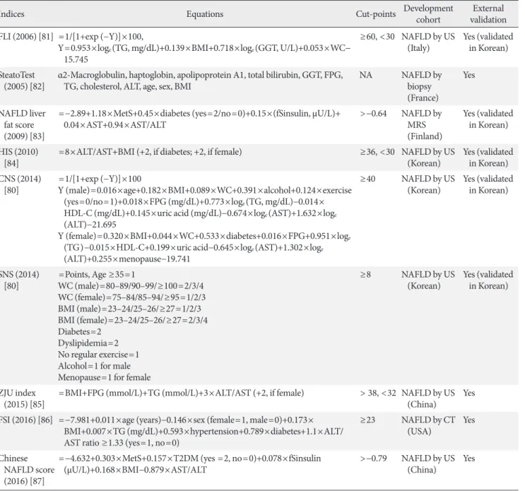
Many noninvasive prediction models have been proposed to detect hepatic steatosis in NAFLD cohorts (Table 3) [80-87].
The majority of models are derived from datasets with fatty liver defined by US. European NAFLD guidelines state that fatty liver index (FLI), SteatoTest (BioPredictive, Paris, France), and NAFLD liver fat score are well-validated models to detect steatosis in the general population [42]. FLI, NAFLD liver fat score, hepatic steatosis index (HSI), and comprehensive/sim-
ple NAFLD scores (CNS/SNS) were externally validated in the Korean population. Depending on population characteristics, cut-off values of these models might be adjusted to improve sensitivity and specificity [80]. Epidemiologic studies with no imaging information can apply these models to calculate an operational definition for hepatic steatosis [88], but prudent interpretation/application of multiple models is needed. How- ever, these biomarker-based indices do not add much diagnos- tic information for clinicians who routinely perform imaging studies such as US for patients with suspected NAFLD.
NASH
Currently there are no well-established prediction models or biomarkers to distinguish NASH from steatosis, despite inten- sive efforts [42]. Circulating levels of cytokeratin-18 (CK-18) fragments, which are released from apoptotic or dead cells, have been extensively investigated as a novel biomarker [89];
however, its clinical utility remains unknown due to low repro- ducibility and measurement issues [90]. Other panels com- bined with novel biomarkers, such as NASH test (BioPredic- tive) [91], NASH diagnostics [92], and OxNASH score [93]
have been introduced. Further research is required to validate their accuracy and practical utility.
Hepatic fibrosis
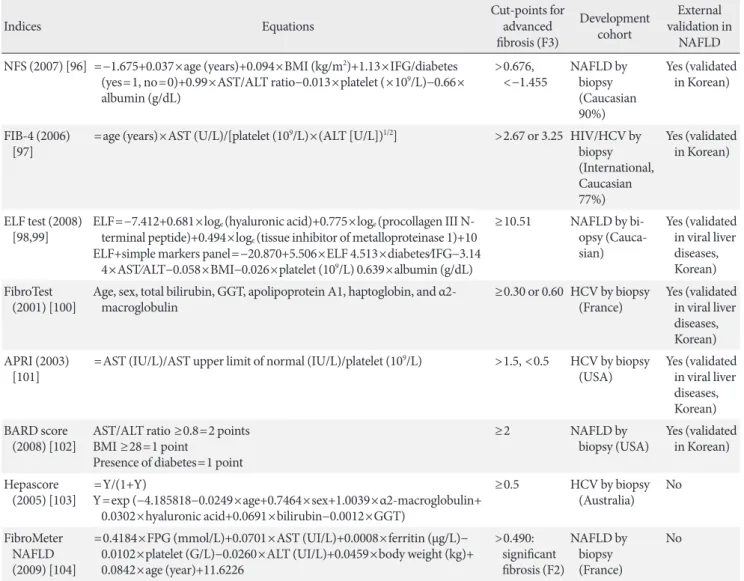
Severity of hepatic fibrosis is the most powerful determinant of long-term outcomes, including mortality [94,95]; thus, nonin- vasive assessment of fibrosis is essential to manage patients with NAFLD. Beyond imaging modalities to estimate hepatic fibrosis, several prediction scores have been developed and validated to identify or exclude advanced fibrosis. Focusing on reliable blood-based predictive models in the NAFLD guide- lines, equations, cut-points for advanced fibrosis (F3), infor- mation about the original development cohort, and external validation in individuals with NAFLD are summarized in Ta- ble 4 [96-104]. This review does not compare diagnostic perfor- mance of these models due to limited space.
Contrary to steatosis or NASH scores, fibrosis prediction models in clinical practice are recommended in the recent NAFLD guidelines [40,42] because some models have achieved acceptable accuracy to detect significant fibrosis and have been widely validated in NAFLD patients. The NAFLD fibrosis score (NFS) [96] and fibrosis-4 (FIB-4) index [97] are current- ly the most studied decision-making systems and are clinically endorsed by both European and United States NAFLD guide-
lines. The National Institute for Health and Care Excellence (NICE, London, United Kingdom) recommended use of the Enhanced Liver Fibrosis (ELF) panel, a commercial diagnostic kit using fibrosis-related biomarkers, for NAFLD manage- ment. These blood-based fibrosis prediction models perform
better when excluding advanced fibrosis versus diagnosing it, due to higher negative predictive values [105]. Therefore, com- bining results from both imaging modalities and biomarker- based scores may be a better approach to identify ambiguous patients at risk for advanced fibrosis or who need liver biopsy.
Table 3. Summary of biomarker-based prediction models to assess hepatic steatosis
Indices Equations Cut-points Development cohort External
validation FLI (2006) [81] =1/[1+exp (−Y)]×100,
Y=0.953×loge (TG, mg/dL)+0.139×BMI+0.718×loge (GGT, U/L)+0.053×WC−
15.745
≥60, <30 NAFLD by US
(Italy) Yes (validated in Korean) SteatoTest
(2005) [82] α2-Macroglobulin, haptoglobin, apolipoprotein A1, total bilirubin, GGT, FPG,
TG, cholesterol, ALT, age, sex, BMI NA NAFLD by
biopsy (France)
Yes
NAFLD liver fat score (2009) [83]
=−2.89+1.18×MetS+0.45×diabetes (yes=2/no=0)+0.15×(fSinsulin, μU/L)+
0.04×AST+0.94×AST/ALT >−0.64 NAFLD by
MRS (Finland)
Yes (validated in Korean) HIS (2010)
[84] =8×ALT/AST+BMI (+2, if diabetes; +2, if female) ≥36, <30 NAFLD by US
(Korean) Yes (validated in Korean) CNS (2014)
[80] =1/[1+exp (−Y)]×100
Y (male)=0.016×age+0.182×BMI+0.089×WC+0.391×alcohol+0.124×exercise (yes=0/no=1)+0.018×FPG (mg/dL)+0.773×loge (TG, mg/dL)−0.014×
HDL-C (mg/dL)+0.145×uric acid (mg/dL)−0.674×loge (AST)+1.632×loge
(ALT)−21.695
Y (female)=0.320×BMI+0.044×WC+0.533×diabetes+0.016×FPG+0.951×loge
(TG)−0.015×HDL-C+0.199×uric acid−0.645×loge (AST)+1.302×loge
(ALT)+0.255×menopause−19.741
≥40 NAFLD by US
(Korean) Yes (validated in Korean)
SNS (2014)
[80] =Points, Age ≥35=1
WC (male)=80–89/90–99/≥100=2/3/4 WC (female)=75–84/85–94/≥95=1/2/3 BMI (male)=23–24/25–26/≥27=1/2/3 BMI (female)=23–24/25–26/≥27=2/3/4 Diabetes=2
Dyslipidemia=2 No regular exercise=1 Alcohol=1 for male Menopause=1 for female
≥8 NAFLD by US
(Korean) Yes (validated in Korean)
ZJU index
(2015) [85] =BMI+FPG (mmol/L)+TG (mmol/L)+3×ALT/AST (+2, if female) > 38, <32 NAFLD by US
(China) Yes
FSI (2016) [86] =−7.981+0.011×age (years)−0.146×sex (female=1, male=0)+0.173×
BMI+0.007×TG (mg/dL)+0.593×hypertension+0.789×diabetes+1.1×ALT/
AST ratio ≥1.33 (yes=1, no=0)
≥23 NAFLD by CT
(USA) Yes
Chinese NAFLD score (2016) [87]
=−4.632+0.303×MetS+0.157×T2DM (yes =2, no=0)+0.078×fSinsulin
(μU/L)+0.168×BMI−0.879×AST/ALT >−0.79 NAFLD by US
(China) Yes
FLA, fatty liver index; TG, triglyceride; BMI, body mass index; GGT, γ-glutamyltransferase; WC, waist circumference; NAFLD, nonalcoholic fatty liver disease; US, ultrasonography; FPG, fasting plasma glucose; ALT, alanine aminotransferase; NA, not available; MetS, metabolic syn- drome; fSinsulin, fasting serum insulin; AST, aspartate aminotransferase; MRS, magnetic resonance spectroscopy; HSI, hepatic steatosis index;
CNS, comprehensive NAFLD score; HDL-C, high density lipoprotein cholesterol; SNS, simple NAFLD score; ZJU, Zhejiang University; FSI, Framingham Steatosis Index; CT, computed tomography; T2DM, type 2 diabetes mellitus.
CONCLUSIONS
Compared to the general population, NAFLD is widely preva- lent in patients with T2DM, and incidence of both metabolic diseases is steadily increasing worldwide. Accumulating evi- dence indicates that T2DM is considered an emerging risk fac- tor for NASH and/or fibrosis and that NAFLD may worsen di- abetes-related health outcomes, such as vascular complica- tions, and mortality. Extensive assessment of NAFLD severity and of cardiometabolic risk profiles is highly recommended for management of patients with T2DM. Although many imaging modalities and biomarker-based prediction models have been
developed to evaluate NAFLD disease status, more investiga- tion is required before their application in routine clinical care.
CONFLICTS OF INTEREST
No potential conflict of interest relevant to this article was re- ported.
ACKNOWLEDGMENTS
Editorial assistance was provided by Caron Modeas.
Table 4. Summary of biomarker-based prediction models to assess hepatic fibrosis
Indices Equations Cut-points for
advanced fibrosis (F3)
Development cohort
External validation in
NAFLD NFS (2007) [96] =−1.675+0.037×age (years)+0.094×BMI (kg/m2)+1.13×IFG/diabetes
(yes=1, no=0)+0.99×AST/ALT ratio−0.013×platelet (×109/L)−0.66×
albumin (g/dL)
>0.676,
<−1.455 NAFLD by biopsy (Caucasian 90%)
Yes (validated in Korean)
FIB-4 (2006)
[97] =age (years)×AST (U/L)/[platelet (109/L)×(ALT [U/L])1/2] >2.67 or 3.25 HIV/HCV by biopsy (International, Caucasian 77%)
Yes (validated in Korean)
ELF test (2008)
[98,99] ELF=−7.412+0.681×loge (hyaluronic acid)+0.775×loge (procollagen III N- terminal peptide)+0.494×loge (tissue inhibitor of metalloproteinase 1)+10 ELF+simple markers panel=−20.870+5.506×ELF 4.513×diabetes⁄IFG−3.14
4×AST⁄ALT−0.058×BMI−0.026×platelet (109/L) 0.639×albumin (g/dL)
≥10.51 NAFLD by bi- opsy (Cauca- sian)
Yes (validated in viral liver diseases, Korean) FibroTest
(2001) [100] Age, sex, total bilirubin, GGT, apolipoprotein A1, haptoglobin, and α2-
macroglobulin ≥0.30 or 0.60 HCV by biopsy
(France) Yes (validated in viral liver diseases, Korean) APRI (2003)
[101] =AST (IU/L)/AST upper limit of normal (IU/L)/platelet (109/L) >1.5, <0.5 HCV by biopsy
(USA) Yes (validated in viral liver diseases, Korean) BARD score
(2008) [102] AST/ALT ratio ≥0.8=2 points BMI ≥28=1 point
Presence of diabetes=1 point
≥2 NAFLD by
biopsy (USA) Yes (validated in Korean) Hepascore
(2005) [103] =Y/(1+Y)
Y=exp (−4.185818−0.0249×age+0.7464×sex+1.0039×α2-macroglobulin+
0.0302×hyaluronic acid+0.0691×bilirubin−0.0012×GGT)
≥0.5 HCV by biopsy (Australia) No FibroMeter
NAFLD (2009) [104]
=0.4184×FPG (mmol/L)+0.0701×AST (UI/L)+0.0008×ferritin (μg/L)−
0.0102×platelet (G/L)−0.0260×ALT (UI/L)+0.0459×body weight (kg)+
0.0842×age (year)+11.6226
>0.490:
significant fibrosis (F2)
NAFLD by biopsy (France)
No
NAFLD, nonalcoholic fatty liver disease; NFS, NAFLD fibrosis score; BMI, body mass index; IFG, impaired fasting glucose; AST, aspartate ami- notransferase; ALT, alanine aminotransferase; FIB-4, fibrosis-4; HIV, human immunodeficiency virus; HCV, hepatitis C virus; ELF, Enhanced Liver Fibrosis; GGT, γ-glutamyltransferase; APRI, AST to platelet ratio index; FPG, fasting plasma glucose.
REFERENCES
1. International Diabetes Federation: IDF Diabetes Atlas: Key Messages. Available from: http://diabetesatlas.org/key- messages.html (cited 2019 Feb 8).
2. Korean Diabetes Association: Diabetes Fact Sheet in Korea 2018. Available from: http://www.diabetes.or.kr/pro/news/
admin.php?category=A&code=admin&number=1615&mod e=view (cited 2019 Feb 8).
3. Tilg H, Moschen AR, Roden M. NAFLD and diabetes melli- tus. Nat Rev Gastroenterol Hepatol 2017;14:32-42.
4. Adams LA, Anstee QM, Tilg H, Targher G. Non-alcoholic fat- ty liver disease and its relationship with cardiovascular disease and other extrahepatic diseases. Gut 2017;66:1138-53.
5. Younossi Z, Tacke F, Arrese M, Sharma BC, Mostafa I, Bugia- nesi E, Wong VW, Yilmaz Y, George J, Fan J, Vos MB. Global perspectives on non-alcoholic fatty liver disease and non-al- coholic steatohepatitis. Hepatology 2018 Sep 4 [Epub]. https://
doi.org/10.1002/hep.30251.
6. Younossi ZM. Non-alcoholic fatty liver disease: a global public health perspective. J Hepatol 2018 Nov 9 [Epub]. https://doi.
org/10.1016/j.jhep.2018.10.033.
7. Park SH, Jeon WK, Kim SH, Kim HJ, Park DI, Cho YK, Sung IK, Sohn CI, Keum DK, Kim BI. Prevalence and risk factors of non-alcoholic fatty liver disease among Korean adults. J Gas- troenterol Hepatol 2006;21(1 Pt 1):138-43.
8. Jeong EH, Jun DW, Cho YK, Choe YG, Ryu S, Lee SM, Jang EC. Regional prevalence of non-alcoholic fatty liver disease in Seoul and Gyeonggi-do, Korea. Clin Mol Hepatol 2013;19:
266-72.
9. Lonardo A, Ballestri S, Marchesini G, Angulo P, Loria P. Non- alcoholic fatty liver disease: a precursor of the metabolic syn- drome. Dig Liver Dis 2015;47:181-90.
10. Browning JD, Szczepaniak LS, Dobbins R, Nuremberg P, Hor- ton JD, Cohen JC, Grundy SM, Hobbs HH. Prevalence of he- patic steatosis in an urban population in the United States: im- pact of ethnicity. Hepatology 2004;40:1387-95.
11. Portillo-Sanchez P, Bril F, Maximos M, Lomonaco R, Bier- nacki D, Orsak B, Subbarayan S, Webb A, Hecht J, Cusi K.
High prevalence of nonalcoholic fatty liver disease in patients with type 2 diabetes mellitus and normal plasma aminotrans- ferase levels. J Clin Endocrinol Metab 2015;100:2231-8.
12. Kim BY, Jung CH, Mok JO, Kang SK, Kim CH. Prevalences of diabetic retinopathy and nephropathy are lower in Korean type 2 diabetic patients with non-alcoholic fatty liver disease. J
Diabetes Investig 2014;5:170-5.
13. Eguchi Y, Hyogo H, Ono M, Mizuta T, Ono N, Fujimoto K, Chayama K, Saibara T; JSG-NAFLD. Prevalence and associat- ed metabolic factors of nonalcoholic fatty liver disease in the general population from 2009 to 2010 in Japan: a multicenter large retrospective study. J Gastroenterol 2012;47:586-95.
14. Anstee QM, Targher G, Day CP. Progression of NAFLD to di- abetes mellitus, cardiovascular disease or cirrhosis. Nat Rev Gastroenterol Hepatol 2013;10:330-44.
15. Ballestri S, Zona S, Targher G, Romagnoli D, Baldelli E, Nas- cimbeni F, Roverato A, Guaraldi G, Lonardo A. Nonalcoholic fatty liver disease is associated with an almost twofold in- creased risk of incident type 2 diabetes and metabolic syn- drome. Evidence from a systematic review and meta-analysis.
J Gastroenterol Hepatol 2016;31:936-44.
16. Kim CH, Park JY, Lee KU, Kim JH, Kim HK. Fatty liver is an independent risk factor for the development of type 2 diabetes in Korean adults. Diabet Med 2008;25:476-81.
17. Sung KC, Kim SH. Interrelationship between fatty liver and insulin resistance in the development of type 2 diabetes. J Clin Endocrinol Metab 2011;96:1093-7.
18. Sung KC, Wild SH, Byrne CD. Resolution of fatty liver and risk of incident diabetes. J Clin Endocrinol Metab 2013;98:3637-43.
19. Yamazaki H, Tsuboya T, Tsuji K, Dohke M, Maguchi H. Inde- pendent association between improvement of nonalcoholic fatty liver disease and reduced incidence of type 2 diabetes.
Diabetes Care 2015;38:1673-9.
20. Bae JC, Han JM, Cho JH, Kwon H, Park SE, Park CY, Lee WY, Oh KW, Kwon S, Park SW, Rhee EJ. The persistence of fatty liver has a differential impact on the development of diabetes:
the Kangbuk Samsung Health Study. Diabetes Res Clin Pract 2018;135:1-6.
21. Sung KC, Jeong WS, Wild SH, Byrne CD. Combined influ- ence of insulin resistance, overweight/obesity, and fatty liver as risk factors for type 2 diabetes. Diabetes Care 2012;35:717-22.
22. Bae JC, Rhee EJ, Lee WY, Park SE, Park CY, Oh KW, Park SW, Kim SW. Combined effect of nonalcoholic fatty liver disease and impaired fasting glucose on the development of type 2 di- abetes: a 4-year retrospective longitudinal study. Diabetes Care 2011;34:727-9.
23. Bae JC, Kim SK, Han JM, Kwon S, Lee DY, Kim J, Park SE, Park CY, Lee WY, Oh KW, Park SW, Rhee EJ. Additive effect of non-alcoholic fatty liver disease on the development of dia- betes in individuals with metabolic syndrome. Diabetes Res Clin Pract 2017;129:136-43.
24. Choi JH, Rhee EJ, Bae JC, Park SE, Park CY, Cho YK, Oh KW, Park SW, Lee WY. Increased risk of type 2 diabetes in subjects with both elevated liver enzymes and ultrasonographically di- agnosed nonalcoholic fatty liver disease: a 4-year longitudinal study. Arch Med Res 2013;44:115-20.
25. Chang Y, Jung HS, Yun KE, Cho J, Cho YK, Ryu S. Cohort study of non-alcoholic fatty liver disease, NAFLD fibrosis score, and the risk of incident diabetes in a Korean population.
Am J Gastroenterol 2013;108:1861-8.
26. Bhatia LS, Curzen NP, Calder PC, Byrne CD. Non-alcoholic fatty liver disease: a new and important cardiovascular risk factor? Eur Heart J 2012;33:1190-200.
27. Ekstedt M, Franzen LE, Mathiesen UL, Thorelius L, Hol- mqvist M, Bodemar G, Kechagias S. Long-term follow-up of patients with NAFLD and elevated liver enzymes. Hepatology 2006;44:865-73.
28. Kwok R, Choi KC, Wong GL, Zhang Y, Chan HL, Luk AO, Shu SS, Chan AW, Yeung MW, Chan JC, Kong AP, Wong VW.
Screening diabetic patients for non-alcoholic fatty liver disease with controlled attenuation parameter and liver stiffness mea- surements: a prospective cohort study. Gut 2016;65:1359-68.
29. Koehler EM, Plompen EP, Schouten JN, Hansen BE, Darwish Murad S, Taimr P, Leebeek FW, Hofman A, Stricker BH, Cas- tera L, Janssen HL. Presence of diabetes mellitus and steatosis is associated with liver stiffness in a general population: the Rotterdam study. Hepatology 2016;63:138-47.
30. McPherson S, Hardy T, Henderson E, Burt AD, Day CP, Anst- ee QM. Evidence of NAFLD progression from steatosis to fi- brosing-steatohepatitis using paired biopsies: implications for prognosis and clinical management. J Hepatol 2015;62:1148- 55.
31. Goh GB, Pagadala MR, Dasarathy J, Unalp-Arida A, Sargent R, Hawkins C, Sourianarayanane A, Khiyami A, Yerian L, Pai RK, Dasarathy S, McCullough AJ. Clinical spectrum of non- alcoholic fatty liver disease in diabetic and non-diabetic pa- tients. BBA Clin 2014;3:141-5.
32. Hossain N, Afendy A, Stepanova M, Nader F, Srishord M, Rafiq N, Goodman Z, Younossi Z. Independent predictors of fibrosis in patients with nonalcoholic fatty liver disease. Clin Gastroenterol Hepatol 2009;7:1224-9.
33. Han E, Lee YH. Non-alcoholic fatty liver disease: the emerg- ing burden in cardiometabolic and renal diseases. Diabetes Metab J 2017;41:430-7.
34. Targher G, Bertolini L, Rodella S, Tessari R, Zenari L, Lippi G, Arcaro G. Nonalcoholic fatty liver disease is independently as-
sociated with an increased incidence of cardiovascular events in type 2 diabetic patients. Diabetes Care 2007;30:2119-21.
35. Targher G, Bertolini L, Rodella S, Zoppini G, Lippi G, Day C, Muggeo M. Non-alcoholic fatty liver disease is independently associated with an increased prevalence of chronic kidney dis- ease and proliferative/laser-treated retinopathy in type 2 dia- betic patients. Diabetologia 2008;51:444-50.
36. Adams LA, Harmsen S, St Sauver JL, Charatcharoenwitthaya P, Enders FB, Therneau T, Angulo P. Nonalcoholic fatty liver disease increases risk of death among patients with diabetes: a community-based cohort study. Am J Gastroenterol 2010;105:
1567-73.
37. Targher G, Pichiri I, Zoppini G, Trombetta M, Bonora E. In- creased prevalence of cardiovascular disease in type 1 diabetic patients with non-alcoholic fatty liver disease. J Endocrinol Invest 2012;35:535-40.
38. Targher G, Mantovani A, Pichiri I, Mingolla L, Cavalieri V, Mantovani W, Pancheri S, Trombetta M, Zoppini G, Chon- chol M, Byrne CD, Bonora E. Nonalcoholic fatty liver disease is independently associated with an increased incidence of chronic kidney disease in patients with type 1 diabetes. Diabe- tes Care 2014;37:1729-36.
39. Byrne CD, Patel J, Scorletti E, Targher G. Tests for diagnosing and monitoring non-alcoholic fatty liver disease in adults.
BMJ 2018;362:k2734.
40. Chalasani N, Younossi Z, Lavine JE, Charlton M, Cusi K, Ri- nella M, Harrison SA, Brunt EM, Sanyal AJ. The diagnosis and management of nonalcoholic fatty liver disease: practice guid- ance from the American Association for the Study of Liver Diseases. Hepatology 2018;67:328-57.
41. Ratziu V, Charlotte F, Heurtier A, Gombert S, Giral P, Bruck- ert E, Grimaldi A, Capron F, Poynard T; LIDO Study Group.
Sampling variability of liver biopsy in nonalcoholic fatty liver disease. Gastroenterology 2005;128:1898-906.
42. European Association for the Study of the Liver (EASL); Euro- pean Association for the Study of Diabetes (EASD); European Association for the Study of Obesity (EASO). EASL-EASD- EASO Clinical Practice Guidelines for the management of non-alcoholic fatty liver disease. J Hepatol 2016;64:1388-402.
43. Schwenzer NF, Springer F, Schraml C, Stefan N, Machann J, Schick F. Non-invasive assessment and quantification of liver steatosis by ultrasound, computed tomography and magnetic resonance. J Hepatol 2009;51:433-45.
44. Szczepaniak LS, Nurenberg P, Leonard D, Browning JD, Rein- gold JS, Grundy S, Hobbs HH, Dobbins RL. Magnetic reso-
nance spectroscopy to measure hepatic triglyceride content:
prevalence of hepatic steatosis in the general population. Am J Physiol Endocrinol Metab 2005;288:E462-8.
45. Dulai PS, Sirlin CB, Loomba R. MRI and MRE for non-inva- sive quantitative assessment of hepatic steatosis and fibrosis in NAFLD and NASH: clinical trials to clinical practice. J Hepa- tol 2016;65:1006-16.
46. Karlas T, Petroff D, Sasso M, Fan JG, Mi YQ, de Ledinghen V, Kumar M, Lupsor-Platon M, Han KH, Cardoso AC, Ferraioli G, Chan WK, Wong VW, Myers RP, Chayama K, Friedrich- Rust M, Beaugrand M, Shen F, Hiriart JB, Sarin SK, Badea R, Jung KS, Marcellin P, Filice C, Mahadeva S, Wong GL, Crotty P, Masaki K, Bojunga J, Bedossa P, Keim V, Wiegand J. Indi- vidual patient data meta-analysis of controlled attenuation pa- rameter (CAP) technology for assessing steatosis. J Hepatol 2017;66:1022-30.
47. Caussy C, Alquiraish MH, Nguyen P, Hernandez C, Cepin S, Fortney LE, Ajmera V, Bettencourt R, Collier S, Hooker J, Sy E, Rizo E, Richards L, Sirlin CB, Loomba R. Optimal thresh- old of controlled attenuation parameter with MRI-PDFF as the gold standard for the detection of hepatic steatosis. Hepa- tology 2018;67:1348-59.
48. Park CC, Nguyen P, Hernandez C, Bettencourt R, Ramirez K, Fortney L, Hooker J, Sy E, Savides MT, Alquiraish MH, Va- lasek MA, Rizo E, Richards L, Brenner D, Sirlin CB, Loomba R. Magnetic resonance elastography vs transient elastography in detection of fibrosis and noninvasive measurement of ste- atosis in patients with biopsy-proven nonalcoholic fatty liver disease. Gastroenterology 2017;152:598-607.
49. Hsu C, Caussy C, Imajo K, Chen J, Singh S, Kaulback K, Le MD, Hooker J, Tu X, Bettencourt R, Yin M, Sirlin CB, Ehman RL, Nakajima A, Loomba R. Magnetic resonance vs transient elastography analysis of patients with nonalcoholic fatty liver disease: a systematic review and pooled analysis of individual participants. Clin Gastroenterol Hepatol 2018 Jun 14 [Epub].
https://doi.org/10.1016/j.cgh.2018.05.059.
50. Tapper EB, Challies T, Nasser I, Afdhal NH, Lai M. The per- formance of vibration controlled transient elastography in a US cohort of patients with nonalcoholic fatty liver disease. Am J Gastroenterol 2016;111:677-84.
51. Saadeh S, Younossi ZM, Remer EM, Gramlich T, Ong JP, Hur- ley M, Mullen KD, Cooper JN, Sheridan MJ. The utility of ra- diological imaging in nonalcoholic fatty liver disease. Gastro- enterology 2002;123:745-50.
52. Hernaez R, Lazo M, Bonekamp S, Kamel I, Brancati FL, Gual-
lar E, Clark JM. Diagnostic accuracy and reliability of ultraso- nography for the detection of fatty liver: a meta-analysis. Hep- atology 2011;54:1082-90.
53. Ryan CK, Johnson LA, Germin BI, Marcos A. One hundred consecutive hepatic biopsies in the workup of living donors for right lobe liver transplantation. Liver Transpl 2002;8:1114- 22.
54. Zhang YN, Fowler KJ, Hamilton G, Cui JY, Sy EZ, Balanay M, Hooker JC, Szeverenyi N, Sirlin CB. Liver fat imaging-a clini- cal overview of ultrasound, CT, and MR imaging. Br J Radiol 2018;91:20170959.
55. Piekarski J, Goldberg HI, Royal SA, Axel L, Moss AA. Differ- ence between liver and spleen CT numbers in the normal adult: its usefulness in predicting the presence of diffuse liver disease. Radiology 1980;137:727-9.
56. Bohte AE, van Werven JR, Bipat S, Stoker J. The diagnostic ac- curacy of US, CT, MRI and 1H-MRS for the evaluation of he- patic steatosis compared with liver biopsy: a meta-analysis.
Eur Radiol 2011;21:87-97.
57. Ito D, Shimizu S, Inoue K, Saito D, Yanagisawa M, Inukai K, Akiyama Y, Morimoto Y, Noda M, Shimada A. Comparison of ipragliflozin and pioglitazone effects on nonalcoholic fatty liver disease in patients with type 2 diabetes: a randomized, 24-week, open-label, active-controlled trial. Diabetes Care 2017;40:1364-72.
58. Shibuya T, Fushimi N, Kawai M, Yoshida Y, Hachiya H, Ito S, Kawai H, Ohashi N, Mori A. Luseogliflozin improves liver fat deposition compared to metformin in type 2 diabetes patients with non-alcoholic fatty liver disease: a prospective random- ized controlled pilot study. Diabetes Obes Metab 2018;20:438- 42.
59. Zheng D, Guo Z, Schroder PM, Zheng Z, Lu Y, Gu J, He X.
Accuracy of MR imaging and MR spectroscopy for detection and quantification of hepatic steatosis in living liver donors: a meta-analysis. Radiology 2017;282:92-102.
60. Yokoo T, Bydder M, Hamilton G, Middleton MS, Gamst AC, Wolfson T, Hassanein T, Patton HM, Lavine JE, Schwimmer JB, Sirlin CB. Nonalcoholic fatty liver disease: diagnostic and fat-grading accuracy of low-flip-angle multiecho gradient-re- called-echo MR imaging at 1.5 T. Radiology 2009;251:67-76.
61. Yokoo T, Serai SD, Pirasteh A, Bashir MR, Hamilton G, Her- nando D, Hu HH, Hetterich H, Kuhn JP, Kukuk GM, Loomba R, Middleton MS, Obuchowski NA, Song JS, Tang A, Wu X, Reeder SB, Sirlin CB; RSNA-QIBA PDFF Biomarker Com- mittee. Linearity, bias, and precision of hepatic proton density
fat fraction measurements by using MR imaging: a meta-anal- ysis. Radiology 2018;286:486-98.
62. Loomba R. Role of imaging-based biomarkers in NAFLD: re- cent advances in clinical application and future research direc- tions. J Hepatol 2018;68:296-304.
63. Cui J, Philo L, Nguyen P, Hofflich H, Hernandez C, Betten- court R, Richards L, Salotti J, Bhatt A, Hooker J, Haufe W, Hooker C, Brenner DA, Sirlin CB, Loomba R. Sitagliptin vs.
placebo for non-alcoholic fatty liver disease: a randomized controlled trial. J Hepatol 2016;65:369-76.
64. Sasso M, Beaugrand M, de Ledinghen V, Douvin C, Marcellin P, Poupon R, Sandrin L, Miette V. Controlled attenuation pa- rameter (CAP): a novel VCTETM guided ultrasonic attenua- tion measurement for the evaluation of hepatic steatosis: pre- liminary study and validation in a cohort of patients with chronic liver disease from various causes. Ultrasound Med Biol 2010; 36:1825-35.
65. Wong VW, Petta S, Hiriart JB, Camma C, Wong GL, Marra F, Vergniol J, Chan AW, Tuttolomondo A, Merrouche W, Chan HL, Le Bail B, Arena U, Craxi A, de Ledinghen V. Validity cri- teria for the diagnosis of fatty liver by M probe-based con- trolled attenuation parameter. J Hepatol 2017;67:577-84.
66. de Ledinghen V, Hiriart JB, Vergniol J, Merrouche W, Bedossa P, Paradis V. Controlled attenuation parameter (CAP) with the XL probe of the Fibroscan(R): a comparative study with the M probe and liver biopsy. Dig Dis Sci 2017;62:2569-77.
67. Wong VW, Vergniol J, Wong GL, Foucher J, Chan AW, Cher- mak F, Choi PC, Merrouche W, Chu SH, Pesque S, Chan HL, de Ledinghen V. Liver stiffness measurement using XL probe in patients with nonalcoholic fatty liver disease. Am J Gastro- enterol 2012;107:1862-71.
68. Imajo K, Kessoku T, Honda Y, Tomeno W, Ogawa Y, Mawatari H, Fujita K, Yoneda M, Taguri M, Hyogo H, Sumida Y, Ono M, Eguchi Y, Inoue T, Yamanaka T, Wada K, Saito S, Nakajima A. Magnetic resonance imaging more accurately classifies ste- atosis and fibrosis in patients with nonalcoholic fatty liver dis- ease than transient elastography. Gastroenterology 2016;150:
626-37.
69. Lee YH, Kim JH, Kim SR, Jin HY, Rhee EJ, Cho YM, Lee BW.
Lobeglitazone, a novel thiazolidinedione, improves non-alco- holic fatty liver disease in type 2 diabetes: its efficacy and pre- dictive factors related to responsiveness. J Korean Med Sci 2017;32:60-9.
70. Karlas T, Petroff D, Wiegand J. Collaboration, not competi- tion: the role of magnetic resonance, transient elastography,
and liver biopsy in the diagnosis of nonalcoholic fatty liver disease. Gastroenterology 2017;152:479-81.
71. Venkatesh SK, Yin M, Ehman RL. Magnetic resonance elas- tography of liver: technique, analysis, and clinical applications.
J Magn Reson Imaging 2013;37:544-55.
72. Sandrin L, Fourquet B, Hasquenoph JM, Yon S, Fournier C, Mal F, Christidis C, Ziol M, Poulet B, Kazemi F, Beaugrand M, Palau R. Transient elastography: a new noninvasive method for assessment of hepatic fibrosis. Ultrasound Med Biol 2003;
29:1705-13.
73. Castera L, Vilgrain V, Angulo P. Noninvasive evaluation of NAFLD. Nat Rev Gastroenterol Hepatol 2013;10:666-75.
74. Castera L, Forns X, Alberti A. Non-invasive evaluation of liver fibrosis using transient elastography. J Hepatol 2008;48:835- 47.
75. Wong VW, Vergniol J, Wong GL, Foucher J, Chan HL, Le Bail B, Choi PC, Kowo M, Chan AW, Merrouche W, Sung JJ, de Lédinghen V. Diagnosis of fibrosis and cirrhosis using liver stiffness measurement in nonalcoholic fatty liver disease.
Hepatology 2010;51:454-62.
76. Friedrich-Rust M, Ong MF, Martens S, Sarrazin C, Bojunga J, Zeuzem S, Herrmann E. Performance of transient elastogra- phy for the staging of liver fibrosis: a meta-analysis. Gastroen- terology 2008;134:960-74.
77. Foucher J, Castera L, Bernard PH, Adhoute X, Laharie D, Ber- tet J, Couzigou P, de Ledinghen V. Prevalence and factors as- sociated with failure of liver stiffness measurement using Fi- broScan in a prospective study of 2114 examinations. Eur J Gastroenterol Hepatol 2006;18:411-2.
78. de Ledinghen V, Wong VW, Vergniol J, Wong GL, Foucher J, Chu SH, Le Bail B, Choi PC, Chermak F, Yiu KK, Merrouche W, Chan HL. Diagnosis of liver fibrosis and cirrhosis using liver stiffness measurement: comparison between M and XL probe of FibroScan(R). J Hepatol 2012;56:833-9.
79. Lee YH, Bang H, Kim DJ. How to establish clinical prediction models. Endocrinol Metab (Seoul) 2016;31:38-44.
80. Lee YH, Bang H, Park YM, Bae JC, Lee BW, Kang ES, Cha BS, Lee HC, Balkau B, Lee WY, Kim DJ. Non-laboratory-based self-assessment screening score for non-alcoholic fatty liver disease: development, validation and comparison with other scores. PLoS One 2014;9:e107584.
81. Bedogni G, Bellentani S, Miglioli L, Masutti F, Passalacqua M, Castiglione A, Tiribelli C. The fatty liver index: a simple and accurate predictor of hepatic steatosis in the general popula- tion. BMC Gastroenterol 2006;6:33.
82. Poynard T, Ratziu V, Naveau S, Thabut D, Charlotte F, Mes- sous D, Capron D, Abella A, Massard J, Ngo Y, Munteanu M, Mercadier A, Manns M, Albrecht J. The diagnostic value of biomarkers (SteatoTest) for the prediction of liver steatosis.
Comp Hepatol 2005;4:10.
83. Kotronen A, Peltonen M, Hakkarainen A, Sevastianova K, Bergholm R, Johansson LM, Lundbom N, Rissanen A, Rid- derstrale M, Groop L, Orho-Melander M, Yki-Jarvinen H.
Prediction of non-alcoholic fatty liver disease and liver fat us- ing metabolic and genetic factors. Gastroenterology 2009;137:
865-72.
84. Lee JH, Kim D, Kim HJ, Lee CH, Yang JI, Kim W, Kim YJ, Yoon JH, Cho SH, Sung MW, Lee HS. Hepatic steatosis index:
a simple screening tool reflecting nonalcoholic fatty liver dis- ease. Dig Liver Dis 2010;42:503-8.
85. Wang J, Xu C, Xun Y, Lu Z, Shi J, Yu C, Li Y. ZJU index: a novel model for predicting nonalcoholic fatty liver disease in a Chi- nese population. Sci Rep 2015;5:16494.
86. Long MT, Pedley A, Colantonio LD, Massaro JM, Hoffmann U, Muntner P, Fox CS. Development and validation of the Framingham Steatosis Index to identify persons with hepatic steatosis. Clin Gastroenterol Hepatol 2016;14:1172-80.
87. Xia MF, Yki-Jarvinen H, Bian H, Lin HD, Yan HM, Chang XX, Zhou Y, Gao X. Influence of ethnicity on the accuracy of non-invasive scores predicting non-alcoholic fatty liver dis- ease. PLoS One 2016;11:e0160526.
88. Lee YH, Jung KS, Kim SU, Yoon HJ, Yun YJ, Lee BW, Kang ES, Han KH, Lee HC, Cha BS. Sarcopaenia is associated with NAFLD independently of obesity and insulin resistance: Na- tionwide surveys (KNHANES 2008-2011). J Hepatol 2015;63:
486-93.
89. Feldstein AE, Wieckowska A, Lopez AR, Liu YC, Zein NN, McCullough AJ. Cytokeratin-18 fragment levels as noninva- sive biomarkers for nonalcoholic steatohepatitis: a multicenter validation study. Hepatology 2009;50:1072-8.
90. Younossi ZM, Loomba R, Anstee QM, Rinella ME, Bugianesi E, Marchesini G, Neuschwander-Tetri BA, Serfaty L, Negro F, Caldwell SH, Ratziu V, Corey KE, Friedman SL, Abdelmalek MF, Harrison SA, Sanyal AJ, Lavine JE, Mathurin P, Charlton MR, Goodman ZD, Chalasani NP, Kowdley KV, George J, Lindor K. Diagnostic modalities for nonalcoholic fatty liver disease, nonalcoholic steatohepatitis, and associated fibrosis.
Hepatology 2018;68:349-60.
91. Poynard T, Ratziu V, Charlotte F, Messous D, Munteanu M, Imbert-Bismut F, Massard J, Bonyhay L, Tahiri M, Thabut D,
Cadranel JF, Le Bail B, de Ledinghen V; LIDO Study Group;
CYTOL study group. Diagnostic value of biochemical mark- ers (NashTest) for the prediction of non alcoholo steato hepa- titis in patients with non-alcoholic fatty liver disease. BMC Gastroenterol 2006;6:34.
92. Younossi ZM, Jarrar M, Nugent C, Randhawa M, Afendy M, Stepanova M, Rafiq N, Goodman Z, Chandhoke V, Baranova A. A novel diagnostic biomarker panel for obesity-related nonalcoholic steatohepatitis (NASH). Obes Surg 2008;18:
1430-7.
93. Alkhouri N, Berk M, Yerian L, Lopez R, Chung YM, Zhang R, McIntyre TM, Feldstein AE, Hazen SL. OxNASH score corre- lates with histologic features and severity of nonalcoholic fatty liver disease. Dig Dis Sci 2014;59:1617-24.
94. Dulai PS, Singh S, Patel J, Soni M, Prokop LJ, Younossi Z, Se- bastiani G, Ekstedt M, Hagstrom H, Nasr P, Stal P, Wong VW, Kechagias S, Hultcrantz R, Loomba R. Increased risk of mor- tality by fibrosis stage in nonalcoholic fatty liver disease: sys- tematic review and meta-analysis. Hepatology 2017;65:1557- 65.
95. Ekstedt M, Hagstrom H, Nasr P, Fredrikson M, Stal P, Kecha- gias S, Hultcrantz R. Fibrosis stage is the strongest predictor for disease-specific mortality in NAFLD after up to 33 years of follow-up. Hepatology 2015;61:1547-54.
96. Angulo P, Hui JM, Marchesini G, Bugianesi E, George J, Far- rell GC, Enders F, Saksena S, Burt AD, Bida JP, Lindor K, Sanderson SO, Lenzi M, Adams LA, Kench J, Therneau TM, Day CP. The NAFLD fibrosis score: a noninvasive system that identifies liver fibrosis in patients with NAFLD. Hepatology 2007;45:846-54.
97. Sterling RK, Lissen E, Clumeck N, Sola R, Correa MC, Mon- taner J, S Sulkowski M, Torriani FJ, Dieterich DT, Thomas DL, Messinger D, Nelson M; APRICOT Clinical Investigators.
Development of a simple noninvasive index to predict signifi- cant fibrosis in patients with HIV/HCV coinfection. Hepatol- ogy 2006;43:1317-25.
98. Guha IN, Parkes J, Roderick P, Chattopadhyay D, Cross R, Harris S, Kaye P, Burt AD, Ryder SD, Aithal GP, Day CP, Rosenberg WM. Noninvasive markers of fibrosis in nonalco- holic fatty liver disease: validating the European Liver Fibrosis Panel and exploring simple markers. Hepatology 2008;47:455- 60.
99. Nobili V, Parkes J, Bottazzo G, Marcellini M, Cross R, New- man D, Vizzutti F, Pinzani M, Rosenberg WM. Performance of ELF serum markers in predicting fibrosis stage in pediatric
non-alcoholic fatty liver disease. Gastroenterology 2009;136:
160-7.
100. Imbert-Bismut F, Ratziu V, Pieroni L, Charlotte F, Benhamou Y, Poynard T; MULTIVIRC Group. Biochemical markers of liver fibrosis in patients with hepatitis C virus infection: a pro- spective study. Lancet 2001;357:1069-75.
101. Wai CT, Greenson JK, Fontana RJ, Kalbfleisch JD, Marrero JA, Conjeevaram HS, Lok AS. A simple noninvasive index can predict both significant fibrosis and cirrhosis in patients with chronic hepatitis C. Hepatology 2003;38:518-26.
102. Harrison SA, Oliver D, Arnold HL, Gogia S, Neuschwander- Tetri BA. Development and validation of a simple NAFLD clinical scoring system for identifying patients without ad-
vanced disease. Gut 2008;57:1441-7.
103. Adams LA, Bulsara M, Rossi E, DeBoer B, Speers D, George J, Kench J, Farrell G, McCaughan GW, Jeffrey GP. Hepascore: an accurate validated predictor of liver fibrosis in chronic hepati- tis C infection. Clin Chem 2005;51:1867-73.
104. Cales P, Laine F, Boursier J, Deugnier Y, Moal V, Oberti F, Hu- nault G, Rousselet MC, Hubert I, Laafi J, Ducluzeaux PH, Lu- nel F. Comparison of blood tests for liver fibrosis specific or not to NAFLD. J Hepatol 2009;50:165-73.
105. Vilar-Gomez E, Chalasani N. Non-invasive assessment of non-alcoholic fatty liver disease: clinical prediction rules and blood-based biomarkers. J Hepatol 2018;68:305-15.