ABSTRACT
Background and Purpose: Prospective memory (PM) has a known relationship with frontal
function, and PM decline has been observed in amnestic mild cognitive impairment (aMCI).
Cerebral small vessel disease, as evidenced by white matter hyperintensities (WMHs), is linked to frontal dysfunction. This study was undertaken to evaluate the relationship between PM decline and WMHs in patients with aMCI.
Methods: Of 74 enrollees with aMCI, 69 completed this prospective study. We compared total
scores and sub-scores of the Prospective and Retrospective Memory Questionnaire (PRMQ) administered at baseline and 3 months later, stratifying patients by degree of WMHs.
Results: A significant decline was seen in PRMQ total scores and PM scores at the 3-month
mark in patients with moderate (vs. mild) degrees of WMHs (−2.8±7.2 vs. 0.2±7.1; p=0.032).
In addition, patients with moderate (vs. mild) degrees of deep WMHs (DWMHs) showed greater PM decline, whereas PM loss in patients with mild, moderate, or severe degrees of periventricular WMHs (PVWMHs) did not differ significantly.
Conclusions: Findings of this study indicate that the burden of WMHs is consistently
implicated in PM deterioration experienced by patients with aMCI, and signifies greater PM decline, especially in instances of extensive DWMHs. Greater attention to the change of PM is therefore needed in aMCI patients with WMHs.
Keywords:
Prospective Memory; Mild Cognitive Impairment; White Matter;
Cerebral Small Vessel Disease
INTRODUCTION
Memory is a 2-pronged function, involving recall of the past and planning for the future.
1Prospective memory (PM), referred to as ‘remembering to remember,’ entails the formation of future action representations, temporary storage of such representations in memory, and their retrieval at future time points.
2PM is pivotal in meeting the challenges of daily living, and is key to autonomy and independence. PM tasks rely on self-initiated retrieval of intentions within specific time frames and involve externally prompted retrieval of information content.
2,3PM is further characterized as time- or event-based recollections.
Time-based PM relates to actions to be taken at specified points in the future or after a
Original Article
Received: Oct 2, 2018 Revised: Nov 3, 2018 Accepted: Nov 20, 2018 Correspondence to Soo Jin Yoon, MD
Department of Neurology, Eulji University School of Medicine, 95 Dunsanseo-ro, Seo-gu, Daejeon 35233, Korea.
E-mail: [email protected]
© 2018 Korean Dementia Association This is an Open Access article distributed under the terms of the Creative Commons Attribution Non-Commercial License (https://
creativecommons.org/licenses/by-nc/4.0/) which permits unrestricted non-commercial use, distribution, and reproduction in any medium, provided the original work is properly cited.
ORCID iDs Bora Yoon
https://orcid.org/0000-0002-1135-3392 Funding
This research was supported by the Original Technology Research Program for Brain Science through the National Research Foundation of Korea (NRF) funded by the Korean government (MSIP) (No.
2014M3C7A1064752), and Eisai Korea Inc.
Conflict of Interest
Although authors received a fund from Eisai, this study itself was an observational study, which was no intentional intervention related to any drug. Therefore, there was no interest and no impact on the results.
Bora Yoon ,
1Sun Young Ryu,
2Soo Jin Yoon
31 Department of Neurology, Konyang University Hospital, College of Medicine, Konyang University, Daejeon, Korea
2 Department of Neurology, Daejeon St. Mary's Hospital, College of Medicine, The Catholic University of Korea, Daejeon, Korea
3Department of Neurology, Eulji University School of Medicine, Daejeon, Korea
Prospective Memory Loss and Related
White Matter Changes in Patients with
Amnestic Mild Cognitive Impairment
Author Contributions
Conceptualization: Yoon B, Ryu SY, Yoon SJ; Data curation: Yoon B, Ryu SY, Yoon SJ; Formal analysis: Yoon B, Ryu SY, Yoon SJ; Investigation: Yoon B, Ryu SY, Yoon SJ;
Methodology: Yoon B, Ryu SY, Yoon SJ; Project administration: Yoon B, Ryu SY, Yoon SJ;
Resources: Yoon B, Ryu SY, Yoon SJ; Validation:
Yoon B, Ryu SY, Yoon SJ; Visualization: Yoon B, Ryu SY, Yoon SJ; Writing - original draft: Yoon B, Ryu SY, Yoon SJ; Writing - review & editing:
Yoon B, Ryu SY, Yoon SJ.
specified period of time, whereas event-based PM pertains to actions triggered by specific events
2; memory being crucial in both instances.
3Thus, successful execution of PM tasks requires not only timely detection of prospectively cued events/target times (prospective component), but also correct retrieval of delayed intention content (retrospective component).
3PM deficits are particularly pronounced in individuals with mild cognitive impairment and Alzheimer's disease (AD),
4-10and several areas of the brain have been identified as PM-related regions. Based on the cumulative evidence of neuroimaging studies to date, the frontal cortex, particularly rostral prefrontal cortex (approximating Brodmann area 10), is primarily implicated in PM.
11-14Cerebral small vessel disease is typically manifested as white matter hyperintensities and lacunes. Past studies have shown that the aggregate or extent of white matter hyperintensities (WMHs) predicts the rate of cognitive decline in mild cognitive impairment (MCI),
15,16and in AD
17there is evidence that WMHs distributed in anterior regions of the brain bear an association with cognitive impairment and markers of AD pathology.
18Many studies have also demonstrated that WMHs negatively correlated with cognitive performance, including processing speed, immediate and delayed memory, executive functions, global cognitive functions
19-23governing mobility,
24,25urinary control,
26,27and activities of daily living (ADL),
28-31in non-demented elderly adults and patients with AD. The link between WMHs and frontal dysfunction has been particularly well established. Consequently, we anticipated that WMHs may impact PM in patients with amnestic mild cognitive impairment (aMCI).
The purpose of this study was to assess PM differences in patients with aMCI, based on degree of WMHs, investigating whether PM changes are worsened by extensive WMHs. We also examined other factors, namely type of WMHs (periventricular vs. deep), acetylcholinesterase inhibitor (AChEI) use, and apolipoprotein E (APOE) genotype, for their effects on declining PM.
METHODS
Study participants
Between December 2011 and January 2014, we recruited consecutive patients with aMCI at 3 memory clinics of university-based hospitals. Although 74 patients were initially enrolled, only 69 patients (93.2%) completed this prospective study. Four of the 5 patients excluded from this analysis were lost to follow-up, and one patient withdrew consent. At baseline, all patients underwent standardized dementia assessments encompassing basic demographics, informant-based histories, collection of past medical histories, physical/
neurologic examinations, comprehensive neuropsychological testing, laboratory diagnostics, and magnetic resonance imaging (MRI) of the brain. APOE genotyping was performed only as permitted by patients. The study protocol was approved by the Institutional Review Board (IRB) of each participating hospital (Konyang University, No. 11-15), and written informed consent was obtained from all patients and their caregivers, after a complete study description was given.
In general, we adhered to diagnostic criteria for aMCI defined by Petersen et al.
32and Winblad
et al.
33as follows: 1) memory complaints; 2) cognitive impairment (at least −1.0 SD below
age- and education-adjusted norms) involving memory and/or ≥1 domain (executive function,
language, or visuospatial ability) on standard neuropsychological tests; 3) normal functional
activities (informant's report of intact ADL); 4) clinical dementia rating (CDR) = 0.5; and 5)
absence of dementia, as defined by Diagnostic and Statistical Manual of Mental Disorders, 4th edition (DSM-IV) criteria. Hemiparesis or clinically evident stroke was grounds for study exclusion, given related physical activity impediments. In addition, we excluded patients with histories of neurologic disorders (e.g., active epilepsy or Parkinson's disease), psychiatric illnesses (e.g., schizophrenia, mental retardation, major depression, or mania), psychotropic medication use, or significant alcohol and/or other substance abuses. Likewise, any candidates whose cognitive deficits were secondary in nature, as indicated by laboratory findings (i.e., complete blood count, blood chemistry, vitamin B
12/folate, syphilis serology, or thyroid function tests) were also excluded. We also excluded patients with large territory infarctions/
hemorrhage in MRI studies of the brain (screening for organic lesions that affect cognitive impairment), and patients displaying high MRI signal abnormalities related to various conditions (brain tumor, radiation injury, hippocampal sclerosis, or multiple sclerosis).
All subjects with aMCI met the Peterson MCI criteria (Petersen et al., 2000)
32as follows: 1) assistant-corroborated subjective memory impairment; 2) memory lower than expected for age and education of subject (within −1.0 SD below norms), confirmed by Korean Mini- Mental State Examination (K-MMSE) and Shiraz Verbal Learning Test (SVLT), especially 20-min delayed recall scores; and 3) no or very minimal impact of memory deficits on subject activities and CDR scores=0.5.
34Clinical and prospective memory assessments
Cognitive functions were assessed by neuropsychologists using the K-MMSE
35and the Seoul Neuropsychological Screening Battery (SNSB).
36The SNSB addresses five specific cognitive domains: attention, visuospatial abilities, language, verbal/visual memory, and frontal executive functions. The 15-item Geriatric Depression Scale (GDS-15),
37Korean Instrumental Activities of Daily Living (K-IADL)
38or Seoul Instrumental Activities of Daily Living
(S-IADL),
39CDR, and CDR Sum of Boxes (CDR-SB) were measured at baseline.
We used the Korean version of the Prospective and Retrospective Memory Questionnaire (PRMQ) (Supplementary Table 1) to assess PM.
40-44PRMQ is a questionnaire that allows patients to gauge and self-report levels of PM and retrospective memory (RM) operant in their daily lives. It consists of 16 questions (eight each on PM and RM) pertaining to memory type, period, and types of clues. Each item is set to a 5-point scale.
MRI acquisition and WMHs assessment
All patients underwent MRI scans under standard conditions. Imaging performed at each
center involved a 3.0T MRI system, referencing anterior-posterior commissure. MRIs were
set to ideal parameters. Axial T2, T1, and fluid-attenuated inversion recovery (FLAIR) images
were generated at 5-mm thickness without gaps. A neurologist and a radiologist (both
on staff at the participating centers) rated degrees of WMHs on FLAIR sequences. In the
event of rating disagreements, central committee members ultimately reached consensus
determinations. WMH-rating scales were developed by the central committee for the Clinical
Research Center for Dementia of South Korea (CREDOS) study, by modifying Fazekas et
al.
45and Scheltens et al.
46scales. The broadest diameters of WMHs around lateral ventricles
(capping or banding of periventricular areas) and depths of WMHs (especially at centrum
semiovale) were evaluated. PVWMHs were rated as P1 (<5 mm), P2 (≥5 mm, <10 mm), or
P3 (≥10 mm); and DWMHs were rated as D1 (<10 mm), D2 (≥10 mm and <25 mm), or D3
(≥25 mm). Periventricular and deep ratings were combined to produce overall ischemic
ratings (mild, moderate, or severe). Combined D1+P1 (D1P1) and D1+P2 (D1P2) ratings were
considered mild. Most other combinations (D2P1, D3P1, D2P2, D3P2, D1P3, and D2P3) were deemed moderate, with D3P3 rated as severe. Inter-rater reliabilities for ratings of PVWMHs (κ=0.595), DWMHs (κ=0.787), and WMHs (κ=0.785) were high, as were intra-rater reliabilities for WMHs ratings (PVWMHs + DWMHs: κ=0.694-0.979). We grouped patients by degree of WMHs as mild, moderate, or severe. A number of studies have already used the same protocol.
30,47-49Statistical analysis
In patients with aMCI stratified by degree of WMHs, we used a paired t-test to compare PRMQ total scores and sub-scores tabulated at baseline and 3 months later. We similarly compared such scores according to type of WMHs, AChEI use, and APOE genotype.
Categorical variables were analyzed using χ
2test or Fisher's exact test, invoking analysis of variance for general characteristics and analysis of covariance for results of neuropsychological testing. The latter was based on ratings of WMHs and status of APOE ε4, once adjusted for age, gender, education, K-MMSE, and CDR-SB, and after investigating multiplicative interaction terms. All computations relied on standard software (SPSS for Windows v20.0; SPSS Inc. [IBM], Chicago, IL, USA), setting statistical significance at p<0.05.
RESULTS
Although 74 participants were registered, only 69 (93.2%) completed the study. One withdrew consent, and four were lost to follow-up. A summary of patient demographics and PRMQ profiles at baseline is shown in Table 1. Patients with WMHs of moderate (vs. mild) degree
Table 1. Baseline clinical characteristics of patient subsets (mild vs. moderate WMHs)Variable Mild WMHs (n=54) Moderate WMHs (n=15) p value
Female 40 (74.1) 11 (73.3) 0.570
Age (yr) 70.9±7.5 74.3±3.4 0.059
Education (yr) 6.7±4.7 6.4±4.7 0.881
Vascular risk factors
Hypertension 17 (31.5) 11 (73.3) 0.008
Diabetes mellitus 10 (18.5) 2 (13.3) 0.139
Hyperlipidemia 18 (33.3) 5 (33.3) 0.723
Heart disease 5 (9.3) 2 (13.3) 0.108
Cholinesterase inhibitor use 23 (42.6) 7 (46.7) 0.285
APOE ε4 carrier 16 (29.6) 4 (26.7) 0.565
K-MMSE 24.4±3.1 23.5±3.7 0.728
CDR-SB 1.6±1.0 1.6±0.7 0.898
GDS-SF 3.9±2.4 2.5±2.1 0.371
PRMQ total score 35.9±10.6 36.0±10.2 0.972
PM score 18.9±6.2 18.1±6.0 0.704
STM 7.5±3.6 8.2±3.4 0.570
LTM 5.9±2.8 6.6±3.5 0.436
SC 6.6±2.9 7.7±3.5 0.244
EC 6.8±3.8 7.5±3.3 0.609
RM score 17.2±5.8 17.9±4.5 0.698
STM 6.7±3.2 8.0±3.2 0.237
LTM 6.4±3.3 7.1±3.7 0.573
SC 6.6±4.5 8.2±4.7 0.308
EC 5.3±2.9 6.5±2.7 0.245
Continuous variables expressed as mean±standard deviation, categorical variables as number (%).
WMHs: white matter hyperintensities, APOE: apolipoprotein E, K-MMSE: Korean Mini-Mental State Examination, CDR-SB: clinical dementia rating sum of boxes, GDS-SF: 15-item Geriatric Depression Scale, PRMQ: Prospective and Retrospective Memory Questionnaire, PM: prospective memory, STM: short-term memory, LTM: long-term memory, SC: self-cued, EC: environmentally-cued, RM: retrospective memory.
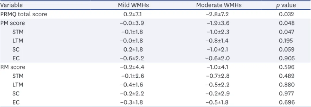
tended to be older and have higher incidences of hypertension. Otherwise, between-group demographic differences were not significant, and there was no statistical difference in baseline PRMQ profiles. Mean changes in total PRMQ scores at 3 months also differed (mild, 0.2±7.1;
moderate, −2.8±7.2), those patients with moderate (vs. mild) degrees of WMHs showing greater decline in PRMQ scores (p=0.032). The same was true of PRMQ PM sub-scores. Patients with moderate (vs. mild) degrees of WMHs demonstrated faster PM decline. However, RM scores of the two groups were not significantly different (Table 2). When further stratified by type of WMHs (DWMHs vs. PVWMHs), PM scores in patients with moderate (vs. mild) degrees of DWMHs showed greater decline. There were no differences in PM among groups stratified by degree of PVWMHs (mild, moderate, or severe) (Table 3), and changes of PRMQ scores in patient groups with mild or moderate degrees of WMHs did not differ significantly in terms of AChEI use or APOE Ɛ4 carrier status (Supplementary Tables 2 and 3).
DISCUSSION
The aim of this study was to document changes in PRMQ-assessed PM displayed by patients with aMCI 3 months after baseline testing. Our results subsequently indicated significant declines in total and PM scores generated by PRMQ at 3 months in patients with moderate
Table 2. Changes in PRMQ scores of patient subsets at 3 months (vs. baseline)Variable Mild WMHs Moderate WMHs p value
PRMQ total score 0.2±7.1 −2.8±7.2 0.032
PM score −0.0±3.9 −1.9±3.6 0.048
STM −0.1±1.8 −1.0±2.3 0.047
LTM −0.0±1.8 −0.8±1.4 0.195
SC 0.2±1.8 −1.0±2.1 0.059
EC −0.6±2.2 −0.6±2.0 0.905
RM score −0.2±4.4 −1.0±4.1 0.596
STM −0.1±2.6 −0.7±2.8 0.489
LTM −0.4±1.6 −0.5±2.2 0.880
SC −0.2±2.2 −0.2±2.9 0.977
EC −0.3±1.8 −0.5±1.8 0.696
All values expressed as mean±standard deviation of each score-based slope, reflecting differences between baseline and follow-up data.
PRMQ: Prospective and Retrospective Memory Questionnaire, WMHs: white matter hyperintensities, PRMQ:
Prospective and Retrospective Memory Questionnaire, PM: prospective memory, STM: short-term memory, LTM:
long-term memory, SC: self-cued, EC: environmentally-cued, RM: retrospective memory.
Table 3. Changes in PRMQ scores at 3 months (vs. baseline) by type and degree of WMHs
Variable DWMHs PVWMHs
Mild Moderate p value Mild Moderate Severe p value
PRMQ total 0.2±7.0 −3.9±9.0 0.018 −0.4±7.8 −1.4±6.2 1.0±4.0 0.807
PM score −0.1±3.8 −2.6±4.5 0.033 −0.5±4.2 −0.1±3.4 0.1±1.9 0.649
STM −0.1±1.8 −1.6±2.8 0.041 −0.4±2.0 0.0±2.0 0.3±0.8 0.336
LTM −0.1±1.8 −0.9±1.7 0.198 −0.3±2.0 0.0±1.3 0.1±0.7 0.518
SC 0.2±1.8 −1.6±2.4 0.078 −0.0±2.0 0.0±2.3 0.1±0.7 0.817
EC −0.5±2.1 −1.0±2.5 0.666 −0.7±2.3 −0.1±1.8 −0.3±1.3 0.481
RM score −0.2±4.4 −1.4±5.0 0.524 −0.3±4.6 −1.3±4.4 0.7±2.7 0.782
STM −0.1±2.5 −1.1±3.5 0.438 −0.1±2.6 −1.0±3.4 0.0±1.5 0.794
LTM −0.4±1.6 −0.4±2.7 0.772 −0.4±1.7 −0.3±1.9 −0.1±1.5 0.678
SC −0.2±2.2 −0.7±3.5 0.865 −0.1±2.3 −1.0±3.2 0.3±0.8 0.974
EC −0.3±1.8 −0.6±1.9 0.459 −0.5±1.9 −0.3±1.3 0.9±0.9 0.076
All values expressed as mean±standard deviation of each score-based slope, reflecting differences between baseline and follow-up data.
PRMQ: Prospective and Retrospective Memory Questionnaire, WMHs: white matter hyperintensities, DWMHs: deep white matter hyperintensities, PVWMHs:
periventricular white matter hyperintensities, PM: prospective memory, STM: short-term memory, LTM: long-term memory, SC: self-cued, EC: environmentally- cued, RM: retrospective memory.
(vs. mild) degrees of WMHs. Similarly, we confirmed that WMHs (especially DWMHs) positively correlated with PM decline, exerting significant negative impact.
As in the present study, previous investigations of WMHs in this setting have regularly demonstrated their negative effects on cognition.
50-52WMHs serve to disrupt neuronal connectivity, impairing cognitive function, and PM loss is sustained in the same manner.
However, perhaps due to inherent heterogeneity, specific ramifications of PVWMHs and DWMHs have yet to be clarified. The impact of WMHs is known to vary in accord with size, location, and extent of lesions. Some researchers have linked heightened loads of PVWMHs with reduced cognitive function in normal aging adults and in patients with MCI or AD
47,53-55; and longitudinal studies of patients with cognitive dysfunction have suggested a predilection for periventricular WM lesions.
52These findings seem plausible, given that periventricular fiber connections convey long-association, interhemispheric, and long-projection fibers, whereas DWMHs convey relatively short-association fibers involved in specific brain regions.
56Unlike the above studies, other efforts have shown a correlation between DWMHs and cognitive functions,
57-60thus supporting our contention that DWMHs (rather than PVWMHs) are preferentially involved in declining PM. It may be that PVWMHs have diffuse consequences, whereas effects of DWMHs are largely confined to frontal-subcortical circuits.
In addition, PM requires higher-level function and is rooted not only in frontal lobe, but also in the temporal lobe, which is chiefly responsible for memory. In contrast to RM, PM has greater bearing on various frontal lobe functions. Thus, by disrupting frontal-subcortical circuits, WMHs (especially extensive DWMHs) promote frontal dysfunction and more rapid progression of PM. As we have shown, the extent of DWMHs seems to correspond with the rapidity of PM decline. Yet, rather than hastily assign more importance to DWMHs or PVWMHs, it is reasonable to conclude that the burden of WMHs is clearly and consistently implicated in loss of PM (rather than RM) experienced by patients with aMCI. Contrary to our expectations, the present analysis yielded no group-wise differences with AChEI use.
However, the 3-month time interval stipulated may not have ensured full drug efficacy, and not everyone was prescribed an AChEI, so this comparison may be premature.
The present study has several acknowledged limitations. First, this was a small, hospital- driven study, not a population-based investigation, creating a potential for bias in patient selection and limiting any generalization of our results. Also, we did not collect detailed information on lacunes, again possibly skewing our results. Furthermore, we did not include patients with severe degrees of DWMHs, and the follow-up period was relatively brief. The overall credibility of our findings could be bolstered by addressing such deficiencies.
In summary, WMHs affect PM, and WMHs are involved in greater PM decline, especially if DWMHs are extensive. Therefore, it is important to pay greater attention to the change of PM in aMCI patients with WMHs.
SUPPLEMENTARY MATERIALS
Supplementary Table 1
Korean version Prospective Retrospective Memory Questionnaire
Click here to viewSupplementary Table 2
Changed scores of PRMQ during 3 months according to AChEI use
Click here to viewSupplementary Table 3
Changed scores of PRMQ during 3 months according to APOE genotype
Click here to viewREFERENCES
1. Schacter DL, Addis DR. The cognitive neuroscience of constructive memory: remembering the past and imagining the future. Philos Trans R Soc Lond B Biol Sci 2007;362:773-786.
PUBMED | CROSSREF
2. Crystal JD, Wilson AG. Prospective memory: a comparative perspective. Behav Processes 2015;112:88-99.
PUBMED | CROSSREF
3. Kliegel M, Ballhausen N, Hering A, Ihle A, Schnitzspahn KM, Zuber S. Prospective memory in older adults: where we are now and what is next. Gerontology 2016;62:459-466.
PUBMED | CROSSREF
4. Dermody N, Hornberger M, Piguet O, Hodges JR, Irish M. Prospective memory impairments in Alzheimer's disease and behavioral variant frontotemporal dementia: clinical and neural correlates. J Alzheimers Dis 2016;50:425-441.
PUBMED | CROSSREF
5. Costa A, Fadda L, Perri R, Brisindi M, Lombardi MG, Caltagirone C, et al. Sensitivity of a time-based prospective memory procedure in the assessment of amnestic mild cognitive impairment. J Alzheimers Dis 2015;44:63-67.
PUBMED | CROSSREF
6. Rabin LA, Chi SY, Wang C, Fogel J, Kann SJ, Aronov A. Prospective memory on a novel clinical task in older adults with mild cognitive impairment and subjective cognitive decline. Neuropsychol Rehabil 2014;24:868-893.
PUBMED | CROSSREF
7. Delprado J, Kinsella G, Ong B, Pike K. Naturalistic measures of prospective memory in amnestic mild cognitive impairment. Psychol Aging 2013;28:322-332.
PUBMED | CROSSREF
8. Zhou T, Broster LS, Jiang Y, Bao F, Wang H, Li J. Deficits in retrospective and prospective components underlying prospective memory tasks in amnestic mild cognitive impairment. Behav Brain Funct 2012;8:39.
PUBMED | CROSSREF
9. Thompson CL, Henry JD, Withall A, Rendell PG, Brodaty H. A naturalistic study of prospective memory function in MCI and dementia. Br J Clin Psychol 2011;50:425-434.
PUBMED | CROSSREF
10. Costa A, Caltagirone C, Carlesimo GA. Prospective memory impairment in mild cognitive impairment: an analytical review. Neuropsychol Rev 2011;21:390-404.
PUBMED | CROSSREF
11. Okuda J, Fujii T, Ohtake H, Tsukiura T, Yamadori A, Frith CD, et al. Differential involvement of regions of rostral prefrontal cortex (Brodmann area 10) in time- and event-based prospective memory. Int J Psychophysiol 2007;64:233-246.
PUBMED | CROSSREF
12. Burgess PW, Scott SK, Frith CD. The role of the rostral frontal cortex (area 10) in prospective memory: a lateral versus medial dissociation. Neuropsychologia 2003;41:906-918.
PUBMED | CROSSREF
13. Volle E, Gonen-Yaacovi G, Costello AL, Gilbert SJ, Burgess PW. The role of rostral prefrontal cortex in prospective memory: a voxel-based lesion study. Neuropsychologia 2011;49:2185-2198.
PUBMED | CROSSREF
14. Burgess PW, Gonen-Yaacovi G, Volle E. Functional neuroimaging studies of prospective memory: what have we learnt so far? Neuropsychologia 2011;49:2246-2257.
PUBMED | CROSSREF
15. Tosto G, Zimmerman ME, Carmichael OT, Brickman AM; Alzheimer's Disease Neuroimaging Initiative. Predicting aggressive decline in mild cognitive impairment: the importance of white matter hyperintensities. JAMA Neurol 2014;71:872-877.
PUBMED | CROSSREF
16. Tosto G, Zimmerman ME, Hamilton JL, Carmichael OT, Brickman AM; Alzheimer's Disease Neuroimaging Initiative. The effect of white matter hyperintensities on neurodegeneration in mild cognitive impairment. Alzheimers Dement 2015;11:1510-1519.
PUBMED | CROSSREF
17. Brickman AM, Provenzano FA, Muraskin J, Manly JJ, Blum S, Apa Z, et al. Regional white matter hyperintensity volume, not hippocampal atrophy, predicts incident Alzheimer disease in the community.
Arch Neurol 2012;69:1621-1627.
PUBMED | CROSSREF
18. Haight TJ, Landau SM, Carmichael O, Schwarz C, DeCarli C, Jagust WJ; Alzheimer's Disease
Neuroimaging Initiative. Dissociable effects of Alzheimer disease and white matter hyperintensities on brain metabolism. JAMA Neurol 2013;70:1039-1045.
PUBMED | CROSSREF
19. Au R, Massaro JM, Wolf PA, Young ME, Beiser A, Seshadri S, et al. Association of white matter hyperintensity volume with decreased cognitive functioning: the Framingham Heart Study. Arch Neurol 2006;63:246-250.
PUBMED | CROSSREF
20. Debette S, Markus HS. The clinical importance of white matter hyperintensities on brain magnetic resonance imaging: systematic review and meta-analysis. BMJ 2010;341:c3666.
PUBMED | CROSSREF
21. Murray ME, Senjem ML, Petersen RC, Hollman JH, Preboske GM, Weigand SD, et al. Functional impact of white matter hyperintensities in cognitively normal elderly subjects. Arch Neurol 2010;67:1379-1385.
PUBMED | CROSSREF
22. Poggesi A, Pantoni L, Inzitari D, Fazekas F, Ferro J, O'Brien J, et al. 2001–2011: a decade of the LADIS (Leukoaraiosis And DISability) study: what have we learned about white matter changes and small-vessel disease? Cerebrovasc Dis 2011;32:577-588.
PUBMED | CROSSREF
23. van der Flier WM, van Straaten EC, Barkhof F, Verdelho A, Madureira S, Pantoni L, et al. Small vessel disease and general cognitive function in nondisabled elderly: the LADIS study. Stroke 2005;36:2116-2120.
PUBMED | CROSSREF
24. Baezner H, Blahak C, Poggesi A, Pantoni L, Inzitari D, Chabriat H, et al. Association of gait and balance disorders with age-related white matter changes: the LADIS study. Neurology 2008;70:935-942.
PUBMED | CROSSREF
25. Moscufo N, Guttmann CR, Meier D, Csapo I, Hildenbrand PG, Healy BC, et al. Brain regional lesion burden and impaired mobility in the elderly. Neurobiol Aging 2011;32:646-654.
PUBMED | CROSSREF
26. Poggesi A, Pracucci G, Chabriat H, Erkinjuntti T, Fazekas F, Verdelho A, et al. Urinary complaints in nondisabled elderly people with age-related white matter changes: the Leukoaraiosis And DISability (LADIS) study. J Am Geriatr Soc 2008;56:1638-1643.
PUBMED | CROSSREF
27. Wakefield DB, Moscufo N, Guttmann CR, Kuchel GA, Kaplan RF, Pearlson G, et al. White matter hyperintensities predict functional decline in voiding, mobility, and cognition in older adults. J Am Geriatr Soc 2010;58:275-281.
PUBMED | CROSSREF
28. Inzitari D, Pracucci G, Poggesi A, Carlucci G, Barkhof F, Chabriat H, et al. Changes in white matter as determinant of global functional decline in older independent outpatients: three year follow-up of LADIS (leukoaraiosis and disability) study cohort. BMJ 2009;339:b2477.
PUBMED | CROSSREF
29. Inzitari D, Simoni M, Pracucci G, Poggesi A, Basile AM, Chabriat H, et al. Risk of rapid global functional decline in elderly patients with severe cerebral age-related white matter changes: the LADIS study. Arch Intern Med 2007;167:81-88.
PUBMED | CROSSREF
30. Moon SY, Na DL, Seo SW, Lee JY, Ku BD, Kim SY, et al. Impact of white matter changes on activities of daily living in mild to moderate dementia. Eur Neurol 2011;65:223-230.
PUBMED | CROSSREF
31. Pantoni L, Poggesi A, Basile AM, Pracucci G, Barkhof F, Chabriat H, et al. Leukoaraiosis predicts hidden global functioning impairment in nondisabled older people: the LADIS (Leukoaraiosis and Disability in the Elderly) study. J Am Geriatr Soc 2006;54:1095-1101.
PUBMED | CROSSREF
32. Petersen RC, Stevens JC, Ganguli M, Tangalos EG, Cummings JL, DeKosky ST. Practice parameter: early detection of dementia: mild cognitive impairment (an evidence-based review). Report of the Quality Standards Subcommittee of the American Academy of Neurology. Neurology 2001;56:1133-1142.
PUBMED | CROSSREF
33. Winblad B, Palmer K, Kivipelto M, Jelic V, Fratiglioni L, Wahlund LO, et al. Mild cognitive impairment-- beyond controversies, towards a consensus: report of the International Working Group on Mild Cognitive Impairment. J Intern Med 2004;256:240-246.
PUBMED | CROSSREF
34. Morris JC. The Clinical Dementia Rating (CDR): current version and scoring rules. Neurology 1993;43:2412-2414.
PUBMED | CROSSREF
35. Kang Y, Na DL, Hahn S. A validity study on the Korean Mini-Mental State Examination (K-MMSE) in dementia patients. J Korean Neurol Assoc 1997;15:300-308.
36. Kang Y, Na DL. Seoul Neuropsychological Screening Battery (SNSB). Seoul: Human Brain Research &
Consulting Co., 2003.
37. Bae JN, Cho MJ. Development of the Korean version of the Geriatric Depression Scale and its short form among elderly psychiatric patients. J Psychosom Res 2004;57:297-305.
PUBMED | CROSSREF
38. Kang SJ, Choi SH, Lee BH, Kwon JC, Na DL, Han SH, et al. The reliability and validity of the Korean Instrumental Activities of Daily Living (K-IADL). J Korean Neurol Assoc 2002;20:8-14.
39. Ku HM, Kim JH, Kwon EJ, Kim SH, Lee HS, Ko HJ, et al. A study on the reliability and validity of Seoul- Instrumental Activities of Daily Living (S-IADL). J Korean Neuropsychiatr Assoc 2004;43:189-199.
40. Crawford JR, Henry JD, Ward AL, Blake J. The Prospective and Retrospective Memory Questionnaire (PRMQ):
latent structure, normative data and discrepancy analysis for proxy-ratings. Br J Clin Psychol 2006;45:83-104.
PUBMED | CROSSREF
41. Crawford JR, Smith G, Maylor EA, Della Sala S, Logie RH. The Prospective and Retrospective Memory Questionnaire (PRMQ): normative data and latent structure in a large non-clinical sample. Memory 2003;11:261-275.
PUBMED | CROSSREF
42. Pyun J, Kang Y, Park J, Kim YJ, Park K, Han IW. A comparison of the prospective memory among college students, normal elderly, and Parkinson's disease patients. Dement Neurocogn Disord 2012;11:95-103.
CROSSREF
43. Piauilino DC, Bueno OF, Tufik S, Bittencourt LR, Santos-Silva R, Hachul H, et al. The Prospective and Retrospective Memory Questionnaire: a population-based random sampling study. Memory 2010;18:413-426.
PUBMED | CROSSREF
44. Smith G, Della Sala S, Logie RH, Maylor EA. Prospective and retrospective memory in normal ageing and dementia: a questionnaire study. Memory 2000;8:311-321.
PUBMED | CROSSREF
45. Fazekas F, Chawluk JB, Alavi A, Hurtig HI, Zimmerman RA. MR signal abnormalities at 1.5 T in Alzheimer's dementia and normal aging. AJR Am J Roentgenol 1987;149:351-356.
PUBMED | CROSSREF
46. Scheltens P, Barkhof F, Leys D, Pruvo JP, Nauta JJ, Vermersch P, et al. A semiquantative rating scale for the assessment of signal hyperintensities on magnetic resonance imaging. J Neurol Sci 1993;114:7-12.
PUBMED | CROSSREF
47. Lee JY, Na DL, Kim SY, Cheong HK, Moon SY, Shim YS, et al. Different associations of periventricular and deep white matter lesions with cognition, neuropsychiatric symptoms, and daily activities in dementia. J Geriatr Psychiatry Neurol 2011;24:84-90.
PUBMED | CROSSREF
48. Ku BD, Na DL, Moon SY, Kim SY, Seo SW, Cheong HK, et al. Neuropsychological correlates of the proportional impact of white matter hyperintensities on mild to moderate dementia: the MRI 300 study.
Dement Geriatr Cogn Disord 2011;31:397-405.
PUBMED | CROSSREF
49. Shim YS, Youn YC, Na DL, Kim SY, Cheong HK, Moon SY, et al. Effects of medial temporal atrophy and white matter hyperintensities on the cognitive functions in patients with Alzheimer's disease. Eur Neurol 2011;66:75-82.
PUBMED | CROSSREF
50. Habes M, Erus G, Toledo JB, Zhang T, Bryan N, Launer LJ, et al. White matter hyperintensities and imaging patterns of brain ageing in the general population. Brain 2016;139:1164-1179.
PUBMED | CROSSREF
51. Tomimoto H. White matter integrity and cognitive dysfunction: radiological and neuropsychological correlations. Geriatr Gerontol Int 2015;15 Suppl 1:3-9.
PUBMED | CROSSREF
52. Bolandzadeh N, Davis JC, Tam R, Handy TC, Liu-Ambrose T. The association between cognitive function and white matter lesion location in older adults: a systematic review. BMC Neurol 2012;12:126.
PUBMED | CROSSREF
53. Kim JH, Hwang KJ, Kim JH, Lee YH, Rhee HY, Park KC. Regional white matter hyperintensities in normal aging, single domain amnestic mild cognitive impairment, and mild Alzheimer's disease. J Clin Neurosci 2011;18:1101-1106.
PUBMED | CROSSREF
54. Luo X, Jiaerken Y, Huang P, Xu XJ, Qiu T, Jia Y, et al. Alteration of regional homogeneity and white matter hyperintensities in amnestic mild cognitive impairment subtypes are related to cognition and CSF biomarkers. Brain Imaging Behav 2018;12:188-200.
PUBMED | CROSSREF
55. Dhamoon MS, Cheung YK, Bagci A, Alperin N, Sacco RL, Elkind MS, et al. Periventricular white matter hyperintensities and functional decline. J Am Geriatr Soc 2018;66:113-119.
PUBMED | CROSSREF
56. de Groot JC, de Leeuw FE, Oudkerk M, van Gijn J, Hofman A, Jolles J, et al. Cerebral white matter lesions and cognitive function: the Rotterdam Scan Study. Ann Neurol 2000;47:145-151.
PUBMED | CROSSREF
57. Clark AL, Sorg SF, Schiehser DM, Luc N, Bondi MW, Sanderson M, et al. Deep white matter
hyperintensities affect verbal memory independent of PTSD symptoms in veterans with mild traumatic brain injury. Brain Inj 2016;30:864-871.
PUBMED | CROSSREF
58. Yamawaki M, Wada-Isoe K, Yamamoto M, Nakashita S, Uemura Y, Takahashi Y, et al. Association of cerebral white matter lesions with cognitive function and mood in Japanese elderly people: a population- based study. Brain Behav 2015;5:e00315.
PUBMED | CROSSREF
59. Soriano-Raya JJ, Miralbell J, López-Cancio E, Bargalló N, Arenillas JF, Barrios M, et al. Deep versus periventricular white matter lesions and cognitive function in a community sample of middle-aged participants. J Int Neuropsychol Soc 2012;18:874-885.
PUBMED | CROSSREF
60. Silbert LC, Nelson C, Howieson DB, Moore MM, Kaye JA. Impact of white matter hyperintensity volume progression on rate of cognitive and motor decline. Neurology 2008;71:108-113.
PUBMED | CROSSREF