Received on November 6, 2012. Revised on November 22, 2012. Accepted on November 27, 2012.
CC This is an open access article distributed under the terms of the Creative Commons Attribution Non-Commercial License (http://creativecommons.org/licenses/by-nc/3.0) which permits unrestricted non-commercial use, distribu- tion, and reproduction in any medium, provided the original work is properly cited.
*Corresponding Author. Hyunah Lee, Office of Biomedical Professors, Samsung Medical Center, Sungkyunkwan University School of Medicine, Seoul, Korea. Tel: 82-2-3410-3455; Fax: 82-2-2148-7379; E-mail: [email protected]
Keywords: Dendritic cell, Lung cancer, Anti-tumor immunity
Abbreviations: DC, dendritic cell; MoDC, monocyte-derived DC; SDC, stem cell-derived DC; LLC, Lewis Lung Carcinoma cells
Dendritic Cell (DC) Vaccine in Mouse Lung Cancer Minimal Residual Model; Comparison of Monocyte-derived DC vs.
Hematopoietic Stem Cell Derived-DC
Soyoung Baek1, Seog Jae Lee2, Myoung Joo Kim1 and Hyunah Lee1*
1Office of Biomedical Professors, Samsung Medical Center, Sungkyunkwan University School of Medicine, Seoul 135-710, 2Department of Thoracic & Cardiovascular Surgery, Jeju National University School of Medicine, Jeju 690-767, Korea
The anti-tumor effect of monocyte-derived DC (MoDC) vac- cine was studied in lung cancer model with feasible but weak Ag-specific immune response and incomplete blocking of tu- mor growth. To overcome this limitation, the hematopoietic stem cell-derived DC (SDC) was cultured and the anti-tumor effect of MoDC & SDC was compared in mouse lung cancer minimal residual model (MRD). Therapeutic DCs were cul- tured from either CD34+ hematopoietic stem cells with GM-CSF, SCF and IL-4 for 14 days (SDC) or monocytes with GM-CSF and IL-4 for 7 days (MoDC). DCs were injected twice by one week interval into the peritoneum of mice that are inoculated with Lewis Lung Carcinoma cells (LLC) one day before the DC injection. Anti-tumor responses and the immune modulation were observed 3 weeks after the final DC injection. CD11c expression, IL-12 and TGF-β secretion were higher in SDC but CCR7 expression, IFN-γ and IL-10 secre- tion were higher in MoDC. The proportion of CD11c+CD8a+ cells was similar in both DC cultures. Although both DC re- duced the tumor burden, histological anti-tumor effect and the frequencies of IFN-γ secreting CD8+ T cells were high- er in SDC treated group than in MoDC. Conclusively, al- though both MoDC and SDC can induce the anti-tumor im- munity, SDC may be better module as anti-tumor vaccine than MoDC in mouse lung cancer.
[Immune Network 2012;12(6):269-276]
INTRODUCTION
The development of anti-tumor immune therapies through the activation of antigen-specific immune function has the advant- age that the side effects can be significantly reduced and the treatment effect of personalized therapy is excellent. Among these, dendritic cells (DCs) have been studied as anti-tumor vaccine since the late 1990’s (1). DCs are the most potent antigen-presenting cells (APCs) and are equipped with the necessary co-stimulatory, adhesion and histocompatibility molecules for initiation of a primary immune response (2-4).
DC-based vaccination trials in human solid cancers have aimed to activate the immune system to recognize and de- stroy tumor cells. The safety and the feasibility of anti-cancer DC vaccines have been confirmed in several clinical trials (5-9). Nevertheless, reports on DC therapy for lung cancer patients were limited (10-12).
Lung cancer is a leading cause of cancer-related death in both men and women worldwide (13). Although various ther- apeutic modalities, including surgical resection, radiation ther- apy, and chemotherapy, have been developed, the disease is rarely curable and the prognosis is dismal, with an overall 5-year survival rate of only 15% (14). Therefore the develop- ment of effective therapies is clearly needed for treatment of lung cancer. In our previous study (15), we verified the possi-
bility of anti-tumor effect of cultured DCs as an adjuvant ther- apy for minimal residual disease state of lung cancer.
Although feasible anti-tumor effect of monocyte-derived DC (MoDC) vaccine was resulted in our previous study (15), antigen specific immune responses were weak with partial blocking of tumor growth. To improve the efficiency of an- ti-tumor effect using DC vaccine for lung cancer, we tested CD34+ hematopoietic stem cell derived DC (SDC) as an alter- native in mouse model in this study. SDC can be applied in many clinical researches because hematopoietic stem cells generate a large number of mature DCs in vitro and able to differentiate into various stages. Ward et al. (16) reported that CD34+ DC generated with GM-CSF, IL-4, Flt-3L and SCF, ca- pable of inducing naive T-cell responses and can be ex- panded to clinically useful numbers. There are reports that CD34+ DCs from core blood have anti-tumor activity (17-19).
Our SDC were generated from CD34+-mouse bone marrow cell and compared to MoDC for their DC characters and in vivo anti-tumor effect.
MATERIALS AND METHODS Animal
Specific pathogen-free female C57BL/6 mice, 5∼6 weeks old, were purchased from the Dae-Han BioLink (EumSung, Korea). The mice were provided with water and food, adlibi- tum and quarantined under 12 h light: 12 h dark photoperiod in the animal care facility of the Samsung Biomedical Resear- ch Institute, Samsung Medical Center, Seoul, Korea. Animal care was performed following the ILAR guideline. The mice were acclimated for at least one week before any experiments were conducted.
Reagents & cell lines
Complete medium (CM) consist of RPMI 1640 with 10% Fetal Bovine Serum, 2 mM glutamine and 100 U/ml penicillin plus 100μg/ml streptomycin (all from GIBCO laboratories, Grand Island, NY, USA). Following antibodies for flow cytometric phenotyping were purchased from BD (SanDiego, CA); fluo- rescein isothiocyanate (FITC)-or phycoerythrin (PE)-labeled monoclonal Abs for MHC I/II, CD11c, CD8a, CD54 and CCR7.
ELISA sets for cytokines including IL-10, IL-12, TGF-β and IFN-γ was purchased from BD (Sunnyvale, CA). For isolation of hematopoietic stem cells, a lineage cell depletion kit was obtained from Miltenyi Biotech Inc (Bergisch Gladbach, Germany). C57BL/6 syngeneic Lewis Lung Carcinoma (LLC)
cell line was purchased from American type culture collection (ATCC) (Rockville, MD, USA). The following hybridoma cell lines were also purchased from ATCC; GK1.5 for anti-L3T4, 53.672 for anti-Lyt-2, RA3 for anti-B220, and J11d for anti-B cells/neutrophils.
Ex vivo generation of DCs
Mononuclear cells (MNCs) from bone marrow were obtained from the tibia and femur of C57BL/6 mouse sacrificed by cer- vical dislocation. The viability of red blood cell (RBC) free-MNCs was routinely demonstrated to be over 95% by try- pan blue exclusion. To separate the myeloid lineage cells, RBC free-MNCs were treated with antibodies against L3T4, Lyt-2, B220, and B cell/neutrophil with complement. Purified cells (1×10⁶/ml) were incubated with GM-CSF and IL-4 (1×10³ units/ml each) at 37oC for 7 d in a humidified CO2 incubator for MoDC differentiation. To generate SDCs, CD34+ cells were isolated from the bone marrow cells by MACS line- age cell depletion kit and cultured with GM-CSF, SCF and IL-4 for 14 days. Eighteen hours before cell harvest, tumor cell lysate (50μg protein/ml) was added to culture media. Tumor cell lysate was prepared from cultured LLC cells by a freez- ing-thawing process that was repeated six times in liquid ni- trogen (−180oC) and an incubator (37oC). Protein quantifica- tion of the tumor cell lysate was performed using centrifuged (1,500 rpm, 15 min) supernatant and the Bradford method (Bio-Rad Protein Assay; Bio-Rad Laboratories, Hercules, CA, USA). Cultured cells were characterized as therapeutic DCs by surface marker phenotyping and cytokine secretion into the culture supernatant.
Tumor inoculation and injection of therapeutic-DCs Syngeneic LLC cells were inoculated intravenously into C57BL/6 mice on day 0. Starting from the day after the tumor cell injection, antigen pulsed therapeutic-DCs were injected twice by one week interval into the peritoneum of mice. This way the minimal residual disease status was simulated by having the tumor cells in the blood without established burdens. Each experimental group was composed of 5∼7 mice and the control group was treated with saline. Animals were sacrificed at three weeks after the final DC injection to observe tumor growth in the lung and to analyze the systemic immune modulation. The treatment schedule is summarized in Fig. 1. Pulmonary tumor burden and IFN-γ secreting CD8+ T cell proportion (ELISPOT) were detected from the spleno- cytes of mice in each group.
Figure 1. Treatment schedule. D-0: LLC cells were inoculated 1×10⁵/mouse i.v. D-1 and 8: In order to minimal residual model, therapeutic-DCs were injected twice by one week interval into the peritoneum of tumor cell inoculated-mice. D-29: Mouse sacrifice and immune monitoring. Each experimental group was composed of 5∼7 mice.
Immunophenotypic analysis of cultured cells by flow cytometry
Phenotype of cultured cells was analyzed by direct immune- fluorescence staining of cell surface antigens using FITC or PE conjugated antibodies against MHC I/II, CD11c, CD8a, CD54 and CCR7. Single cells of cultured DC were incubated with fluorescence-labeled surface antibodies in PBS with 0.1%
sodium azide and 1% FBS (PBS-CS) for 40 min at 4oC. Within 1 h of the antibody labeling, cells in the 500μl PBS-CS were analyzed by flow cytometry (FACS Vantage; BD, CA, USA).
Measurement of cytokine secretion from DCs The concentrations of IL-10, IL-12, TGF-β and IFN-γ in cul- ture supernatant of DC were measured using commercial ELISA kits (BD, Sunnyvale, CA).
Mouse lung observation (In vivo anti-tumor effect of DC vaccine)
Three weeks after the last DC injection, tumor bearing mice were sacrificed. In order to investigate in vivo anti-tumor ef- fect of DC vaccine, lung with heart of the mice were eliminated. To quantify the effect on the tumor establishment and growth, tumor lesions bigger than 2 mm were counted under a microscope (×100). Then these lungs were paraffin embedded and stained with H&E to observe the histo-patho-
logical alterations. Histological observation was performed under the microscope (Leica DM 3000). The picture was pre- sented with ×100 magnification.
Analysis of IFN-γ producing cells using enzyme-linked immunospot (ELISPOT) assay
The ELISPOT assay was adopted to detect and enumerate in- dividual cells that secrete IFN-γ protein in vitro upon ex- posure to antigen. ELISPOT IFN-γ assay kit were purchased from AID (AID, Strassberg, Germany) and assays were per- formed according to the manufacturer’s instruction. In brief, using magnetic bead cell separation system (MACSTM Miltenyi Biotec, Germany), CD3+ T cells were purified from the sple- nocytes (2×10⁵ cells) and stimulated in vitro with tumor ly- sate on a 96-well pre-coated plate. The plate was incubated for 20 h at 37oC with 5% CO2. After washing, each well was added with detection antibody and was incubated for 2 h at room temperature. The plate was incubated with alkaline phosphatase conjugate and developed with BCIP/NBT sub- strate solution. Visible spots were enumerated using an auto- mated AID ELISPOT reader (AID, Strassberg, Germany) and the default program.
Statistical analysis
The analyzed values are reported as the mean value±stand- ard error (SE). Each experimental group was composed of 5∼7 mice, and the same experimental protocol was repeated at least two times with similar results. Statistical significance was determined by two-tailed Student’s t-test. p-values<0.05 were interpreted to represent statistically significant differen- ces.
RESULTS
Characterizations of generated DC
Generated therapeutic DCs were verified through cytokine se- cretion and phenotype expression. From the SDC supernatant, immune-stimulatory cytokine IL-12 was measured significantly higher level than MoDC (424.82±87.2 vs. 73.51±6.15 pg/ml for SDC vs. MoDC group, 574.38±191 vs. 66.06±2.38 pg/ml for SDC/lysate vs. MoDC/lysate group respectively) (Fig. 2).
On the other hand, in the MoDC supernatant, IFN-γ as an im- mune-stimulatory cytokine were determined significantly high- er level than SDC (399.79±105.59 vs. 2,601.31±680.13 pg/ml for SDC vs. MoDC group, 352.69±21.19 vs. 2,090.26±329.86 pg/ml for SDC/lysate vs. MoDC/lysate group respectively) (Fig.
(A) (B)
(C) (D)
Figure 2. Characterization of cultured DCs. DCs cultured from either CD34+ cells with GM-CSF, SCF and IL-4 for 14 days (SDC) or antibody- panned monocytes with GM-CSF and IL-4 for 7 days (MoDC). Each DC type was pulsed with LLC lysate (SDC/lysate and MoDC/lysate). Cytokines IL-12, IL-10, IFN-γ and TGF-β produced into the culture supernatant were measured by ELISA and expressed as pg/ml/1×106 cells. Statistical significance (p<0.05) was compared by asterisks between the SDC and MoDC. Statistical differences (p<0.05) between the lysate pulsed DCs were expressed by + signs.
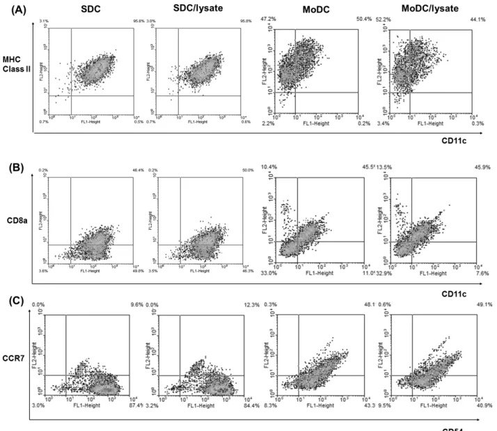
2). Although secretion of immune-inhibitory cytokine IL-10 or TGF-β was observed from each DC group (IL-10 in MoDC group vs. TGF-β in SDC group), there were amounts of im- mune-stimulatory cytokines which are considered as counter- balance. Over 90% MoDC & SDC were expressed MHC I/II molecules (Fig. 3A) (The data of MHC I does not shown).
However, a DC marker CD11c expression was prominent in SDC (Table I). The proportion of CD11c+CD8a+ cell, known as Th1-inducible DC subset was similar in two DC types (Table I) (Fig. 3B). Although the CD54 expression was similar in both DCs, CCR7 expression was prominent in MoDC (Table I) suggesting the better migratory function of MoDC (Fig. 3C). The characters of both SDC and MoDC revealed that both DCs could induce anti-tumor immunity with differ- ent mechanisms.
In vivo anti-tumor effect of DCs
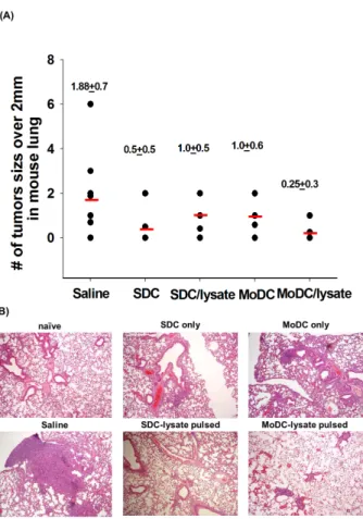
To quantify the effect on the tumor establishment and growth, we eliminated lung with heart of the mice and observed tu- mor lesions under a microscope. After the DC treatment in LLC injected mice, countable tumor burdens (over than 2 mm under a ×100 microscope) were decreased compared to sal- ine treated group (Fig. 4A). However, no statistical sig- nificance was found between the MoDC and SDC as well as with/without tumor lysate group. Despite the fact that statisti- cally significant differences through eye observation were not found, histological alterations were observed from the H&E stained lung sections (Fig. 4B). In DC treated group, a sig- nificant amount of tumor was reduced. Moreover, SDC treat- ed group pulsed with LLC lysate shows definite tumor regression. This H&E staining shows SDC/lysate induce more powerful anti-tumor immunity than MoDC/lysate in mouse lung cancer.
Figure 3. Immunogenic phenotypes of cultured DCs were analyzed by flow cytometry. Dendritic cell marker CD11c as well as MHC II, CD8a and CCR7 with CD54 were measured to characterize and differentiate the cultured SDC and MoDC.
Table I. Phenotypes of cultured DCs
Type of DC % of cell surface markers
MHC II CD11c CD8a CD11c/CD8a CCR7 CD54
SDC 98.9 96.3 46.6 46.4 9.6 97.0
SDC/lysate 98.8 96.4 50.2 50.0 12.3 96.7
MoDC 97.6 50.6 55.9 45.5 49.7 91.4
MoDC/lysate 96.3 44.4 59.4 45.9 49.7 90.0
Figure 4. Anti-tumor effect of DCs for LLC formed in lung. Tumor bearing mice were sacrificed after three weeks from the last DC injection. (A) Circle represents number of tumor size over 0.2 mm in a mouse lung. Number and red line show mean value±standard error (SE). Same value represents one circle. Each experimental group was composed of 5∼7 mice. (B) Pulmonary tissues of tumor-bearing mice were H&E stained.
Figure 5. Induction of tumor antigen-specific IFN-secreting cells.
Tumor antigen-specific alterations in immunological parameters were analyzed with splenic lymphocytes obtained from the tumor-bearing mice treated with DC vaccine. As an effector molecule of therapeutic response, the proportion of IFN-secreting CD8+ T cells was observed by ELISPOT assay. Asterisk indicate the statistical significance (p
<0.05) compared with MoDC.
Ag-specific T cell immune response
To confirm SDC/lysate induce more systemic anti-tumor im- munity than MoDC/lysate, we checked effector T cells in splenic lymphocytes from LLC bearing mice. To measure the anti-tumor effector T cells, ELISPOT assay counting IFN-γ se- creting CD8+ T cells was operated (Fig. 5). The frequency of IFN-γ secreting CD8+ T cells in SDC treated group was significantly higher than that in MoDC treated group (58.7±
8.3 vs. 11.7±1.8 spots for SDC/lysate vs. MoDC/lysate treat- ed group, respectively). It shows SDC/lysate induce naïve T cells to effector T cells which lead to regress residual tumor in mouse lung. The therapeutic responses were associated with induction of IFN-γ secreting CD8+ T cells.
DISCUSSION
In spite of the aggressive treatment with surgery, radiation, and chemotherapy, the long-term survival for patients with lung cancer still remains low. Even patients with early stage disease often succumb to lung cancer due to the development of metastasis, indicating the need for effective approaches for the systemic therapy of this condition (20). Based on the anti- gen specificity of the immune system and the safety profile of cancer vaccines, the effective immunotherapy would be an ideal adjuvant, following initial clinical responses to definitive therapy (21). Even though there have been numerous clinical trials for various types of cancer, there are few DC vaccine trials in patients with lung cancer, and many aspects related to the immunotherapy are unknown. Our previous study sug- gests the possible anti-tumor effect of cultured DCs as an ad- juvant therapy for minimal residual disease state of lung can- cer (15). Nevertheless, MoDC vaccine in previous study shows weak antigen-specific immune response and incomplete tu- mor regression. In order to increase the efficiency of anti-tu- mor response using DC therapy, we introduced other culture methods as alternatives. Hematopoietic stem cell (HSC) as an origin of DC culture has advantage that the culture yield can be increased by more than 30 to 60 times depending on the culture protocol and/or incubation period. Also the HSC has the potential to differentiate into the various type of cell.
Authors reported the HSC-derived DC vaccine effect on the renal cell carcinoma and breast cancer patients (7). No report for the effects of SDC on lung cancer treatment was found.
The characters of SDC cultured in this protocol allow expect-
ations that SDC may work to induce the anti-tumor immunity, since these cells produce significantly more IL-12 and lower IL-10 than MoDC (Fig. 2). Unlike SDC, cultured MoDC pro- duced significantly higher IFN-γ and lower TGF-β than SDC (Fig. 2) which also may work as anti-tumor effector by differ- ent mechanism. Indeed, the proportion of cells expressing CD11c and CD8a, an immunogenic DC subset inducing Th1 response in mouse (22,23), was similar in both DC culture (Fig. 3) with similar response to inhibiting tumor growth (Fig.
4A). One day after inoculation of LLC cells, therapeutic-DCs were injected twice by one week interval into the peritoneum of tumor cell injected mice. Thus the effect of DC could be analyzed as an adjuvant treated after removing the tumor burden. Although DC vaccines inhibit the tumor growth in the lung, there were no statistical significances between the effect of MoDC and SDC when we observed tumor burden over 2 mm size under the microscope (Fig. 4A). This phenom- enon was considered because we missed the presence of tumor under 2 mm size. For this reason, we observed lung tissue through H&E staining to determine histo-pathological changes (Fig. 4B). The entire DC treated group revealed the remark- able tumor regression. Affirmatively, no small tumorigenic changes other than countable tumors were observed in the tumor lysate- pulsed SDC treated lung. Also SDC significantly induced antigen-specific IFN-γ secreting CD8+ T cells than MoDC did (Fig. 5). Collectively, although both MoDC and SDC can induce the anti-tumor response, SDC considered as efficient inducer of anti-tumor immunity than MoDC in the mouse lung cancer.
ACKNOWLEDGEMENTS
This study was mainly supported by the grant from the Korean Ministry of Knowledge Economy (#10021360) and in part by Samsung Biomedical Research Institute grants (#SBRI C-A8-501-2).
CONFLICTS OF INTEREST
The authors declare that they have no competing interests.
REFERENCES
1. Steinman, R. M. 1991. The dendritic cell system and its role in immunogenicity. Annu. Rev. Immunol. 9: 271-296.
2. Banchereau, J. and R. M. Steinman. 1998. Dendritic cells and
the control of immunity. Nature 392: 245-252.
3. Paglia, P., C. Chiodoni, M. Rodolfo, and M. P. Colombo.
1996. Murine dendritic cells loaded in vitro with soluble pro- tein prime cytotoxic T lymphocytes against tumor antigen in vivo. J. Exp. Med. 183: 317-322.
4. Palucka, K. and J. Banchereau. 1999. Dendritic cells: a link between innate and adaptive immunity. J. Clin. Immunol. 19:
12-25.
5. Paczesny, S., J. Banchereau, K. M. Wittkowski, G. Saracino, J. Fay, and A. K. Palucka. 2004. Expansion of melanoma-spe- cific cytolytic CD8+ T cell precursors in patients with meta- static melanoma vaccinated with CD34+ progenitor-derived dendritic cells. J. Exp. Med. 199: 1503-1511.
6. Kim, J. H., Y. Lee, Y. S. Bae, W. S. Kim, K. Kim, H. Y.
Im, W. K. Kang, K. Park, H. Y. Choi, H. M. Lee, S. Y. Baek, H. Lee, H. Doh, B. M. Kim, C. Y. Kim, C. Jeon, and C. W.
Jung. 2007. Phase I/II study of immunotherapy using autolo- gous tumor lysate-pulsed dendritic cells in patients with meta- static renal cell carcinoma. Clin. Immunol. 125: 257-267.
7. Baek, S., C. S. Kim, S. B. Kim, Y. M. Kim, S. W. Kwon, Y. Kim, H. Kim, and H. Lee. 2011. Combination therapy of renal cell carcinoma or breast cancer patients with dendritic cell vaccine and IL-2: results from a phase I/II trial. J. Transl.
Med. 9: 178.
8. Ragde, H., W. A. Cavanagh, and B. A. Tjoa. 2004. Dendritic cell based vaccines: progress in immunotherapy studies for prostate cancer. J. Urol. 172: 2532-2538.
9. Iwashita, Y., K. Tahara, S. Goto, A. Sasaki, S. Kai, M. Seike, C. L. Chen, K. Kawano, and S. Kitano. 2003. A phase I study of autologous dendritic cell-based immunotherapy for pa- tients with unresectable primary liver cancer. Cancer Immu- nol. Immunother. 52: 155-161.
10. Hirschowitz, E. A., T. Foody, R. Kryscio, L. Dickson, J.
Sturgill, and J. Yannelli. 2004. Autologous dendritic cell vac- cines for non-small-cell lung cancer. J. Clin. Oncol. 22: 2808- 2815.
11. Hege, K. M. and D. P. Carbone. 2003. Lung cancer vaccines and gene therapy. Lung Cancer 41 Suppl 1: S103-113.
12. Fong, L., Y. Hou, A. Rivas, C. Benike, A. Yuen, G. A. Fisher, M. M. Davis, and E. G. Engleman. 2001. Altered peptide ligand vaccination with Flt3 ligand expanded dendritic cells for tumor immunotherapy. Proc. Natl. Acad. Sci. U.S.A. 98: 8809-8814.
13. Parkin, D. M., F. Bray, J. Ferlay, and P. Pisani. 2005. Global cancer statistics, 2002. CA Cancer J. Clin. 55: 74-108.
14. Spira, A. and D. S. Ettinger. 2004. Multidisciplinary manage- ment of lung cancer. N. Engl. J. Med. 350: 379-392.
15. Lee, S. J., M. J. Kim, S. H. In, S. Baek, and H. Lee. 2005.
Immunocell therapy for lung cancer: dendritic cell based ad- juvant therapy in mouse lung cancer model. Immune Netw.
5: 36-44.
16. Ward, K. A., L. A. Stewart, and A. P. Schwarer. 2006.
CD34+-derived CD11c+++ BDCA-1++ CD123++ DC:
expansion of a phenotypically undescribed myeloid DC1 pop- ulation for use in adoptive immunotherapy. Cytotherapy 8:
130-140.
17. Encabo, A., P. Solves, E. Mateu, P. Sepúlveda, F. Carbonell- Uberos, and M. D. Miñana. 2004. Selective generation of dif- ferent dendritic cell precursors from CD34+ cells by inter-
leukin-6 and interleukin-3. Stem Cells 22: 725-740.
18. Guo, G., S. Chen, J. Zhang, L. Luo, J. Yu, H. Dong, H. Xu, Z. Su, and L. Wu. 2005. Antitumor activity of a fusion of esophageal carcinoma cells with dendritic cells derived from cord blood. Vaccine 23: 5225-5230.
19. Xu, R. L., Y. Tang, P. L. Ogburn, K. Malinowski, S. Madaje- wicz, F. Santiago-Schwarz, and Q. Fan. 2004. Implication of delayed TNF-alpha exposure on dendritic cell maturation and expansion from cryopreserved cord blood CD34+ hema- topoietic progenitors. J. Immunol. Methods 293: 169-182.
20. Baleeiro, R. B., L. B. Anselmo, F. A. Soares, C. A. Pinto, O. Ramos, J. L. Gross, F. Haddad, R. N. Younes, M. Y.
Tomiyoshi, P. C. Bergami-Santos, and J. A. Barbuto. 2008.
High frequency of immature dendritic cells and altered in situ
production of interleukin-4 and tumor necrosis factor-alpha in lung cancer. Cancer Immunol. Immunother. 57: 1335-1345.
21. Hirschowitz, E. A., T. Foody, G. E. Hidalgo, and J. R.
Yannelli. 2007. Immunization of NSCLC patients with anti- gen-pulsed immature autologous dendritic cells. Lung Cancer 57: 365-372.
22. Moser, M. and K. M. Murphy. 2000. Dendritic cell regulation of TH1-TH2 development. Nat. Immunol. 1: 199-205.
23. Martín, P., G. M. del Hoyo, F. Anjuère, S. R. Ruiz, C. F.
Arias, A. R. Marín, and C. Ardavín. 2000. Concept of lym- phoid versus myeloid dendritic cell lineages revisited: both CD8alpha(−) and CD8alpha(+) dendritic cells are generated from CD4(low) lymphoid-committed precursors. Blood 96:
2511-2519.