Introduction
Inadequate bone volume due to alveolar bone resorption is often associated with pneumatization of the maxillary sinus, causing clinical problems for implant placement in the posterior maxilla.1 Successful implant placement in the posterior maxilla in such cases requires the bone height to be increased via alveolar bone augmentation,
which is often performed by maxillary sinus floor aug- mentation. Augmentation of the maxillary sinus floor using autogenous bone was first described by Boyne and James in 1980 for implant placement.2 It still remains a safe and predictable surgical procedure for augmenta- tion of the alveolar ridge in atrophic posterior maxillae.3 However, sufficient knowledge about sinus anatomy and its vascular supply, as well as a thorough preoperative as- sessment, are key factors in the success of this treatment modality.
The infraorbital artery and the posterior superior alveo- lar artery(PSAA) are the branches of the maxillary artery that provide blood supply to the lateral sinus walls and the sinus floor membrane.4 To avoid traumatizing these
Prevalence and location of the posterior superior alveolar artery using cone-beam computed tomography
Maryam Tehranchi1, Ferial Taleghani1,*, Shahriar Shahab2, Arash Nouri3
1Department of Periodontology, Faculty of Dentistry, Shahed University, Tehran, Iran
2Department of Oral and Maxillofacial Radiology, Faculty of Dentistry, Shahed University, Tehran, Iran
3Nouriʼs Dental Clinic, Tehran, Iran
ABSTRACT
Purpose: Insufficient knowledge of the anatomy of the maxillary sinuses prior to sinus graft surgery may lead to perioperative or postoperative complications. This study sought to characterize the position of the posterior superior alveolar artery(PSAA) within the maxillary sinuses using cone-beam computed tomography(CBCT).
Materials and Methods: A total of 300 patients with edentulous posterior maxillae, including 138 females and 162 males with an age range of 33-86 years, who presented to a radiology clinic between 2013 and 2015 were enrolled in this retrospective cross-sectional study. The distance from the inferior border of the PSAA to the alveolar crest according to the residual ridge classification by Lekholm and Zarb, the distance from the PSAA to the nasal septum and zygomatic arch, and the diameter and position of the PSAA were all assessed on patients’ CBCT scans. The data were analyzed using the Mann-Whitney test and the t-test.
Results: The PSAA was detected on the CBCT scans of 87% of the patients; it was located beneath the sinus membrane in 47% of cases and was intraosseous in 47% of cases. The diameter of the artery was between 1 and 2mm in most patients(72%). The mean diameter of the artery was 1.29±0.39mm, and the mean distances from the PSAA to the zygomatic arch, nasal septum, and alveolar crest were 22.59±4.89mm, 26.51±3.52mm, and 16.7±3.96mm, respectively.
Conclusion: The likelihood of detecting the PSAA on CBCT scans is high; its location is intraosseous or beneath the sinus membrane in most patients. Determining the exact location of the PSAA on CBCT scans preoperatively can help prevent it from being damaged during surgery.(Imaging Sci Dent 2017; 47: 39-44)
KEY WORDS: Maxillary Sinus; Artery; Cone-Beam Computed Tomography
Copyright ⓒ 2017 by Korean Academy of Oral and Maxillofacial Radiology
This is an Open Access article distributed under the terms of the Creative Commons Attribution Non-Commercial License(http://creativecommons.org/licenses/by-nc/3.0) which permits unrestricted non-commercial use, distribution, and reproduction in any medium, provided the original work is properly cited.
Imaging Science in Dentistry·pISSN 2233-7822 eISSN 2233-7830
*This study was partially supported by the Vice Chancellor for Research of Shahed University.
Received September 21, 2016; Revised November 28, 2016; Accepted December 13, 2016
*Correspondence to : Prof. Ferial Taleghani
Department of Periodontology, Faculty of Dentistry, Shahed University, Vesal Str.
Italia Str. No 39 PC 14177, Teheran, Iran
Tel) 98-912-1460485, Fax) 98-218-8967618, E-mail) [email protected]
arteries and subsequent perioperative bleeding, locating their exact position is imperative prior to sinus floor aug- mentation surgery.5 The use of a 3-dimensional imaging technique to visualize the anatomy of the maxillary sinus can be very helpful in this respect.6 Several factors can affect the selection of the radiographic technique for each patient. These factors include cost, availability, patient ra- diation dose, and case type.1
Cone-beam computed tomography(CBCT) was intro- duced to dentistry in the late 1990s. It is a digital imaging modality that provides accurate information about the mor- phology of bone and the location of anatomical landmarks such as the PSAA.7,8 A lower patient radiation dose is a major advantage of CBCT over computed tomography (CT).9
Several studies have assessed the anatomy of the max- illary sinuses using panoramic radiography and 3-dimen- sional radiographic modalities such as CT and CBCT.
However, considering the clinical significance of this topic, the lack of adequate information in this regard, and the controversial results of previous studies on the exact position of the PSAA in the Iranian population, this study sought to assess the prevalence and location of the PSAA and its relation to the alveolar ridge, nasal septum, and zygomatic arch.
Materials and Methods
This retrospective cross-sectional study was conducted in the Periodontology Department of Shahed University during 2014 and 2015. The study protocol was approved by the Ethics Committee of Shahed University.
The CBCT scans used in this study were obtained using a NewTom VG apparatus(QR srl, Verona, Italy), set at a tube voltage of 110kVp and a tube current of 13.11 to 20.18mA. In our study group, we performed 2 types of scans: one a with 0.3-mm voxel size for a larger field of view(160×110mm) and the other with a 0.24-mm voxel size for a smaller field of view(120×70mm). The axial thickness was either 0.24mm or 0.3mm(isotropic voxel reconstruction). The slice thickness of the multiplanar re- construction images was 1mm.
A total of 300 CBCT scans of 138 females and 162 males between 33 and 86 years of age, who had present- ed to the radiology clinic of our institution between 2013 and 2015, were randomly selected. The inclusion criteria were: 1) the availability of CBCT scans of the posterior maxilla with maxillary molar and premolar teeth missing in at least one quadrant, 2) visibility on the CBCT scans
of up to 2cm above the maxillary sinus roof, and 3) the absence of motion or scattering artifacts.
Images showing changes in the morphology of the si- nus walls due to trauma or pathological conditions were excluded from the study. The posterior superior alveolar canal along the posterolateral wall of the maxillary sinus was assessed on the coronal sections using NNT Worksta- tion version 4.5 software(QR srl, Verona, Italy) and the data were recorded in charts for each patient.
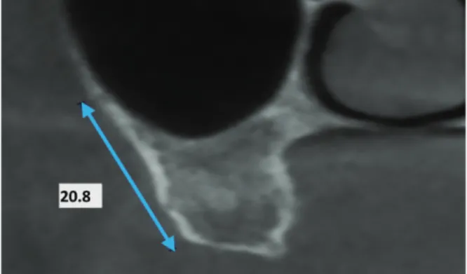
The PSAA was assessed in terms of the following fac- tors: 1) Distance from the inferior border of PSAA to the alveolar crest: the closest distance to the edentulous ridge was measured(Fig. 1). 2) Diameter of the PSAA: since the artery was visible in more than one area and consid- ering the importance of the diameter of the artery in the occurrence of perioperative bleeding, the greatest diam- eter of the PSAA was measured. According to the PSAA diameter, 3 groups were defined: <1mm, 1-2mm, and
>2mm. 3) Position of the PSAA: according to a study
Fig. 1. Distance from the lower border of the artery to the alveolar crest.
Fig. 2. Coronal view of the maxillary sinus, showing the intraosse- ous artery.
by Ilgüy et al. in 2013,6 the PSAA was divided into 3 cat- egories in terms of its position: (1) intraosseous(Fig. 2), (2) beneath the membrane(Fig. 3), and (3) over the exter- nal cortex of the lateral sinus wall(Fig. 4). The software used in the current study had high accuracy and enabled the classification of PSAA into the abovementioned 3 groups. 4) Distance from the PSAA to the zygomatic arch (Fig. 5): considering the extent of the zygomatic arch, the most anterior and inferior point of the zygomatic arch
was marked first, and then the closest distance from this point to the PSAA was measured using the 3-dimension- al distance measurement feature of the CBCT software.
5) Distance from the PSAA to the nasal septum(Fig. 5):
the closest distance from the PSAA to the nasal septum was measured. Considering the variable diameter of the nasal septum in different areas, its thickness at the center was used as a reference. 6) Determination of the type of residual alveolar ridge: the type of edentulous ridge was classified according to the classification by Lekholm and Zarb in 198510 based on the amount of resorption.
The CBCT scans were evaluated by a dentist trained by an oral and maxillofacial radiologist for this purpose. To determine the reliability and reproducibility of the find- ings(intrarater agreement), the CBCT scans were evaluat- ed again by the same observer 2 weeks later.
The data were analyzed using SPSS version 22.0(IBM Corp., Armonk, NY, USA). Descriptive statistics(mean, variance, standard deviation, and range) were calculat- ed for the variables. The t-test was used to compare the mean values of quantitative variables between males and females and also for the comparison of relative measure- ments. The non-parametric Mann-Whitney test was appli-
Fig. 4. Coronal view of the maxillary sinus, showing the artery(ar-
row), which runs over the external cortex of the lateral sinus wall. Fig. 5. Distance from the artery to the zygomatic arch(A) and to the nasal septum(B).
Table 1. Position of the posterior superior alveolar artery based on sex and type of edentulism
Not detectable Intraosseous Beneath the membrane On the external cortex of
the lateral sinus wall P value Males
Females 18(12.5%)
21(13.5%) 69(47.9%)
54(34.7%) 48(33.3%)
75(48.0%) 9(6.2%)
6(3.8%) <0.05
Complete edentulism
Partial edentulism 27(25.7%)
12(6.1%) 30(28.6%)
93(47.7%) 48(45.7%)
75(38.5%) 0(0%)
15(7.7%) <0.05
Fig. 3. Coronal view of the maxillary sinus, showing the artery (arrow), which is below the membrane.
ed to compare the mean values of qualitative variables between the 2 groups. The level of significance was set at P<.05.
Results
The mean age of the patients was 62.4±10.6 years.
The PSAA was detected in the CBCT scans of 87% of the patients; it was located beneath the membrane in 47% of patients, while it was intraosseous in 47% of patients. The PSAA was located on the external cortex of the lateral si- nus wall in 6% of the patients.
The diameter of PSAA was 1-2mm in most patients (74.8%), >2mm in 4.5%, and <1mm in 20.7%. The results of the parametric t-test revealed a significant dif- ference in the diameter of the PSAA between males and females(P<.05). The mean diameter of the PSAA was 1.38±0.35mm in males and 1.2±0.37mm in females.
The results of the non-parametric Mann-Whitney test showed a significant difference in the position of the PSAA between males and females(P<.05). The position of the PSAA was intraosseous in 47.9% of males, while it was beneath the membrane in 48% of females(Table 1).
A significant difference was noted in the prevalence of the PSAA between patients with complete and partial edentulism, and the likelihood of not detecting the PSAA on the CBCT scans of patients with complete edentulism was higher(25.7%) than for patients with partial edentu- lism(6.1%); this difference was statistically significant
(P<.05, Table 1).
The mean distance from the PSAA to the zygomatic arch was 25.59±4.89mm, and the difference in this respect between males and females was not significant(P>.05).
The mean distance from the inferior border of PSAA to the alveolar crest was 16.7±3.96mm, and the difference in this respect between males and females was statistical- ly significant(P<.05).
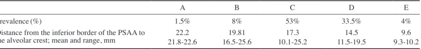
The mean distance from the PSAA to the nasal septum was 26.51±3.52mm, and a significant difference existed in this respect between males and females(P<.05, Table 2). According to Lekholm and Zarb’s classification, most patients had type C ridges(53%), followed by type D (33.5%). Based on the type of residual ridge, the distance from the inferior border of the PSAA to the alveolar crest was measured, and it was found that smaller edentulous ridges were associated with lower values of this distance (Table 3).
Discussion
Maxillary sinus floor augmentation is a highly predic- table method for the successful placement of dental im- plants in atrophic posterior maxillae.3 Knowledge of the anatomy of the region is especially important for the success of this treatment modality. In the current study, we assessed the prevalence and position of PSAA using CBCT scans. The PSAA was detected on the CBCT scans of 87% of the patients, which is similar to the preva- lence reported by Ilgüy et al.(89.3%).6 The prevalence of PSAA in the current study was higher than that reported by Elian et al.(52.9%)11 and Mardinger et al.(55%)12; this difference may be due to differences in the methodology of these studies and the sample size and definitions of the PSAA on images, because Solar et al.4 and Rosano et al.13 demonstrated that an endosseous anastomosis of the PSAA and infraorbital artery was present in 100% of ca- daveric specimens. This finding indicates that not detect- ing the PSAA on CBCT or CT scans does not necessar- ily mean that it is absent. It may not be visible due to its
Table 2. Descriptive statistics related to the PSAA based on sex (mean±standard deviation)
Females Males
Distance from the PSAA to the
zygomatic arch, mm 25.54±4.01 25.63±5.69
Distance from the inferior border
of PSAA to the alveolar crest, mm 15.94±4.06 17.50±3.69*
Distance from the PSAA to the
nasal septum, mm 25.92±3.71 27.40±3.20*
*: P<.05
PSAA, posterior superior alveolar artery.
Table 3. Assessment of the type of residual ridge according to the classification of Lekholm and Zarb10
A B C D E
Prevalence(%) 1.5% 8% 53% 33.5% 4%
Distance from the inferior border of the PSAA to
the alveolar crest; mean and range, mm 22.2
21.8-22.6 19.81
16.5-25.6 17.3
10.1-25.2 14.5
11.5-19.5 9.6
9.3-10.2 PSAA: posterior superior alveolar artery.
small diameter.12
In the current study, the PSAA was present beneath the membrane as often as it had an intraosseous location (47%), but in a small percentage of patients, the PSAA was detected on the external cortex of the lateral sinus wall(6%). The frequency of the presence of the PSAA on the external cortex was 5.7% in a study by Güncü et al.,14 and 5.2% in a study by Ilgüy et al.6; these values are close to our obtained value.
No significant difference between males and females in the prevalence of PSAA was found in our study, which is in agreement with the results of Ilgüy et al.6 However, Kim et al.15 reported a higher prevalence in males(64%). The difference between their results and ours may be due to differences in the male-to-female ratio.
In the current study, the position of the PSAA signifi- cantly varied in males and females. The same finding was reported by Kim et al.16 However, this difference was not significant in the study by Ilgüy et al.6 This discrepancy may likewise be attributed to differences in the male-to- female ratio.
In our study, the PSAA had a moderate diameter(1-2 mm) in most patients(72%); this finding accords with the results of Güncü et al.(1.3mm)14 and Ella et al.(1.2 mm).5 In the study by Ilgüy et al.,6 the diameter of the PSAA was <1 mm in most cases(68.9%). In the study by Mardinger et al.,12 the number of patients with a PSAA diameter <1mm and between 1 and 2mm was the same.
This variability in the results may be attributed to racial differences in the study populations. In the current study, the diameter of the PSAA in males was greater than in fe- males(P<.05), which is in accordance with the results of Güncü et al.14
Our study showed that the PSAA was not detected in a higher percentage of completely edentulous patients than partially edentulous subjects(25.7% versus 6.1%). Monje et al.17 concluded that the mean lateral wall thickness was smaller in fully edentulous patients than in partially eden- tulous subjects. Sinus pneumatization and a reduction in lateral sinus wall thickness often occur in patients with complete edentulism. Thus, it is assumed that the impres- sion of the PSAA canal on the lateral sinus wall is less visible in these patients due to the reduced thickness of the sinus wall.
To determine the position of the PSAA and its distance from the zygomatic arch, the distance from the inferior border of the PSAA to the alveolar crest and the distance from the PSAA to the nasal septum were measured. The
mean distance from the PSAA to the zygomatic arch was 25.59±4.89mm; the difference in this respect between males and females was not significant. The mean distance from the PSAA to the nasal septum was 26.51±3.52mm;
the difference in this respect between males and females was statistically significant. To the best of our knowledge, no previous study has evaluated the distance from the PSAA to the zygomatic arch or to the nasal septum. By using these anatomical landmarks and measurements, the approximate location of the PSAA can be estimated pri- or to surgery when CBCT may not be available. Conse- quently, damage to the PSAA and subsequent complica- tions can be prevented.
The mean distance from the inferior border of the PSAA to the alveolar crest was 16.7±3.96mm, which is close to the values reported in other studies. This distance was re- ported to be 17mm by Ilgüy et al.,6 18±4.9mm by Güncü et al.,14 16.4mm by Elian et al.,11 16.9mm by Mardinger et al.,12 and 18.9mm by Kim et al.16 The variability in the results may be explained by differences in the vertical height of the alveolar ridge among individuals.
During the preparation of the lateral window in the lat- eral sinus wall, there is a high risk of injury to the PSAA.
The height of the residual alveolar ridge is an important factor determining the approximate position of the PSAA.
In groups A and B according to the classification pro- posed by Lekholm and Zarb, the distance from the PSAA to the alveolar crest is more than 16mm. In groups C and D, the distance from the PSAA to the alveolar crest is more than 10mm. In group E, the ridge is severely atro- phic; thus, this distance is often less than 10mm. The results of Mardinger et al.12 are similar to our findings in this respect. With the reduction in size of the residual alveolar ridge from class A to E, the distance from the PSAA to the alveolar crest decreased as well. Thus, it is recommended that the superior border of the osteotomy in classes A to C should be placed 15-16mm above the alve- olar bone crest to prevent trauma to the PSAA. In ridges with more severe resorption, as in classes D and E, care must be taken not to damage the PSAA.
In conclusion, the likelihood of detecting the PSAA on CBCT scans is high. Its location is intraosseous or be- neath the sinus membrane in most patients, with a mean distance of 16.7±3.96mm to the alveolar crest. Thus, in surgical procedures on severely resorbed ridges, deter- mining the exact location of the PSAA on CBCT scans preoperatively can help prevent it from being damaged during surgery.
References
1. Newman MG, Takei HH, Klokkevold PR, Carranza FA. Car- ranza’s clinical periodontology. 12th ed. St. Louis, MO: Saun- ders; 2015.
2. Boyne PJ, James RA. Grafting of the maxillary sinus floor with autogenous marrow and bone. J Oral Surg 1980; 38: 613- 3. Pjetursson BE, Tan WC, Zwahlen M, Lang NP. A systematic 6.
review of the success of sinus floor elevation and survival of implants inserted in combination with sinus floor elevation. J Clin Periodontol 2008; 35(8Suppl): 216-40.
4. Solar P, Geyerhofer U, Traxler H, Windisch A, Ulm C, Watzek G. Blood supply to the maxillary sinus relevant to sinus floor elevation procedures. Clin Oral Implants Res 1999; 10: 34-44.
5. Ella B, Sedarat C, Noble Rda C, Normand E, Lauverjat Y, Siberchicot F, et al. Vascular connections of the lateral wall of the sinus: surgical effect in sinus augmentation. Int J Oral Maxillofac Implants 2008; 23: 1047-52.
6. Ilgüy D, Ilgüy M, Dolekoglu S, Fisekcioglu E. Evaluation of the posterior superior alveolar artery and the maxillary sinus with CBCT. Braz Oral Res 2013; 27: 431-7.
7. Arai Y, Tammisalo E, Iwai K, Hashimoto K, Shinoda K. De- velopment of a compact computed tomographic apparatus for dental use. Dentomaxillofac Radiol 1999; 28: 245-8.
8. Mozzo P, Procacci C, Tacconi A, Martini PT, Andreis IA. A new volumetric CT machine for dental imaging based on the cone-beam technique: preliminary results. Eur Radiol 1998; 8:
1558-64.
9. Ludlow JB, Ivanovic M. Comparative dosimetry of dental CBCT devices and 64-slice CT for oral and maxillofacial ra- diology. Oral Surg Oral Med Oral Pathol Oral Radiol Endod
2008; 106: 106-14.
10. Lekholm U, Zarb GA. Patient selection and preparation. In:
Brånemark PI, George AZ, Albrektsson T. Tissue-integrated prostheses: osseointegration in clinical dentistry. Chicago, IL:
Quintessence; 1985. p. 199-209.
11. Elian N, Wallace S, Cho SC, Jalbout ZN, Froum S. Distribu- tion of the maxillary artery as it relates to sinus floor augmen- tation. Int J Oral Maxillofac Implants 2005; 20: 784-7.
12. Mardinger O, Abba M, Hirshberg A, Schwartz-Arad D. Prev- alence, diameter and course of the maxillary intraosseous vas- cular canal with relation to sinus augmentation procedure: a radiographic study. Int J Oral Maxillofac Surg 2007; 36: 735- 13. Rosano G, Taschieri S, Gaudy JF, Del Fabbro M. Maxillary 8.
sinus vascularization: a cadaveric study. J Craniofac Surg 2009; 20: 940-3.
14. Güncü GN, Yildirim YD, Wang HL, Tözüm TF. Location of posterior superior alveolar artery and evaluation of maxillary sinus anatomy with computerized tomography: a clinical study.
Clin Oral Implants Res 2011; 22: 1164-7.
15. Kim JH, Ryu JS, Kim KD, Hwang SH, Moon HS. A radio- graphic study of the posterior superior alveolar artery. Implant Dent 2011; 20: 306-10.
16. Kim MJ, Jung UW, Kim CS, Kim KD, Choi SH, Kim CK, et al. Maxillary sinus septa: prevalence, height, location, and morphology. A reformatted computed tomography scan analy- sis. J Periodontol 2006; 77: 903-8.
17. Monje A, Catena A, Monje F, Gonzalez-Garcia R, Galin- do-Moreno P, Suarez F, et al. Maxillary sinus lateral wall thickness and morphologic patterns in the atrophic posterior maxilla. J Periodontol 2014; 85: 676-82.