ABSTRACT
Purpose: Papillary thyroid carcinoma (PTC) has a high rate of lateral neck node metastases, and completeness of surgical resection is an important determinant of outcomes. The
appropriate extent of therapeutic lateral neck dissection remains controversial. This study aims to access the impact of lateral neck node dissection of levels II to V in a large patient series.
Methods: A retrospective review of the clinical charts and hospital records of 778 consecutive patients who had metastatic PTC and who underwent unilateral cervical lymph node dissection at a single institution between 1999 January and 2009 December.
Results: A total of 489 modified radical neck dissection (MRND) (levels II–V) and 289 internal jugular node dissection (IJND) (levels II–IV) were performed in 778 patients. There were no differences in clinicopathological findings except for the retrieved and metastatic lateral lymph nodes (LLNs). The multiple-level lymph node (LN) metastasis was more prevalent in the MRND group, and the distribution of metastasis lymph node levels was significantly different in both group. The recurrence rate and disease-free survival were similar in the 2 groups. In multivariate analysis, female sex, tumor size, and multi-level LLN metastasis were independent predictors recurrence. Postoperative complications were similar in MRND and IJND group.
Conclusion: IJND achieves favorable postoperative results in PTC with lateral neck node metastasis patients, and level V metastasis/recurrence incidence is low. Therefore, the extent of lateral neck node dissection, whether IJND or MRND, can be considered for patients according to the simultaneous metastasis level and the tumor size.
Keywords: Thyroid carcinoma; Neck dissection; Recurrence
INTRODUCTION
The incidence of papillary thyroid carcinoma (PTC), the most common malignancy originating from thyroid gland, is increasing worldwide. In Korea, the annual incidence of thyroid cancer rose from 6.3 cases per 100,000 in 1999 to 51.1 cases per 100,000 in 2017, which represents a 22.4% annual increase over the last 10 years (1).
Original Article
Received: Sep 19, 2020 Revised: Sep 22, 2020 Accepted: Sep 22, 2020 Correspondence to Kee-Hyun Nam
Department of Surgery, Yonsei University College of Medicine, 50 Yonsei-ro, Seodaemun-gu, Seoul 03722, Korea.
E-mail: [email protected]
Copyright © 2020. Korean Association of Thyroid and Endocrine Surgeons; KATES This is an Open Access article distributed under the terms of the Creative Commons Attribution Non-Commercial License (https://
creativecommons.org/licenses/by-nc/4.0/).
ORCID iDs Cho Rok Lee
https://orcid.org/0000-0001-7848-3709 Jin Kyong Kim
https://orcid.org/0000-0001-5121-8462 Sang-Wook Kang
https://orcid.org/0000-0001-5355-833X Jandee Lee
https://orcid.org/0000-0003-4090-0049 Jong Ju Jeong
https://orcid.org/0000-0002-4155-6035 Kee-Hyun Nam
https://orcid.org/0000-0002-6852-1190 Woong Youn Chung
https://orcid.org/0000-0002-0291-8048 Conflicts of Interest
No potential conflict of interest relevant to this article was reported.
Cho Rok Lee , Jin Kyong Kim , Sang-Wook Kang , Jandee Lee , Jong Ju Jeong , Kee-Hyun Nam , Woong Youn Chung
Department of Surgery, Yonsei University College of Medicine, Seoul, Korea
Is the Internal Jugular Node Dissection
without Level V Sufficient in Patients
with Papillary Thyroid Carcinoma with
Lateral Neck Node Metastasis?
Authors Contributions
Conceptualization: Cho Rok Lee, Kee-Hyun Nam; Data curation: Jin Kyong Kim, Sang- Wook Kang, Jandee Lee, Jong Ju Jeong;
Formal analysis: Jin Kyong Kim, Sang-Wook Kang, Jandee Lee, Jong Ju Jeong; Supervision:
Woong Youn Chung; Validation: Woong Youn Chung; Writing - original draft: Cho Rok Lee;
Writing - review & editing: Kee-Hyun Nam.
PTC has a good prognosis, but it has a high incidence of lymph node (LN) metastasis, occurring in 30%–80% in central neck and in 10%–30% in lateral neck (2-5). In cases of lateral lymph node metastasis (LNM) from PTC, lateral neck node dissection (LND) for clinically evident metastasis is the treatment of choice (6). Since the first description of radical neck dissection (RND) in head and neck cancer patients was published by Crile in 1906 (7), a systemic en bloc resection of the lymphatics of the neck has been an important modality to control of regional metastasis. During the past 100 years, the extent of LND in head and neck cancer has evolved from a single, radical operation to a variety of operations that are selected according to the extent of the metastatic tumor in the neck and to the location of the primary tumor.
Although American Thyroid Association (ATA) guideline recommended that therapeutic lateral neck compartmental LN dissection should be performed for patients with biopsy- proven metastatic lateral cervical lymphadenopathy (8). LND should include level II, III, IV, and V, formal modified radical neck dissection (MRND) has more risk of injury of spinal accessory nerve (SAN) and sensory nerve branches such as greater auricular, cervical cutaneous, and supraclavicular nerves (9,10). Therefore, controversy remains among surgeons about whether it is necessary to dissect level V LNs in all PTC patients with LNM.
Currently, most surgeons recommend formal MRND, sparing the SAN, internal jugular vein, and the sternocleidomastoid muscle (11), while some surgeons support internal jugular node dissection (IJND) in low-risk PTC with LNM (level II–IV, but not level V) cautiously to decrease complications after neck dissection (12-14). Until now, few comparative studies have been done on long-term follow-up outcomes after LND with or without level V LN (15- 17). Hence, in this study, we evaluated and compared the surgical outcomes of IJND without level V and MRND in patients with PTC with LNM to determine if the extent of lateral LN dissection affected recurrence and survival rates.
MATERIALS AND METHODS
We retrospectively reviewed the medical records of 1,136 consecutive patients with PTC and LNM who underwent bilateral total thyroidectomy with central compartment neck dissection (CCND) and ipsilateral LND from January 1999 to December 2009 in the Department of Surgery, Severance Hospital, Korea. Among these patients, 358 were excluded due to insufficient medical records, short follow-up periods with less than 2 years, non-PTC, non- proven lateral neck node metastasis in the final pathology report, and synchronous distant metastasis at the initial diagnosis. We also excluded PTC with bilateral LN metastasis and patients who underwent LND due to lateral neck node recurrence after initial bilateral total thyroidectomy. Therefore, a total of 778 patients were finally enrolled in the study.
Patients were classified 2 groups as those who had performed formal MRND and IJND, retrospectively. The MRND group comprised 489 patients and IJND group 289 patients. The indications of IJND were small tumor size, single LLN metastasis, not palpable or suspicious metastasis LN in US or CT in level V. We compared the clinicopathological characteristics, surgical outcomes, such as operation time, hospital stays, and number of retrieved LNs, postoperative complications, recurrence rate and pattern, and disease-free survival (DFS) in the 2 groups.
All patients were diagnosed as having PTC by preoperative ultrasonography (US)-guided fine needle aspiration biopsy (FNAB). Staging neck ultrasonography (US) and a neck computed tomography (CT) scan were performed to evaluate preoperative clinical stages (18).
All study subjects had clinically palpable LNNs or a lateral LN suspected of a metastasis by preoperative US staging. The LNM was confirmed by FNAB for the node, and thyroglobulin (Tg) measurement in the wash-out of needles used for FNAB (19) was performed to determine whether the patient needed therapeutic LND. Cervical LN levels were defined using the Memorial Sloan Kettering Cancer Center nomenclature, where level I represented submental and submandibular group; level II, III and IV represented upper, mid, and lower jugular groups, respectively; and level V represented the posterior triangular group (20).
The extent of LN dissection for unilateral MRND routinely performed at our institution includes CCND and LND (II, III, IV, and V), whereas the SND procedure included CCND and LND (II, III, and IV) without the dissection of level V. During surgery, the cervical LN levels present in dissected specimens were classified individually by the supervising surgeon.
After surgery, all patients were treated with levothyroxine to suppress thyroid-stimulating hormone. All patients received high-dose (150 mCi) radioactive iodine (RI) therapy without a diagnostic 131I whole-body scan in order to avoid a “stunning effect” (i.e., decreased uptake by a thyroid remnant of 131I after diagnostic administration of 131I) (21). RI with 100–150 mCi was administered 8–12 weeks after TT (demand for RI in Korea delayed RI therapy in some cases), when each patient was in hypothyroidism after levothyroxine had been withdrawn for 4 weeks and a low-iodine diet had been maintained for 2 weeks. Patients received written instructions and were assisted by a dietician. The 131I whole-body scan was taken on the second day after RI treatment. We regularly followed all patients by neck US and serum Tg at intervals of 3 or 6 months to examine whether there were any findings to indicate local recurrence. Either chest x-ray or CT scan was also performed once per year to detect potential lung metastases. Recurrence-free intervals were defined as the periods from the date of initial surgery to the date at which recurrence was diagnosed by neck US or CT plus cytological examination (when necessary).
This study was approved by the Institutional Review Board of Yonsei University College of Medicine.
Statistical analysis was performed using SPSS statistical software (version 23.0, SPSS, Chicago, IL, USA). Continuous, quantitative data are expressed as the mean±standard deviation, and categorical, qualitative data as frequencies and percentages. The 2 groups were compared using χ2 test, the Mann-Whitney U test, the Student's t test, or Fisher's exact test for qualitative or quantitative variables, as appropriate. For multivariate analysis for the risk factors for recurrence, multivariate logistic analysis and the Cox-hazard regression model were employed. The Kaplan-Meier method with log rank test was used to calculate survival rates between 2 groups. P values <0.05 were considered statistically significant.
RESULTS
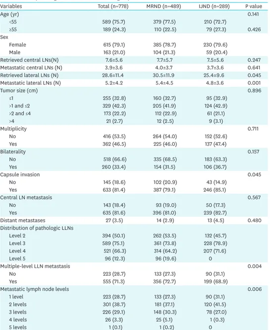
The clinical characteristics of both groups are shown in Table 1. The 2 groups were well matched for age and sex ratio (P=NS). There were no significant differences in clinical findings between the 2 groups, except for number of retrieved and pathologically proven metastatic lateral LNs. The numbers of retrieved lateral LNs (30.5±11.9 vs. 25.4±9.6, P=0.045) and pathologically proven metastatic lateral LNs (5.4±4.5 vs. 4.8±3.6, P=0.001) were significantly greater in the MRND group. In addition, there no significant differences in
terms of tumor size, multiplicity, bilaterality, capsule invasion, and the central LN metastasis.
Incidence of multiple-level LN metastases was higher in MRND group, and the distribution of metastasis LN levels was significantly different in both group.
The median follow up period were 94.3±37.6 months. During follow-up, local or distant recurrence developed in 84 patients, consisting of 57 (11.7%) in MRND and 27 (9.3%) in IJND group. The 10-year DFS was 88.3% for MRND and 90.3% for IJND group, and 15-year DFS was 88.0% in MRND and 89.5% in IJND group. The mean recurrence-free interval was 44.1±33.6 months for MRND and 47.3±37.1 months for IJND group (P=0.694). No significant differences between the 2 groups were found regarding DFS (P=0.283) and recurrence rate (P=0.188) (Table 2).
Table 1. Clinicopathologic characteristics of the 2 groups
Variables Total (n=778) MRND (n=489) IJND (n=289) P value
Age (yr) 0.141
<55 589 (75.7) 379 (77.5) 210 (72.7)
≥55 189 (24.3) 110 (22.5) 79 (27.3) 0.426
Sex
Female 615 (79.1) 385 (78.7) 230 (79.6)
Male 163 (21.0) 104 (21.3) 59 (20.4)
Retrieved central LNs(N) 7.6±5.6 7.7±5.7 7.5±5.6 0.247
Metastatic central LNs (N) 3.9±3.6 4.0±3.7 3.7±3.6 0.641
Retrieved lateral LNs (N) 28.6±11.4 30.5±11.9 25.4±9.6 0.045
Metastatic lateral LNs (N) 5.2±4.2 5.4±4.5 4.8±3.6 0.001
Tumor size (cm) 0.896
≤1 255 (32.8) 160 (32.7) 95 (32.9)
>1 and ≤2 329 (42.3) 205 (41.9) 124 (42.9)
>2 and ≤4 173 (22.2) 112 (22.9) 61 (21.1)
>4 21 (2.7) 12 (2.5) 9 (3.1)
Multiplicity 0.711
No 416 (53.5) 264 (54.0) 152 (52.6)
Yes 362 (46.5) 225 (46.0) 137 (47.4)
Bilaterality 0.157
No 518 (66.6) 335 (68.5) 183 (63.3)
Yes 260 (33.4) 154 (31.5) 106 (36.7)
Capsule invasion 0.045
No 145 (18.6) 102 (20.9) 43 (14.9)
Yes 633 (81.4) 387 (79.1) 246 (85.1)
Central LN metastasis 0.567
No 143 (18.4) 93 (19.0) 50 (17.3)
Yes 635 (81.6) 396 (81.0) 239 (82.7)
Distant metastases 27 (3.5) 14 (2.9) 13 (4.5) 0.480
Distribution of pathologic LLNs
Level 2 394 (50.1) 262 (53.5) 132 (45.7)
Level 3 589 (75.1) 361 (73.8) 228 (78.9)
Level 4 521 (66.3) 314 (64.2) 207 (71.6)
Level 5 96 (12.3) 96 (19.6) 0
Multiple-level LLN metastasis 0.004
No 223 (28.7) 133 (27.3) 90 (31.1)
Yes 555 (71.3) 356 (72.7) 199 (68.9)
Metastatic lymph node levels 0.006
1 level 223 (28.7) 133 (27.3) 90 (31.1)
2 levels 301 (38.7) 181 (37.1) 120 (41.5)
3 levels 226 (29.1) 148 (30.3) 78 (27.0)
4 levels 26 (3.3) 25 (5.1) 1 (0.3)
5 levels 1 (0.1) 1 (0.2) 0
MRND = modified radical neck dissection; IJND = internal jugular node dissection; LN = lymph node; LLN = lateral lymph node.
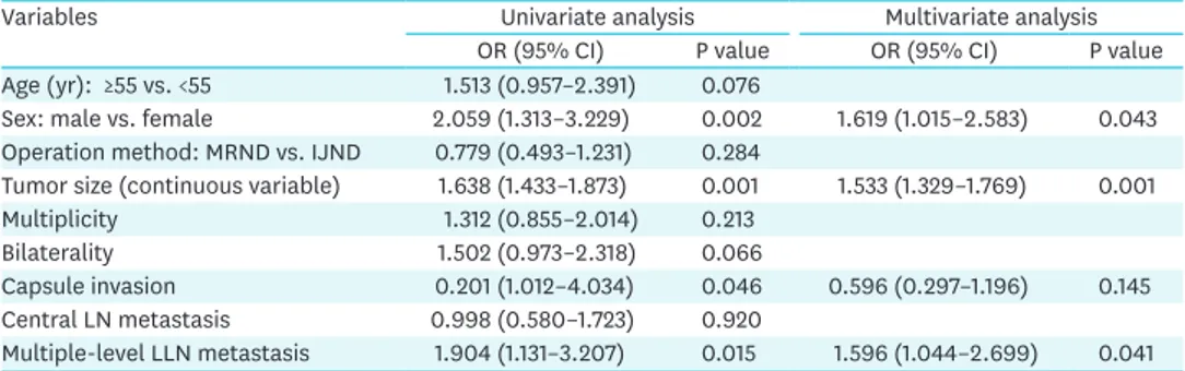
The risk factors for recurrence were evaluated using univariate and multivariate logistic regression analyses for all study populations. The univariate analysis showed that female gender, tumor size, capsule invasion, and multiple-level LLN metastasis were risk factors for recurrence, whereas age group divided by 55 years of age, operation method (MRND vs.
IJND), multiplicity, bilaterality, and central LN metastasis were not significantly associated with recurrence. However, when these variables were included in multivariate logistic regression models, female gender, tumor size, and multiple-level LLN metastasis were risk factors for recurrence. The extent of LND, the main concern of this study, was not significantly associated with recurrence (Table 3).
In analysis of recurrence patterns, 7 patients in the MRND group had distant recurrence in lung and 55 patients had local recurrences that were mostly observed in level IV, followed by level III. In the IJND group, 7 patients had distant recurrence in lung and 1 patient died of simultaneous lung and bone recurrence. Twenty-four patients had local recurrences, the most common site being level IV, followed level III. There were 4 patients in MRND and 1 patient in IJND diagnosed with recurrence in level V (Table 4).
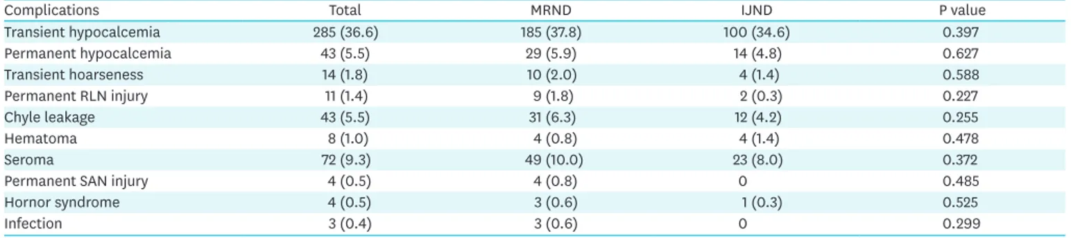
Perioperative complications were compared between groups (Table 5). No significant differences in the frequency of complications were found. The incidence of permanent SAN injury was 0.8% in the MRND group and 0% in the IJND group.
DISCUSSION
Although lymphatic metastasis does not affect overall survival in PTC patients, there is a general consensus that cervical LN metastasis is the most significant prognostic factor for locoregional recurrences (22-24). This uncertainty of the prognostic impact of LN metastasis on overall survival and risk of postoperative morbidity after formal MRND contributes to the continuing debate about whether it is mandatory to dissect level V LNs in all PTC patients with lateral LN metastases.
Table 2. The treatment outcomes of the 2 groups
Variables Total (n=778) MRND (n=489) IJND (n=289) P value
5-year disease free survival 95.9% 93.6% 90.9% 0.283
10-year disease free survival 91.8% 88.3% 90.3% 0.283
15-year disease free survival 89.2% 88.0% 89.5% 0.283
Recurrence rate 10.8% (84/778) 11.7% (57/489) 9.3% (27/289) 0.188
Recurrence-free intervals 45.2±34.6 44.1±33.6 47.3±37.1 0.694
MRND = modified radical neck dissection; IJND = internal jugular node dissection.
Table 3. Cox regression analysis of the relationship between clinicopathologic factors and recurrence
Variables Univariate analysis Multivariate analysis
OR (95% CI) P value OR (95% CI) P value
Age (yr): ≥55 vs. <55 1.513 (0.957–2.391) 0.076
Sex: male vs. female 2.059 (1.313–3.229) 0.002 1.619 (1.015–2.583) 0.043 Operation method: MRND vs. IJND 0.779 (0.493–1.231) 0.284
Tumor size (continuous variable) 1.638 (1.433–1.873) 0.001 1.533 (1.329–1.769) 0.001
Multiplicity 1.312 (0.855–2.014) 0.213
Bilaterality 1.502 (0.973–2.318) 0.066
Capsule invasion 0.201 (1.012–4.034) 0.046 0.596 (0.297–1.196) 0.145
Central LN metastasis 0.998 (0.580–1.723) 0.920
Multiple-level LLN metastasis 1.904 (1.131–3.207) 0.015 1.596 (1.044–2.699) 0.041 OR = odds ratio; CI = confidence interval; LN = lymph node; LLN = lateral lymph node; MRND = modified radical neck dissection; IJND = internal jugular node dissection.
Dissection of level V is associated with postoperative morbidities, such as SAN dysfunction, that decrease patient's quality of life (QOL) postoperatively. The SAN must be identified and retracted to clear away LNs during the dissection of level V. Patten et al. (25) reported that most patients who underwent RND for head and neck cancer experienced pain, weakness, shoulder droop, and movement disability because the SAN had been sacrificed. Kupferman et al. (10) reported that even though the SAN preserved, postoperative shoulder dysfunction occurred in 27% of patients who underwent RND as a result of excessive retraction or ischemia. Also not to be underestimated is the impact on QOL that transection of the cervical rootlets incurs, often resulting in neck numbness or neuropathic pain (26). In our study, permanent SAN injuries were very rare events, with rates of 0% in group I and 1.2% in group II. This finding could be explained by all LND procedures having been performed by an experienced surgeon with a good anatomical knowledge of the SAN course.
Decision on the extent of LND, which is necessary for the treatment of regional metastases from PTC, should be based on predictable drainage patterns. The anterolateral group of nodes (level II, III and IV) is at the greatest risk of LNM, with level III nodes being the most frequently involved (27,28). In our previous study, we found that level IIb and V LNs were rarely metastasized and, therefore, did not necessarily require aggressive dissection. Preoperative nodal evaluation for LNM in PTC patients is essential in determining the inclusion of level V in a LND. Although the LNM could be detected by palpation (5), high resolution US has been widely used for preoperative nodal staging (29,30). Thereafter, the LNM was confirmed by FNAB for the node. In addition, the cystic LNM from PTC can be diagnosed by Tg measurement in the wash-out of needles used for FNAB (19). In our study, all patients who underwent LND underwent preoperative US staging to determine if clinically negative LN in level V.
Table 4. Distribution of recurrence in the 84 patients in 2 groups
Variables Total (n=84) MRND (n=57) IJND (n=27)
Local recurrence (single or multiple)
Total 79 (10.8%) 55 (11.7%) 24 (9.3%)
Recur side Ipsilateral 46 Contralateral 33 Ipsilateral 34 Contralateral 21 Ipsilateral 11 Contralateral 13
Level I 1 1
Level II 12 11 8 7 4 4
Level III 8 20 7 12 1 8
Level IV 13 23 8 16 5 7
Level V 3 2 2 2 1 0
Level VI 21 13 18 9 3 4
Level VII 4 1 4 0 0 1
Distant recurrence
Lung 14 7 7
Lung and bone 1 0 1
MRND = modified radical neck dissection; IJND = internal jugular node dissection.
Table 5. Comparison of perioperative complications in 2 groups
Complications Total MRND IJND P value
Transient hypocalcemia 285 (36.6) 185 (37.8) 100 (34.6) 0.397
Permanent hypocalcemia 43 (5.5) 29 (5.9) 14 (4.8) 0.627
Transient hoarseness 14 (1.8) 10 (2.0) 4 (1.4) 0.588
Permanent RLN injury 11 (1.4) 9 (1.8) 2 (0.3) 0.227
Chyle leakage 43 (5.5) 31 (6.3) 12 (4.2) 0.255
Hematoma 8 (1.0) 4 (0.8) 4 (1.4) 0.478
Seroma 72 (9.3) 49 (10.0) 23 (8.0) 0.372
Permanent SAN injury 4 (0.5) 4 (0.8) 0 0.485
Hornor syndrome 4 (0.5) 3 (0.6) 1 (0.3) 0.525
Infection 3 (0.4) 3 (0.6) 0 0.299
MRND = modified radical neck dissection; IJND = internal jugular node dissection; RLN = recurrent laryngeal nerve; SAN = spinal accessory nerve.
Consequently, we found that the frequency of LNM for level II and V and the presence of multi- level LNM were significantly higher in the MRND group than the IJND group, suggesting our strategy of the LND based on preoperative nodal evaluation using US could be justified.
Currently, for PTC patients with LLN metastasis, most surgeons prefer formal MRND (level II–
V) to gain local control because some studies have demonstrated that high metastatic rates in level V nodes from 25% to 60% (11,13,27). On the other hand, in several recent studies, authors have recommended a selective approach to LND in PTC patients with LNM. Turanli (12) reported in a comparative study of 61 patients who underwent SND or MRND that the type of dissection was not related to DFS, overall survival, and local recurrence. Kandil et al. (31) reported that extensive neck dissection such as MRND among PTC patients did not result in an improved survival benefit compared with no LN dissection or SND. Caron et al. (13) reported that recurrence at levels I and V were uncommon in 106 PTC patients with LNM. They concluded that levels I and V did not require resection unless there is clinical or radiological evidence of disease. Our data regarding treatment outcomes correspond with these previous studies (12,31), suggesting that there was no difference between IJND and MRND groups regarding DFS and recurrence rate. Furthermore, multivariate analysis of this study revealed that the type of LND was not significantly associated with recurrence, and female gender, tumor size, and multiple-level LLN metastasis were independent risk factors for recurrence. Our findings indicate that the local recurrence after complete nodal dissection regardless of the type of LND is still related to aggressive factors like tumor size and LLN metastasis levels, which are known to be important prognostic factors in PTC.
The local recurrence rate after LND was reported to be 8%–32% (12,22). The present study showed of recurrence rate of 10.8% (11.7% in the MRND group and 9.3% in the IJND group) in all the groups compared with previous studies. This reason is that the IJND (level II–IV) performed in this study apply the same comprehensive anatomic dissection as the MRND and harvesting metastases cleared all lymphatic tissue at each level. Therefore, our results showed a significant reduction in the rate of recurrence. At our institute, a berry-picking procedure is not accepted for the surgical treatment of PTC nodal metastases, because it was reported to increase death rates due to recurrence in PTC patients (32).
In assessing recurrence at level V in patients who underwent IJND without level V, our results, which are in accordance with those of the study by Caron and Clark (6) that showed 5 recurrence of total at level V, revealed that only 1 recurrence in level V occurred in the IJND group. The present study may be confronted with debate over the possibility of existence of occult level-V metastases in the IJND group, because Roh et al. (33) reported metastases detected only on postoperative pathological examination (pN1b) were present in a number of level–V specimens (16%–20%). Furthermore, recent studies to identify predictors of level V LLN metastasis demonstrated that simultaneous metastases to level II, III and IV, tumor multifocality, perineural invasion, and macroscopic extranodal extension were significant predictors in PTC patients with nodal metastases (11,34,35). However, these studies could be criticized because of the lack of long-term outcomes according to the type of LND. On the other hand, there are data suggesting that occult LLN metastasis or recurrence does not affect overall survival in PTC patients. Ito et al. (36) reported that pN1b did not independently affect the cause specific survival of patients. Noguchi and Murakami (37) reported that at least 75% of patients with PTC have occult LN metastases, but only about 20% become clinically evident, which means pN1b rarely developed sufficiently to be evident. In addition to these studies, our data support that patients with a clinically
negative level-V LN, although they might have occult LN metastases, would be expected to have lower recurrence rate.
This study has several limitations. First, the patient group divided not from the randomized control design but the operation method already performed and the study could have some inevitable features. Even though the clinicopathologic characteristics were not markedly different between the 2 groups, the IJND was performed on selected patients.
Second, due to the long-term period of data enrollment, different radiologists and surgeons were involved in the evaluation and management of patients, resulting in possible inter-observer variability in the interpretation of metastasis.
Despite these limitations, our study had several strong points. First, this study has a relatively long-term follow-up period. Second, 778 cases of N1b patients were analyzed, which makes it a large study regarding the extent of lateral neck dissection.
In conclusion, IJND achieves favorable postoperative results in PTC with lateral neck node metastasis patients, and level V metastasis and recurrence incidence was low. Therefore, the extent of lateral neck node dissection, whether IJND or MRND, can be considered for patients according to the simultaneous metastasis level and the tumor size.
REFERENCES
1. Hong S, Won YJ, Park YR, Jung KW, Kong HJ, Lee ES, et al. Cancer statistics in Korea: incidence, mortality, survival, and prevalence in 2017. Cancer Res Treat 2020;52:335-50.
PUBMED | CROSSREF
2. Kim E, Park JS, Son KR, Kim JH, Jeon SJ, Na DG. Preoperative diagnosis of cervical metastatic lymph nodes in papillary thyroid carcinoma: comparison of ultrasound, computed tomography, and combined ultrasound with computed tomography. Thyroid 2008;18:411-8.
PUBMED | CROSSREF
3. Mazzaferri EL, Jhiang SM. Long-term impact of initial surgical and medical therapy on papillary and follicular thyroid cancer. Am J Med 1994;97:418-28.
PUBMED | CROSSREF
4. Mirallié E, Sagan C, Hamy A, Paineau J, Kahn X, Le Néel JC, et al. Predictive factors for node involvement in papillary thyroid carcinoma. Univariate and multivariate analyses. Eur J Cancer 1999;35:420-3.
PUBMED | CROSSREF
5. Moley JF, Wells SA. Compartment-mediated dissection for papillary thyroid cancer. Langenbecks Arch Surg 1999;384:9-15.
PUBMED | CROSSREF
6. Caron NR, Clark OH. Papillary thyroid cancer: surgical management of lymph node metastases. Curr Treat Options Oncol 2005;6:311-22.
PUBMED | CROSSREF
7. Crile G. Landmark article Dec 1, 1906: excision of cancer of the head and neck. With special reference to the plan of dissection based on one hundred and thirty-two operations. By George Crile. JAMA 1987;258:3286-93.
PUBMED | CROSSREF
8. Haugen BR, Alexander EK, Bible KC, Doherty GM, Mandel SJ, Nikiforov YE, et al. 2015 American Thyroid Association Management Guidelines for adult patients with thyroid nodules and differentiated thyroid cancer: the American Thyroid Association Guidelines Task Force on thyroid nodules and differentiated thyroid cancer. Thyroid 2016;26:1-133.
PUBMED | CROSSREF
9. Stack BC Jr, Ferris RL, Goldenberg D, Haymart M, Shaha A, Sheth S, et al. American Thyroid Association consensus review and statement regarding the anatomy, terminology, and rationale for lateral neck dissection in differentiated thyroid cancer. Thyroid 2012;22:501-8.
PUBMED | CROSSREF
10. Kupferman ME, Patterson DM, Mandel SJ, LiVolsi V, Weber RS. Safety of modified radical neck dissection for differentiated thyroid carcinoma. Laryngoscope 2004;114:403-6.
PUBMED | CROSSREF
11. Kupferman ME, Weinstock YE, Santillan AA, Mishra A, Roberts D, Clayman GL, et al. Predictors of level V metastasis in well-differentiated thyroid cancer. Head Neck 2008;30:1469-74.
PUBMED | CROSSREF
12. Turanli S. Is the type of dissection in lateral neck metastasis for differentiated thyroid carcinoma important? Otolaryngol Head Neck Surg 2007;136:957-60.
PUBMED | CROSSREF
13. Caron NR, Tan YY, Ogilvie JB, Triponez F, Reiff ES, Kebebew E, et al. Selective modified radical neck dissection for papillary thyroid cancer-is level I, II and V dissection always necessary? World J Surg 2006;30:833-40.
PUBMED | CROSSREF
14. Lee CR, Nam KH. Lateral neck node dissection in differentiated thyroid carcinoma. Korean J Endocr Surg 2014;14:1-6.
CROSSREF
15. Battoo AJ, Sheikh ZA, Thankappan K, Mir AW, Haji AG, Level V. Level V clearance in neck dissection for papillary thyroid carcinoma: a need for homogeneous studies. Int Arch Otorhinolaryngol 2018;22:449-54.
PUBMED | CROSSREF
16. Kim SK, Park I, Hur N, Lee JH, Choe JH, Kim JH, et al. Should level V be routinely dissected in N1b papillary thyroid carcinoma? Thyroid 2017;27:253-60.
PUBMED | CROSSREF
17. Javid M, Graham E, Malinowski J, Quinn CE, Carling T, Udelsman R, et al. Dissection of levels II through V is required for optimal outcomes in patients with lateral neck lymph node metastasis from papillary thyroid carcinoma. J Am Coll Surg 2016;222:1066-73.
PUBMED | CROSSREF
18. Park JS, Son KR, Na DG, Kim E, Kim S. Performance of preoperative sonographic staging of papillary thyroid carcinoma based on the sixth edition of the AJCC/UICC TNM classification system. AJR Am J Roentgenol 2009;192:66-72.
PUBMED | CROSSREF
19. Uruno T, Miyauchi A, Shimizu K, Tomoda C, Takamura Y, Ito Y, et al. Usefulness of thyroglobulin measurement in fine-needle aspiration biopsy specimens for diagnosing cervical lymph node metastasis in patients with papillary thyroid cancer. World J Surg 2005;29:483-5.
PUBMED | CROSSREF
20. Patel KN, Shah JP. Neck dissection: past, present, future. Surg Oncol Clin N Am 2005;14:461-77.
PUBMED | CROSSREF
21. Morris LF, Waxman AD, Braunstein GD. Thyroid stunning. Thyroid 2003;13:333-40.
PUBMED | CROSSREF
22. McConahey WM, Hay ID, Woolner LB, van Heerden JA, Taylor WF. Papillary thyroid cancer treated at the Mayo Clinic, 1946 through 1970: initial manifestations, pathologic findings, therapy, and outcome. Mayo Clin Proc 1986;61:978-96.
PUBMED | CROSSREF
23. Mazzaferri EL. Papillary thyroid carcinoma: factors influencing prognosis and current therapy. Semin Oncol 1987;14:315-32.
PUBMED
24. Akslen LA. Prognostic importance of histologic grading in papillary thyroid carcinoma. Cancer 1993;72:2680-5.
PUBMED | CROSSREF
25. Patten C, Hillel AD. The 11th nerve syndrome. Accessory nerve palsy or adhesive capsulitis? Arch Otolaryngol Head Neck Surg 1993;119:215-20.
PUBMED | CROSSREF
26. Inoue H, Nibu K, Saito M, Otsuki N, Ishida H, Onitsuka T, et al. Quality of life after neck dissection. Arch Otolaryngol Head Neck Surg 2006;132:662-6.
PUBMED | CROSSREF
27. Sivanandan R, Soo KC. Pattern of cervical lymph node metastases from papillary carcinoma of the thyroid. Br J Surg 2001;88:1241-4.
PUBMED | CROSSREF
28. Lee J, Sung TY, Nam KH, Chung WY, Soh EY, Park CS. Is level IIb lymph node dissection always necessary in N1b papillary thyroid carcinoma patients? World J Surg 2008;32:716-21.
PUBMED | CROSSREF
29. Cooper DS, Doherty GM, Haugen BR, Kloos RT, Lee SL, Mandel SJ, et al. Management guidelines for patients with thyroid nodules and differentiated thyroid cancer. Thyroid 2006;16:109-42.
PUBMED | CROSSREF
30. Cooper DS, Doherty GM, Haugen BR, Kloos RT, Lee SL, Mandel SJ, et al. Revised American Thyroid Association management guidelines for patients with thyroid nodules and differentiated thyroid cancer.
Thyroid 2009;19:1167-214.
PUBMED | CROSSREF
31. Kandil E, Friedlander P, Noureldine S, Islam T, Tufano RP. Impact of extensive neck dissection on survival from papillary thyroid cancer. ORL J Otorhinolaryngol Relat Spec 2011;73:330-5.
PUBMED | CROSSREF
32. Noguchi M, Kumaki T, Taniya T, Segawa M, Nakano T, Ohta N, et al. Impact of neck dissection on survival in well-differentiated thyroid cancer: a multivariate analysis of 218 cases. Int Surg 1990;75:220-4.
PUBMED
33. Roh JL, Kim JM, Park CI. Lateral cervical lymph node metastases from papillary thyroid carcinoma:
pattern of nodal metastases and optimal strategy for neck dissection. Ann Surg Oncol 2008;15:1177-82.
PUBMED | CROSSREF
34. Lim YC, Choi EC, Yoon YH, Koo BS. Occult lymph node metastases in neck level V in papillary thyroid carcinoma. Surgery 2010;147:241-5.
PUBMED | CROSSREF
35. Shim MJ, Roh JL, Gong G, Choi KJ, Lee JH, Cho SH, et al. Preoperative detection and predictors of level V lymph node metastasis in patients with papillary thyroid carcinoma. Br J Surg 2013;100:497-503.
PUBMED | CROSSREF
36. Ito Y, Miyauchi A, Jikuzono T, Higashiyama T, Takamura Y, Miya A, et al. Risk factors contributing to a poor prognosis of papillary thyroid carcinoma: validity of UICC/AJCC TNM classification and stage grouping. World J Surg 2007;31:838-48.
PUBMED | CROSSREF
37. Noguchi S, Murakami N. The value of lymph-node dissection in patients with differentiated thyroid cancer. Surg Clin North Am 1987;67:251-61.
PUBMED | CROSSREF