J Appl Biol Chem (2015) 58(1), 1−3 http://dx.doi.org/10.3839/jabc.2015.001
Online ISSN 2234-7941 Print ISSN 1976-0442 Short Communication: Bioactive Materials
2-Hydroxyquinoline and Its Structural Analogs Show Antidiabetic Effects against α-Amylase and α-Glucosidase
Hwa-Won Lee · Hoi-Seon Lee*
Received: 29 May 2014 / Accepted: 24 June 2014 / Published Online: 31 March 2015
© The Korean Society for Applied Biological Chemistry 2015
Abstract This study investigated the inhibitory activities of 2- hydroxyquinoline and its analogs against α-glucosidase and α- amylase. Based on the IC50 values of 2-hydroxyquinoline analogs tested against α-glucosidase and α-amylase, 2-hydroxyquinoline had potent inhibitory activity (64.4 and 130.5µg/mL, respectively), while 2-methyl-8-hydroxyquinoline showed weakly inhibitory activity (90.7 and 215.4µg/mL, respectively). 2-Methylquinoline demonstrated no activity against α-glucosidase and α-amylase. In conclusion, 2-hydroxyquinoline analogs, with the existence of a methyl group and hydroxyl on quinoline, can be useful as a new diabetes treatment.
Keywords 2-hydroxyquinoline · α-amylase · α-glucosidase · anti- diabetic activity
Diabetes mellitus is a major chronic disease induced by an unsuitable balance of blood glucose, having a significant impact on health (Hu et al., 2008). Diabetes can be divided into type I and type II (Jeong et al., 2012; Lee et al., 2012a). Type I diabetes is an insulin-dependent disease, while type II diabetes (adult type diabetes mellitus) is a non-insulin-dependent disease that accounts for approximately 90% of all diabetes mellitus cases (Toshiro et al., 2001; Lee et al., 2012b). Although type I can effectively be controlled by the regulation of insulin, finding a useful therapy for
the management of type II is difficult (Yang et al., 2011). In this regard, effective methods for hypoglycemic action are needed (Choi et al., 2008). One of the methods of therapy is to decrease the hyperglycemia by delaying the absorption of glucose, accomplished by inhibition of starch breakdown enzymes such as α-amylase and α-glucosidase (Bhandari et al., 2008). Therefore, control of α-amylase and α-glucosidase during hyperglycemia can be a significant approach in the management of high blood glucose connected to type II diabetes (Lee, 2005; Wang et al., 2010; Jeong et al., 2012). Plant-derived materials had been used as alternative materials due to their bioactive substances (Lee and Ahn, 1998; Lee, 2002; Yang et al., 2002). In addition, previous studies have reported that plant-derived materials possess α- amylase and α-glucosidase inhibitory effects (Lee, 2005; Jeong et al., 2012). In this regard, the antidiabetic activities of 2- hydroxyquinoline and its structural analogs against α-amylase and α-glucosidase were evaluated.
Acarbose, 2-hydroxyquinoline, 2-methylquinoline, and 2-methyl- 8-hydroxyquinoline were provided from Sigma-Aldrich (USA).
All chemicals were of reagent grade. The activities of 2- hydroxyquinoline and its structural analogs were evaluated against α-amylase and α-glucosidase. The α-amylase inhibitory activity was assayed following the procedure described by Wang et al.
(2010) and Jeong et al. (2012), with small modifications. The enzyme solution (6.25 U/mL) was laid by dissolving α-amylase (Sigma Co., USA) in 0.5 M Tris-HCl buffer (pH 6.9). The starch azure (8 mg) was suspended in 0.5 M Tris-HCl buffer containing 0.01 M calcium chloride, and was soaked in boiling water for 5 min followed by preincubation at 37oC for 10 min. The enzyme solution (100µL) and test sample (100 µL) in 50% dimethyl sulfoxide were mixed in a 96-well plate. After 10 min, 50% acetic acid (50µL) was added to stop the response. The absorbance of the reactants was determined at 595 nm with a microplate reader (Model ASYS UVM 340, Biochrom Ltd., UK). The inhibition percentage (%) was calculated using the following equation:
Inhibition (%)=[1− (sample absorbance/control absorbance)] × 100. α-Glucosidase inhibitory activity was tested according to the H.-W. Lee · H.-S. Lee
Department of Bioenvironmental Chemistry and Institute of Agricultural Science & Technology, College of Agriculture & Life Science, Chonbuk National University, Jeonju 561-756, Republic of Korea
*Corresponding author (H.-S. Lee: [email protected])
This is an Open Access article distributed under the terms of the Creative Commons Attribution Non-Commercial License (http://creativecommons.
org/licenses/by-nc/3.0/) which permits unrestricted non-commercial use, distribution, and reproduction in any medium, provided the original work is properly cited.
2 J Appl Biol Chem (2015) 58(1), 1−3
procedure described by Shinde et al. (2008) and Jeong et al.
(2012), with a slight modification. The formation of p-nitrophenol was measured using α-glucosidase after response with p- nitrophenyl-α-D-glucopyranoside (NPG). The enzyme solution (0.6 U) was prepared by dissolving α-glucosidase in 0.1 M phosphate buffer (pH 7.0) containing 2 g/L bovine serum albumin and 0.2 g/L sodium azide. The enzyme solution (50µL) and sample (10µL) dissolved in dimethyl sulfoxide were mixed, and placed in a 96-well plate. After 15 min, 5 mM NPG (50µL) in the identical buffer was added, and the mixture was incubated for 10 min at 37oC. After 10 min, 0.1 M Sodium carbonate was added to stop the reaction. The absorbance of the reactants was measured at 405 nm using a microplate reader (Model ASYS UVM 340, Biochrom Ltd.). Inhibition percentage (%) was calculated using the equation above for amylase activity. Acarbose was used as a positive control in all experiments. All tests were performed in triplicate. The IC50 values were calculated using the logarithmic regression analysis.
The inhibitory effects of 2-hydroxyquinoline and its structural analogs (2-methylquinoline, 2-methyl-8-hydroxyquinoline) against α-amylase and α-glucosidase are presented in Table 1. Including the information for the acarbose positive control, the IC50 value of 2-hydroxyquinoline demonstrated the strongest inhibitory activity against α-glucosidase (64.4±1.7 µg/mL), followed by acarbose (66.5±1.5µg/mL), and 2-methyl-8-hydroxyquinoline (90.7±2.5 µg/mL). In contrast, 2-methylquinoline was found to have no activity against α-glucosidase. Based on the IC50 values of acarbose, 2-hydroxyquinoline and its structural against α- amylase, 2-hydroxyquinoline (130.5±2.2µg/mL) had potent α- amylase inhibitory activity, followed by acarbose (180.6±1.3µg/
mL), and 2-methyl-8-hydroxyquinoline (215.4±1.4µg/mL) (Table 1). Like for α-glucosidase, 2-methylquinoline also showed no activity against α-amylase.
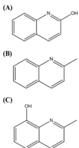
To confirm a structure-activity relationship and to determine the role of functional groups for the antidiabetic effects of 2- hydroxyquinoline analogs, its structural analogs were evaluated based on their antidiabetic activities. The importance of the functional groups was estimated by comparing IC50 values. 2- Hydroxyquinoline, which was the hydroxyl-containing quinoline
(Fig. 1), showed strong inhibitory effects for both α-amylase and α-glucosidase. According to previous studies, the combination with hydroxyl can be an essential content for antidiabetic activity (Miura et al., 1996). On the other hand, 2-methyl-8-hydroxyquinoline, which adds a methyl group to the hydroxyquinoline (Fig. 1), exhibited hypoglycemic effects comparable to acarbose. 2- Methylquinoline, which introduced the methyl group in quinoline (Fig. 1), had no antidiabetic activity. Similarly, it has been reported that compounds combined with methyl groups can have reduced hypoglycemic activity (Matsuda et al., 1998). Based upon these results, the inhibitory effects against α-amylase and α- glucosidase can be varied by adding a functional group into hydroxyquinoline.
On the basis of the Material Safety Data sheet provided by Sigma-Aldrich (2013), the oral lethal dose of 2-hydroxyquinoline was not reported for mammals. Furthermore, 2-hydroxyquinoline has been not reported the side effects on human’s body. Taken together with this result, 2-hydroxyquinoline derivatives could be used as a new diabetes treatment.
References
Bhandari MR, Nilubon JA, Gao H, and Kawabata J (2008) α-Glucosidase and α-amylase inhibitory activities of Nepalese medicinal herb Pakhanbhed (Bergenia ciliate, Haw.). Food Chem 106, 247−52.
Choi HJ, Jeong YK, Kang DO, and Joo WH (2008) Inhibitory effects of four solvent fractions of Alnus firma on α-amylase and α-glucosidase. J Life Sci 18, 1005−10.
Hu W, Jung MJ, Heo SI, and Wang MH (2008) Antioxidant and antidiabetic activities of aralia elata seeds. J Appl Biol Chem 51, 111−6.
Jeong EY, Cho KS, and Lee HS (2012) α-Amylase and α-glucosidase inhibitors isolated from Triticum aestivum L. sprouts. J Korean Soc Appl Biol Chem 55, 47−51.
Lee HS (2002) Tyrosinase inhibitors of Pulsatilla cernua root-derived materials. J Agric Food Chem 50, 1400−03.
Lee HS (2005) Cuminaldehyde: aldose reductase and α-glucosidase inhibitor derived from Cuminum cyminum L. seeds. J Agric Food Chem 53, 2446−50.
Lee HS and Ahn YJ (1998) Growth-inhibiting effects of cinnamomum cassia bark-derived materials on human intestinal bacteria. J Agric Food Chem Table 1 α-Glucosidase and α-amylase inhibitory activities of 2-
hydroxyquioline derivatives
Samples
Anti-diabetic activity α-glucosidase
inhibition IC50 (µg/mL)a
α-amylase inhibition IC50 (µg/mL)
2-Hydroxyquinoline 64.4±1.7 130.5±2.2
2-Methylquinoline NIc NI
2-Methyl-8-hydroxyquinoline 90.7±2.5 215.4±1.4
Acarboseb 66.5±1.5 180.6±1.3
aIC50 values calculated from regression lines, using five different concen- trations tested in triplicate.
bAcarbose was used as a positive control.
cNI: No inhibition at a concentration of 1,000 µg/mL.
Fig. 1 Structures of 2-hydroxyquinoline derivatives; (A) 2-hydroxy- quinoline; (B) 2-methylquinoline; (C) 2-methyl-8-hydroxyquinoline.
J Appl Biol Chem (2015) 58(1), 1−3 3
46, 8−12.
Lee SH, Lee JK, and Kim IH (2012a) Trends and perspectives in the development of antidiabetic drugs for Type 2 diabetes mellitus. Korean J Microbiol Biotechnol 3, 180−5.
Lee YJ, Lee YJ, Yoon JJ, Lee SM, Kim HY, Shin SH et al. (2012b) Anti- diabetic and anti-inflammatory effects of water extract of Ligustrum japonicum Leaves in db/db mouse. Kor J Herbology 27, 107−14.
Matsuda H, Li Y, Murakami T, Matsumura N, Yamahara J, and Yoshikawa M (1998) Antidiabetic principles of natural medicines. III. Structure-related inhibitory activity and action mode of oleanolic acid glycosides on hypoglycemic activity. Europe pubmed central 46, 1399−403.
Miura T, Nishiyama Y, Ichimaru M, Moriyasu M, and Kato A (1996) Hypoglycemic activity and structure-activity relationship of iridoidal glycosides. Europe pubmed central 19, 160−1.
Shinde J, Taldone T, Barietta M, Kunaparaju N, Hu B, and Kumar S (2008)
α-Glucosidase inhibitory activity of Syzygium cumini (Linn.) skeels seed kernel in vitro and in goto-kakizake (GK) rats. Carbohydr Res 343, 1278−81.
Toshiro M, Tetsuya U, Tomoyuki O, Koichi S, Norihiko T, and Kiyoshi M (2001) Alpha-glucosidase inhibitory action of natural acylated anthocyanins. 1. survey of natural pigments with potent inhibitory activity. J Agric Food Chem 49, 1948−51.
Wang H, Du YJ, and Song HC (2010) α-Glucosidase and α-amylase inhibitory activities of guava leaves. Food Chem 123, 6−13.
Yang JY, Jeong EY, Kim DK, and Lee HS (2011) Antioxidative and α- glucosidase inhibitory effects in Triticum aestivum sprouts treated to chilling temperature. J Korean Soc Appl Biol Chem 54, 644−8.
Yang YC, Lee SG, Lee HK, Kim MK, Lee SH, and Lee HS (2002) A piperidine amide extracted from Piper longum L. fruit shows activity against Aedes aegypti mosquito larvae. J Agric Food Chem 50, 3765−7.