Traditionally, adhesive capsulitis (AC) of the shoulder has been regarded as a self-limiting condition without signifi- cant long-term sequelae, lasting 18 or 30 months. How- ever, several studies have reported long-term residual mo- tion restriction and persisting symptoms in AC.1,2) Reeves1) reported that 50% of patients were still experiencing pain
or stiffness of the shoulder at a mean of 7 years from the onset of the condition, although only 11% reported func- tional limitation. To decrease time to recovery and im- prove the outcomes, a variety of regimens have been used for the treatment of AC, which include nonsteroidal anti- inflammatory drugs, local intra-articular steroid injection, physiotherapy, hydrodilation, manipulation under anes- thesia (MUA), and arthroscopic capsular release (ACR).3-8) Among these regimens, MUA has been the long- standing treatment for refractory AC. Numerous studies have reported this approach as a safe and effective treat- ment for reducing the duration of symptoms in patients with AC3,6,9-15); however, others have stated that MUA has no advantages compared with conservative treatment.16-18) Also, the potential complications associated with this
Early Clinical Outcomes of Manipulation under Anesthesia for Refractory Adhesive Capsulitis:
Comparison with Arthroscopic Capsular Release
Du-Han Kim, MD, Kwang-Soon Song, MD, Byung-Woo Min, MD, Ki-Choer Bae, MD, Young-Jae Lim, MD
#, Chul-Hyun Cho, MD
Department of Orthopedic Surgery, Dongsan Medical Center, Keimyung University School of Medicine, Daegu, Korea
Background: The purpose of this study was to compare early clinical outcomes of manipulation under anesthesia (MUA) and ar- throscopic capsular release (ACR) in patients with refractory adhesive capsulitis (AC).
Methods: Thirty AC patients who underwent MUA (MUA group) were included. As a control group, thirty AC patients who under- went ACR (ACR group) were matched for age and sex with the MUA group. Visual analog scale (VAS) pain score, American shoul- der and Elbow Surgeons (ASES) score, and range of motion (ROM) were evaluated preoperatively and at 3, 6, and 12 months after procedure.
Results: Both groups had significant improvements in the VAS pain score, ASES score, and ROM at 12 months after procedure.
VAS pain score and ASES score were significantly better in the MUA group than in the ACR group at 3 months after procedure.
Mean forward flexion was significantly greater in the MUA group than in the ACR group at 3 months after procedure. Mean exter- nal rotation and internal rotation were significantly greater in the MUA group than in the ACR group at 3, 6, and 12 months after procedure. Two patients required additional steroid injections at 3 and 6 months after MUA because of recurrent stiffness with pain.
Conclusions: Compared with ACR, MUA provided equivalent clinical outcomes in the early period after procedure. Our study sug- gests that MUA is a useful option to be considered as treatment for refractory AC before choosing ACR.
Keywords: Shoulder, Adhesive capsulitis, Manipulation, Arthroscopy, Capsular release
Copyright © 2020 by The Korean Orthopaedic Association
This is an Open Access article distributed under the terms of the Creative Commons Attribution Non-Commercial License (http://creativecommons.org/licenses/by-nc/4.0) which permits unrestricted non-commercial use, distribution, and reproduction in any medium, provided the original work is properly cited.
Clinics in Orthopedic Surgery • pISSN 2005-291X eISSN 2005-4408 Received February 28, 2019; Accepted April 26, 2019
Correspondence to: Chul-Hyun Cho, MD
Department of Orthopedic Surgery, Dongsan Medical Center, Keimyung University School of Medicine, 56 Dalseong-ro, Jung-gu, Daegu 41931, Korea Tel: +82-53-250-8028, Fax: +82-53-250-7205
E-mail: [email protected]
#Current affiliation: Department of Orthopedic Surgery, The Open Hospital, Daegu, Korea
procedure (e.g., recurrent stiffness, fractures, dislocation, brachial plexus injury, and rotator cuff tear) have been documented.8,19,20)
Because of recent advances in arthroscopic tech- niques, ACR has shown promising results comparable to those of other treatment options.21-24) Although both MUA and ACR appear to be effective treatments in patients with refractory AC, it is unclear whether there is a difference in the clinical effectiveness of MUA compared to ACR. No comparative studies have evaluated clinical outcomes of both procedures. The aims of AC treatment are to elimi- nate pain and to recover range of motion (ROM) as soon as possible. Because it is important to report early clini- cal outcomes after procedures in addition to final clini- cal outcomes, we focused on the early response to MUA.
Therefore, the purpose of this study was to compare early clinical outcomes between MUA and ACR in patients with AC. We hypothesized that MUA and ACR would have equivalent clinical outcomes.
METHODS
Subjects
The protocol of this study was reviewed and approved by Institutional Review Board of Dongsan Medical Cen- ter (IRB No. 2018-09-003). Written informed consents were obtained. Thirty-one patients underwent MUA for refractory AC in a single institution between 2016 and 2017. One patient was excluded from this study because she received ACR due to worse results at 3 months after MUA. In the MUA group, there were 21 female and nine male patients with a mean age of 54.5 years (range, 43–74 years). The dominant arm was affected in 19 patients.
Eleven patients had a history of diabetes mellitus. The mean duration of symptoms was 12.1 months (range, 4–40 months). From the pool of patients who underwent ACR between 2007 and 2015, 30 patients who were matched for age and sex with the MUA group were included as a con- trol group.
The inclusion criteria included a diagnosis of AC, defined as limitation of motion by greater than 50% in at least two planes (compared to the unaffected shoulder), absence of intrinsic or extrinsic shoulder disease con- firmed by magnetic resonance imaging (MRI) or ultraso- nography, and unsuccessful nonoperative management (e.g., medications, steroid injections, or physical therapy) for at least 3 months. Exclusion criteria were secondary AC with a rotator cuff tear, calcific tendinitis, osteoarthritis, inflammatory arthritis, and postsurgical, posttraumatic, or cervical disc disorder.
Manipulation under Anesthesia
A propofol anesthetic was administered by manual mask ventilation with the patient in the supine position without any special muscle relaxant. All procedures were performed by a single surgeon (CHC). With the scapular stabilized to the posterior part of the chest, the shoulder was moved into forward elevation in the sagittal plane first, then into abduction, by applying gentle pressure to break the adhe- sions. Subsequently, external and internal rotations were performed in three different grades of abduction (0°, 45°, and 90°). To minimize the risks of humeral fractures, all manipulations were performed with the use of a short lever arm. In all cases, under fluoroscopic guidance, an 18-gauge spinal needle was inserted at the glenohumeral joint. The needle was joined to a connection containing an iodinated contrast medium, and the agent was injected to confirm the exact intra-articular location of the needle.
After the position of the needle was confirmed, a mixture of 1mL triamcinolone (40 mg of methylprednisolone ac- etate), 10 mL 1% lidocaine, and 20 mL saline solution was injected to the capsule.16)
Arthroscopic Capsular Release
With the patient under general anesthesia, we assessed the ROM of the shoulder and then performed the same protocol of MUA. Afterwards, patients were placed in the lateral decubitus position, and we started a standard ar- throscopic glenohumeral examination through the poste- rior portals. After confirmation of capsular thickening or synovial hypertrophy, we performed synovial ablation and capsular release by using an electrocautery and a shaver.
The sequential capsular release began below the biceps tendon origin and superior capsule, the rotator interval, and coracohumeral ligament up to the base of the cora- coid process, the anterior capsule, and the inferior capsule involving both the anterior and posterior bands of the inferior glenohumeral ligament. Finally, the posterior cap- sule was released through the anterior viewing portal and posterior working portal. The operation was finished after intra-articular injection of 1-mL triamcinolone (40 mg of methylprednisolone acetate).22)
Postoperative Rehabilitation
Both groups received the same postoperative rehabilita- tion protocol. All patients were engaged in a rehabilitation program including pendulum exercises and immediate passive ROM exercises after procedure.
Assessment of Clinical Outcomes
All patients were evaluated during a 12-month follow-up
period. The evaluation of clinical outcomes was conducted by an independent research coordinator (EJJ). The visual analog scale (VAS) pain score and American Shoulder and Elbow Surgeons (ASES) score were assessed. ROM includ- ing forward flexion, external rotation with the arm at the side, and internal rotation at the back was also assessed.
For statistical analysis of internal rotation, we converted values into contiguously numbered groups: T1 through T12 into 1 through 12; L1 through L5 into 13 through 17;
sacrum into 18; and buttock into 19. Assessments were performed preoperatively at 3, 6, and 12 months after pro- cedure.
Statistical Analysis
IBM SPSS ver. 23.0 (IBM Corp., Armonk, NY, USA) was used for data analysis. Sample size was calculated by us- ing the difference of ASES scores between the two groups at 3 months after procedure. To obtain the large effect size of 0.85, a minimum of 30 patients for each group was required (two-sided α error of 0.05 and β error of 0.15).
To determine the significance of differences between the groups, we used the chi-square test, Fisher exact test, and Mann-Whitney U-test. To evaluate the serial changes in outcome measurements including the VAS pain score, ASES score, and ROMs, we used the Mann-Whitney U-
test and repeated-measures analysis of variance. A p < 0.05 was considered to represent a statistically significant dif- ference.
RESULTS
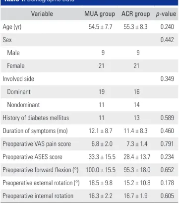
Demographics of patients are summarized in Table 1.
There were no significant differences between the two groups in terms of mean age, sex, affected side, presence of diabetes mellitus, duration of symptoms, preoperative VAS pain score, ASES score, and ROMs (p > 0.05).
VAS pain score and ASES score significantly improved in both groups during the serial follow-up period (p <
0.001). There were no statistically significant differences between the two groups in most of the assessed clinical scores. However, the mean VAS pain score in the MUA group was significantly lower than that in the ACR group at 3 months after procedure (1.6 vs. 3.4, p < 0.001), and the ASES score in the MUA group was higher than in the ACR group at 3 months after procedure (80.3 vs. 66.1, p < 0.001) (Figs. 1 and 2).
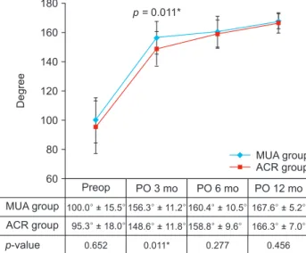
There were no significant differences between the two groups in terms of preoperative forward flexion, exter- nal rotation, and internal rotation (p > 0.05). Both groups had significant improvement in the ROMs of the shoulder joint at the final follow-up compared with the preoperative ROMs (p < 0.001). In the MUA group, mean forward flex- ion was significantly greater than that in the ACR group at Table 1. Demographic Data
Variable MUA group ACR group p-value
Age (yr) 54.5 ± 7.7 55.3 ± 8.3 0.240
Sex 0.442
Male 9 9
Female 21 21
Involved side 0.349
Dominant 19 16
Nondominant 11 14
History of diabetes mellitus 11 13 0.589
Duration of symptoms (mo) 12.1 ± 8.7 11.4 ± 8.3 0.460 Preoperative VAS pain score 6.8 ± 2.0 7.3 ± 1.4 0.791 Preoperative ASES score 33.3 ± 15.5 28.4 ± 13.7 0.234 Preoperative forward flexion (°) 100.0 ± 15.5 95.3 ± 18.0 0.652 Preoperative external rotation (°) 18.5 ± 9.8 15.2 ± 10.8 0.178 Preoperative internal rotation 16.3 ± 2.2 16.7 ± 1.9 0.605 Values are presented as mean ± standard deviation.
MUA: manipulation under anesthesia, ACR: arthroscopic capsular release, VAS: visual analog scale, ASES: American shoulder and elbow surgeons.
10
8
6
4
2
Preop PO 3 mo PO 6 mo PO 12 mo 1.6 + 1.4
3.4 + 2.0
<0.001*
0
MUA group ACR group
MUA group ACR group p-value
2.1 + 2.1 2.0 + 1.5 0.802 6.8 + 2.0
7.3 + 1.4 0.791
1.2 + 1.7 1.6 + 1.7
0.18 p < 0.001*
Score
Fig. 1. Serial changes in visual analog scale (VAS) pain score in the manipulation under anesthesia (MUA) and arthroscopic capsular release (ACR) groups. The mean VAS pain score in the MUA group was significantly lower than that in the ACR group at 3 months after procedure (*). Values are presented as mean ± standard deviation. Preop:
preoperative, PO: postoperative.
3 months after procedure (156.3° vs. 148.6°, p = 0.011) (Fig.
3). Mean external rotation and internal rotation were sig- nificantly greater than those in the ACR group at 3 months (60.6° vs. 38.2°, p < 0.001 and 11.0 vs 14.4, p < 0.001, re-
spectively), 6 months (66.2° vs 51.0°, p < 0.001 and 10.4 vs. 12.3, p = 0.015, respectively), and 12 months (73.0° vs.
61.3°, p < 0.001 and 9.0 vs. 10.7, p = 0.042, respectively) after procedure (Figs. 4 and 5).
No serious complications including instability, iat-
100
80
60
40
20
Preop PO 3 mo PO 6 mo PO 12 mo 80.3 + 15.9
66.1 + 17.3
<0.001*
0
MUA group ACR group
p-value 0.883
33.3 + 15.5 28.4 + 13.7
0.234
89.1 + 14.4 84.7 + 15.2
0.146 MUA group ACR group p < 0.001*
79.7 + 16.3 79.3 + 14.6
Score
Fig. 2. Serial changes in American Shoulder and Elbow Surgeons (ASES) score in the manipulation under anesthesia (MUA) and arthroscopic capsular release (ACR) groups. The mean ASES score in the MUA group was significantly higher than that in the ACR group at 3 months after procedure (*). Values are presented as mean ± standard deviation. Preop:
preoperative, PO: postoperative.
180
160
140
120
100
80
Preop PO 3 mo PO 6 mo PO 12 mo 156.3 + 11.2
148.6 + 11.8 0.011*
60
MUA group ACR group
p-value 0.277
100.0 + 15.5
0.652
167.6 + 5.2 166.3 + 7.0
0.456 MUA group ACR group p =0.011*
160.4 + 10.5 158.8 + 9.6 95.3 + 18.0
Degree
Fig. 3. Serial changes in forward flexion in the manipulation under anesthesia (MUA) and arthroscopic capsular release (ACR) groups.
The mean forward flexion in the MUA group was significantly greater than that in the ACR group at 3 months after procedure (*). Values are presented as mean ± standard deviation. Preop: preoperative, PO:
postoperative.
80
Preop PO 3 mo PO 6 mo PO 12 mo
<0.001*
60
40
20
MUA group ACR group
p-value 0.178
MUA group ACR group p < 0.001*
18.5 + 9.8 60.6 + 11.0 66.2 + 9.7 73.0 + 9.3 61.3 + 10.2 51.0 + 12.3
38.2 + 13.4 15.2 + 10.8
0
<0.001* <0.001*
p < 0.001* p < 0.001*
Degree
Fig. 4. Serial changes in external rotation in the manipulation under anesthesia (MUA) and arthroscopic capsular release (ACR) groups. The mean external rotation in the MUA group was significantly greater than that in the ACR group at 3, 6, and 12 months after procedure (*). Values are presented as mean ± standard deviation. Preop: preoperative, PO:
postoperative.
(Buttock) 19
(L5) 17
(L3) 15
(L1) 13
(T11) 11
(T9) 9
(T7) 7
Preop PO 3 mo PO 6 mo PO 12 mo
<0.001*
MUA group ACR group p-value
16.3 + 2.2
0.605
MUA group ACR group
11.0 + 3.5 10.4 + 3.1 9.0 + 2.8 10.7 + 2.8 12.3 + 1.9
14.4 + 1.3 16.7 + 1.9
0.015* 0.042*
p < 0.001*
p = 0.015*
p = 0.042*
Fig. 5. Serial changes in internal rotation at the back in the manipulation under anesthesia (MUA) and arthroscopic capsular release (ACR) groups.
The mean internal rotation in the MUA group was significantly greater than that in the ACR group at 3, 6, and 12 months after procedure (*).
Values are presented as mean ± standard deviation. Preop: preoperative, PO: postoperative.
rogenic fracture, brachial plexus injury, and infection were encountered in either group. In the MUA group, however, two patients required additional intra-articular steroid injection at 3 and 6 months after MUA for recurrent stiff- ness with pain. After reinjection, these patients achieved satisfactory results at the final follow-up.
DISCUSSION
The most important finding of this study is that compared with ACR, MUA provided equivalent clinical outcomes without major complications in the early period after procedure. In particular, the MUA group achieved earlier restoration of ROM during the follow-up period after the procedure. We think there are two reasons for the above results. First, MUA may obviate the unnecessary surgical damage caused by arthroscopic surgery. Even though we had two patients who needed additional steroid injec- tion because of recurrence, most patients who underwent MUA had a good outcome without complications. Second, although there was no significant difference in preopera- tive clinical scores between groups, the VAS score was slightly higher and ROM was more limited in the ACR group than in the MUA group. These worse preoperative conditions may have affected postoperative results.
Numerous studies have reported that MUA in pa- tients with refractory AC produces overall satisfactory clinical outcomes.3,6 9-15) Dodenhoff et al.3) documented that the mean Constant score of 37 patients with MUA rose from 24 to 63 at 3 weeks and to 67 at 3 months. Over- all, 94% of patients were satisfied with the procedure. They recommended the use of MUA in refractory AC to restore early ROM and improve early function. Tsvieli et al.12) con- cluded that MUA results in dramatic early improvement in ROM and functional outcomes with high satisfaction. The proponents of MUA emphasized that the major role of MUA is to shorten this time span and to achieve an early pain-free functional ROM in the shoulder. They also as- serted satisfactory maintenance of results in the long-term follow-up.
On the other hand, several studies have reported that MUA confers no advantages compared to conserva- tive treatment.16-18) Kivimaki et al.17) compared the effects between MUA with home exercise and home exercise alone in the treatment of AC. At 6 weeks, 3, 6, and 12 months, both groups did not differ at any period of the follow-up in terms of working ability and pain. They con- cluded that MUA in patients with AC did not confer any additional benefits compared with a home exercise pro- gram. Furthermore, the potential complications associated
with MUA (e.g., recurrent stiffness, fractures, dislocation, brachial plexus injury, and rotator cuff tear) have been reported.8,19,20) Jacobs et al.16) recommended the use of ste- roid injection as an optimal treatment option for AC and reported the relative ease and safety of steroid with disten- sion injections.
ACR has been well known as an effective option that can be applied to patients who have failed conserva- tive treatment.21-24) De Carli et al.23) showed that compared to an intra-articular steroid injection, an ACR with MUA provided significant improvement at 6-week follow-up.
Cvetanovich et al.22) reported that ACR for idiopathic AC provided significant early and lasting improvement in ROM, excellent functional outcomes, and low revision and complication rates. Although MUA and ACR both ap- pear to be effective treatments capable of providing a rapid improvement in patients with refractory AC, it is unclear whether there is a difference in the clinical effectiveness of MUA compared to ACR. According to a systematic review of 22 clinical trials, which included 989 patients, there was no clear difference in shoulder ROM or patient-reported outcomes when comparing MUA to ACR for the treat- ment of refractory AC.5) They concluded that the quality of evidence was low and the data available demonstrated a few benefits for ACR instead of, or in addition to, an MUA.In the present study, we confirmed that compared with ACR, MUA provided equivalent clinical outcomes in the early period after procedure. MUA did not result in any complications in our cohort, except in two pa- tients who required additional steroid injection at 3 and 6 months after MUA because of recurrent stiffness with pain. ACR is a costly inpatient procedure, whereas MUA can be carried out as a outpatient procedure. In light of our results, we believe that MUA can shorten the time re- quired for recovery in patients with refractory AC, thereby reducing the economic and social burden produced by the lengthy disability.
This study has several limitations. It is a retrospec- tive, comparative study with a relatively small sample size.
Indications, especially the history of treatment, may have been different between MUA and ACR because this is a not prospective, randomized, controlled study and we did not routinely perform specific image testing such as MRI or ultrasonography on patients. One patient from the MUA group did not achieve improvement in ROM at 3 months and had subsequent arthroscopic release, and this patient was not included in our series. These factors might have affected serial comparison of clinical outcomes after procedure. Although the data of the study group were col-
lected prospectively, the data of the control group were collected retrospectively. Nonetheless, the control group was matched according to sex and age, which could have minimized selective bias. Prospective, randomized, con- trolled trials are needed to compare the clinical, patient- reported, and cost outcomes of using either MUA or ACR to treat refractory AC.
In conclusion, compared with ACR, MUA offered equivalent clinical outcomes in the early period after the procedure. MUA in patients with refractory AC can be a simple and safe procedure to improve shoulder symptoms and function within a short period of time. It can be con- sidered as a useful treatment option before choosing ACR
CONFLICT OF INTEREST
No potential conflict of interest relevant to this article was reported.
ACKNOWLEDGEMENTS
We thank Eun-Ji Jeon and Ye-Ji Kim (Keimyung Univer- sity Dongsan Hospital) for their support with data collec- tion.
Support was received from the National Research Foundation of Korea, funded by the Korean govern- ment (grant No. 2017R1D1A1B03035113 and 2014R 1A5A2010008).
REFERENCES
1. Reeves B. The natural history of the frozen shoulder syn- drome. Scand J Rheumatol. 1975;4(4):193-6.
2. Shaffer B, Tibone JE, Kerlan RK. Frozen shoulder: a long- term follow-up. J Bone Joint Surg Am. 1992;74(5):738-46.
3. Dodenhoff RM, Levy O, Wilson A, Copeland SA. Manipu- lation under anesthesia for primary frozen shoulder: effect on early recovery and return to activity. J Shoulder Elbow Surg. 2000;9(1):23-6.
4. Duke O, Zecler E, Grahame R. Anti-inflammatory drugs in periarthritis of the shoulder: a double-blind, between- patient study of naproxen versus indomethacin. Rheumatol Rehabil. 1981;20(1):54-9.
5. Grant JA, Schroeder N, Miller BS, Carpenter JE. Compari- son of manipulation and arthroscopic capsular release for adhesive capsulitis: a systematic review. J Shoulder Elbow Surg. 2013;22(8):1135-45.
6. Mun SW, Baek CH. Clinical efficacy of hydrodistention with joint manipulation under interscalene block compared with intra-articular corticosteroid injection for frozen shoulder: a prospective randomized controlled study. J Shoulder Elbow Surg. 2016;25(12):1937-43.
7. Ryans I, Montgomery A, Galway R, Kernohan WG, McKane R. A randomized controlled trial of intra-articular triam- cinolone and/or physiotherapy in shoulder capsulitis. Rheu- matology (Oxford). 2005;44(4):529-35.
8. Uppal HS, Evans JP, Smith C. Frozen shoulder: a systematic review of therapeutic options. World J Orthop. 2015;6(2):
263-8.
9. Flannery O, Mullett H, Colville J. Adhesive shoulder capsu- litis: does the timing of manipulation influence outcome?
Acta Orthop Belg. 2007;73(1):21-5.
10. Saito T, Sasanuma H, Iijima Y, et al. Short-term clinical re- sults of frozen shoulder treated with shoulder manipulation under ultrasound-guided cervical nerve root block at outpa- tient setting: a case series. J Orthop Sci. 2017;22(2):275-80.
11. Thomas WJ, Jenkins EF, Owen JM, et al. Treatment of frozen shoulder by manipulation under anaesthetic and injection: does the timing of treatment affect the outcome? J Bone Joint Surg Br. 2011;93(10):1377-81.
12. Tsvieli O, Atoun E, Consigliere P, et al. Manipulation under anaesthetic for frozen shoulder using Codman’s paradox:
a safe and early return of function. Int Orthop. 2018;42(2):
339-44.
13. Vastamaki H, Varjonen L, Vastamaki M. Optimal time for manipulation of frozen shoulder may be between 6 and 9 months. Scand J Surg. 2015;104(4):260-6.
14. Vastamaki H, Vastamaki M. Motion and pain relief remain 23 years after manipulation under anesthesia for frozen shoulder. Clin Orthop Relat Res. 2013;471(4):1245-50.
15. Wang JP, Huang TF, Ma HL, Hung SC, Chen TH, Liu CL.
Manipulation under anaesthesia for frozen shoulder in patients with and without non-insulin dependent diabetes mellitus. Int Orthop. 2010;34(8):1227-32.
16. Jacobs LG, Smith MG, Khan SA, Smith K, Joshi M. Manipu- lation or intra-articular steroids in the management of ad- hesive capsulitis of the shoulder? A prospective randomized trial. J Shoulder Elbow Surg. 2009;18(3):348-53.
17. Kivimaki J, Pohjolainen T, Malmivaara A, et al. Manipula- tion under anesthesia with home exercises versus home ex- ercises alone in the treatment of frozen shoulder: a random-
ized, controlled trial with 125 patients. J Shoulder Elbow Surg. 2007;16(6):722-6.
18. Quraishi NA, Johnston P, Bayer J, Crowe M, Chakrabarti AJ.
Thawing the frozen shoulder: a randomised trial compar- ing manipulation under anaesthesia with hydrodilatation. J Bone Joint Surg Br. 2007;89(9):1197-200.
19. Magnussen RA, Taylor DC. Glenoid fracture during ma- nipulation under anesthesia for adhesive capsulitis: a case report. J Shoulder Elbow Surg. 2011;20(3):e23-6.
20. Woods DA, Loganathan K. Recurrence of frozen shoulder after manipulation under anaesthetic (MUA): the results of repeating the MUA. Bone Joint J. 2017;99(6):812-7.
21. Castellarin G, Ricci M, Vedovi E, et al. Manipulation and arthroscopy under general anesthesia and early rehabilita-
tive treatment for frozen shoulders. Arch Phys Med Rehabil.
2004;85(8):1236-40.
22. Cvetanovich GL, Leroux TS, Bernardoni ED, et al. Clinical outcomes of arthroscopic 360° capsular release for idio- pathic adhesive capsulitis in the lateral decubitus position.
Arthroscopy. 2018;34(3):764-70.
23. De Carli A, Vadala A, Perugia D, et al. Shoulder adhesive capsulitis: manipulation and arthroscopic arthrolysis or in- tra-articular steroid injections? Int Orthop. 2012;36(1):101- 6.
24. Ogilvie-Harris DJ, Biggs DJ, Fitsialos DP, MacKay M. The resistant frozen shoulder: manipulation versus arthroscopic release. Clin Orthop Relat Res. 1995;(319):238-48.