Vol. 2, No. 2, pp 74~80, 2008
Received Jun 16, 2008; 1st revised Jul 27, 2008; 2nd revised Aug 25, 2008; 3rd revised Oct 20, 2008; accepted Oct 20, 2008 Corresponding author:Seung-Woo Suh, MD
Department of Orthopedics, Korea University Guro Hospital,
# 80, Guro-Dong, Guro-Gu, Seoul, Korea 152-703.
Tel: +82-2-865-1541, Fax: +82-2-867-1145, E-mail: [email protected]
Comparison of Apical Axial Derotation between Adolescent Idiopathic and Neuromuscular Scoliosis with Pedicle
Screw Instrumentation.
Hitesh N Modi, Seung-Woo Suh, S Srinivasalu, Satyen Mehta, and Jae-Hyuk Yang
Scoliosis Research Institute, Department of Orthopedics, Korea University Guro Hospital, Seoul, Korea S
Sttuuddyy DDeessiiggnn:: A retrospective study.
P
Puurrppoossee:: To compare outcomes of apical derotation with pedicle screws in idiopathic and neuromuscular scoliosis.
O
Ovveerrvviieeww ooff LLiitteerraattuurree:: No information about apical derotation in neuromuscular scoliosis with pedicle screws is available.
M
Meetthhooddss:: We performed deformity correcting surgery using pedicle screw constructs on 12 adolescent idiopathic scoliosis (AIS) patients (mean age 14.1 years) and 16 neuromuscular (NMS) scoliosis patients (mean age 16.5 years). Preoperative, post- operative, and final follow-up radiographs were analyzed for Cobb’s angle and pelvic obliquity, while apical rotation was measured on CT scans using the Aaro-Dahlborn method.
R
Reessuullttss:: For AIS, the mean preoperative Cobb’s angle, pelvic obliquity, and apical rotation values were 57.3�, 2.8�, and 20.4�, respectively, and postoperatively they were 16.8�, 1.1�and 14.7�, respectively, showing significant correction. For NMS, the mean preoperative Cobb’s angle, pelvic obliquity, and apical rotation values were 75.6�, 13.7�, and 42.9�, respectively, and postoperatively they were 27.1�, 5.8�, and 34.1�, respectively, also showing significant correction. There were no significant differences between AIS and NMS patients (Cobb’s angle p=0.306, pelvic obliquity p=0.887 and apical derotation p=0.113�.
There were no differences in curve severity in the three groups (AIS, NMS �80�and NMS �80�); or the correction of apical rotation (p=0.25), although less correction was achieved in the Cobb’s angle in the �80 (NMS group (p=0.04).
C
Coonncclluussiioonn:: Apical axial derotation can be achieved with posterior only pedicle screw fixation in neuromuscular scoliosis without anterior release, with comparable results in idiopathic scoliosis.
Keywords: Idiopathic scoliosis and neuromuscular scoliosis; Posterior only pedicle screw fixation; CT scan; Apical derotation.
Introduction
Scoliosis is a three dimensional deformity1 with coronal angulations and axial rotation that need to be corrected simultaneously. Harrington rods produce poor control over rotational deformity2,3, and other instrumentation can better correct rotational deformity with coronal angulations. In scoliosis, the most profound rotation is observed in apical vertebrae4, although other vertebrae are also rotated, with
treatment generally improving all vertebrae. Pedicle screws can best correct both deformities simultaneously and were chosen for this study.
Idiopathic scoliosis can be treated with different types of instrumentation for correcting vertebral rotation and the correction of Cobb’s angle. In contrast, neuromuscular scol- iosis, a rigid deformity, is not generally treated surgically to correct axial rotation. Here, we compared post-operative apical axial derotation in adolescent idiopathic and neuro- muscular scoliosis treated with posterior pedicle screw fixa-
tion using CT scans.
Material and Methods
Twelve patients with adolescent idiopathic scoliosis (AIS) and 16 patients with neuromuscular scoliosis were chosen for our study. All patients received surgery at our hospital in 2005 or 2006 with posterior only pedicle screw fixation followed by correction and fusion.
For adolescent idiopathic scoliosis group (group A), the mean patient age (9 females and 3 males) at the time of operation was 14.1 years (Table 1). Nine patients had a major thoracic curve while 3 had major thoracolumbar curves. We excluded the patients who had double curves to maintain uniformity of the study. The average preoperative Cobb’s angle was 57.3�, with flexibility of 38%.
For the neuromuscular scoliosis group, the mean age of all the patients (5 cerebral palsy (CP), 6 Duchenne muscular dystrophy (DMD) and 5 spinal muscular atrophy (SMA)) was 16.5 years. Only patients with a single curve were con- sidered for this study to reduce errors, because derotation of double curves improves one rotation angle and worsens the other. Out of 16 patients included in this group, there were 11 thoracolumbar curves, 3 lumbar curves, and 2 thoracic curves. We have divided neuromuscular patients into two groups according to curve severity; group B (curve <80。, 8 patients) with average Cobb angle of 55.5 (and group C (curve >80。, 8 patients) with an average Cobb angle of 95.7。(Table 2). Results were analyzed by idiopathic and neuromuscular scoliosis, as well as curve severity. Preoper- atively, all the patients received a radiogram, CT scan, and pulmonary function test per our standard protocol.
All patients received surgery from a single spine surgeon with posterior only pedicle screw fixation, followed by cor- rection and fusion with or without rib hump excision depending upon post-fixation appearance. During the opera- tion, after full exposure using the standard posterior approach, pedicle screws were inserted bilaterally with free- hand technique at all the levels and facet joints were thor- oughly destroyed, including the apical and the adjacent lev- els to facilitate maximum rotational correction. Pre-con- toured rods were then inserted over the pedicle screws bilat- erally, followed by a standard derotation maneuver5,6with or without in situ contouring of rods on both sides simultane- ously. The rods were fixed by tightening the screw caps.
Decortications of posterior laminae and posterior fusion were accomplished with bone grafts mixed with allografts.
Multiple-layer wound closure was then performed and two drainage tubes inserted. All the patients underwent radi- ograms and CT scans, which were stored in our computer- ized PACS system with preoperative data, once they were hemodynamically stable and their drains were removed.
We calculated the coronal angulations by Cobb’s angle7 and pelvic obliquity along a horizontal line on radiograms, while apical vertebral rotation was calculated on a CT scan with the Aaro-Dahlborn method4,8 from the mid-sagittal plane (Fig. 1). A well-trained, experienced spine fellow, familiar with all the techniques, performed all the calcula- tions. We analyzed the preoperative and immediate postop- erative Cobb’s angle and apical axial derotation as average correction and percentages of correction according to curve severity and disease groups (group A, B and C) using analy- sis of variance (ANOVA) tests.
The correction in Cobb’s angle, pelvic obliquity, and api- cal axial derotation were compared with a paired t-test for
Fig. 1. Measurement of axial rotation in the mid-sagittal plane CT scan (A) preoperative and (B) postoperative.
A B
all groups. A P value of less than 0.05 was considered sig- nificant.
Results
The average follow-up was 26 months, ranging from 15 to 35 months. For the idiopathic scoliosis group, the aver- age preoperative Cobb’s angle, pelvic obliquity, and apical axial rotation were 57.3。(range, 46~80。), 2.8。(range 0~8。) and 20.4。(range 9~36。), respectively. Postoperative-
ly, the average Cobb’s angle, pelvic obliquity, and apical axial rotation were 16.8。, 1.1。, and 14.7。respectively.
After surgery, the average correction was 71.4% (p<0.0001, paired t-test) for postoperative Cobb’s angle, 51.1%
(p=0.006, paired t-test) for pelvic obliquity, and 31.3%
(p=0.002, paired t-test) for apical axial rotation (Table 1).
Similarly for the neuromuscular scoliosis group, the aver- age preoperative Cobb’s angle, pelvic obliquity, and apical axial rotation were 75.6。(range, 40~112。), 13.7。(range 1~27。) and 42.9。(range 15~72。), respectively. Postopera- tively, the average Cobb’s angle, pelvic obliquity, and api-
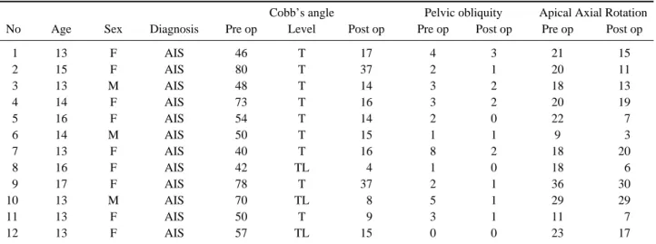
Table 1. Pre and post operative Cobb’s angle, pelvic obliquity and apical axial rotation for AIS group.
Cobb’s angle Pelvic obliquity Apical Axial Rotation
No Age Sex Diagnosis Pre op Level Post op Pre op Post op Pre op Post op
11 13 F AIS 46 T 17 4 3 21 15
12 15 F AIS 80 T 37 2 1 20 11
13 13 M AIS 48 T 14 3 2 18 13
14 14 F AIS 73 T 16 3 2 20 19
15 16 F AIS 54 T 14 2 0 22 17
16 14 M AIS 50 T 15 1 1 9 13
17 13 F AIS 40 T 16 8 2 18 20
18 16 F AIS 42 TL 14 1 0 18 16
19 17 F AIS 78 T 37 2 1 36 30
10 13 M AIS 70 TL 18 5 1 29 29
11 13 F AIS 50 T 19 3 1 11 17
12 13 F AIS 57 TL 15 0 0 23 17
Abbreviations: AIS: Adolescent idiopathic scoliosis; T: thoracic apex; TL: thoracolumbar apex.
Table 2. Pre and post operative Cobb’s angle, pelvic obliquity and apical axial rotation for NMS group.
Cobb’s angle Pelvic obliquity Apical Axial Rotation
No Age Sex Diagnosis Pre op Level Post op Pre op Post op Pre op Post op
11 16 F DCP 164 TL 27 15 13 40 30
12 22 M DCP 140 LL 17 18 14 34 20
13 19 M DCP 152 TL 16 16 12 32 39
14 23 M DCP 165 LT 18 11 16 15 15
15 21 M DCP 108 TL 39 15 15 60 50
16 14 M DMD 100 TL 38 17 15 58 46
17 17 M DMD 181 TL 59 22 17 72 57
18 14 M DMD 140 LL 19 11 14 23 19
19 12 M DMD 166 LL 28 26 12 41 29
10 16 M DMD 183 TL 36 16 12 48 44
11 10 M DMD 146 TL 13 10 18 20 17
12 19 F SMA 171 LT 15 14 14 21 17
13 28 F SMA 192 TL 55 27 12 54 40
14 13 F SMA 112 TL 24 20 16 59 46
15 18 F SMA 108 TL 30 10 13 47 40
16 13 F SMA 182 TL 41 11 16 63 18
Abbreviations: CP: Cerebral palsy; DMD: Duchene muscular dystrophy; SMA: spinal muscular atrophy; T: thoracic apex; TL: thora- columbar apex; L: Lumbar apex.
cal axial rotation were 27.1。, 5.8。, and 34.1。, respectively.
After surgery, the average correction was 65.1% (p<0.0001, paired t-test) for Cobb’s angle, 49.3% (p=0.0008, paired t- test) for pelvic obliquity, and 18.3% (p=0.0003, paired t- test) for apical axial rotation (Table 2).
The correction rates were not different in the idiopathic and neuromuscular groups (Cobb’s angle p=0.306, pelvic obliquity p=0.887 and apical derotation p=0.113; unpaired t-test), despite less correction in apical rotation overall in neuromuscular scoliosis. Clinically, all patients exhibited postoperative improvement in walking ability, cosmetic appearance, and/or sitting balance, which improved quality of life.
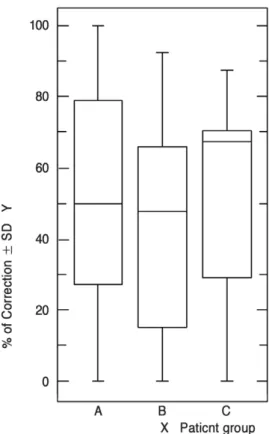
The correction in Cobb’s angle was significantly different in the three groups (p=0.04, ANOVA test) (Fig. 2) while correction in pelvic obliquity (p=0.79, ANOVA test) (Fig. 3) and apical derotation (p=0.25, ANOVA test) (Fig. 4) were not. Additionally we also compared the effect of correction in three diseases of NMS group: CP (cerebral palsy); DMD (Duchenne muscular dystrophy) and SMA (spinal muscular atrophy). The three different disease types, CP, DMD, and
SMA, did not show differences in Cobb’s angle (p=0.54), pelvic obliquity (p=0.10) or apical derotation (p=0.46) by ANOVA.
Discussion
Although, the apical vertebra shows maximum rotation9, the upper and lower end vertebrae also exhibit rotational components in scoliosis. The vertebral and inter-vertebral axial rotation10indicates the severity and rigidity of the scol- iosis curve. Surgical success depends on the correction of the Cobb’s angle, rotational angle, and translation of the vertebrae. Apical rotation outcomes are typically investigat- ed in idiopathic scoliosis11,12in the literature, with no report- ed outcomes in the neuromuscular group, probably because of the relatively small number of cases.
Here we have compared apical axial derotation in neuro- muscular and adolescent idiopathic scoliosis groups.
Although different methods can measure the axial rotation of vertebrae12,13,14,15, computerized tomography is the most
Fig. 2. Graph for analysis for Cobb’s angle. The X-axis denotes scoliosis groups: A:AIS group, B: NMS <80。, and C;
NMS >80。. The Y-axis denotes % of correction in Cobb’s angle with SD.
Fig. 3. Graph for analysis for pelvic obliquity. The X-axis denotes scoliosis groups: A:AIS group, B: NMS <80。, and C;
NMS >80。. The Y-axis denotes % of correction in pelvic obliquity with SD.
accurate. Since the pioneering work of Aaro-Dahlborn4,8in 1980 to measure axial rotation on CT scans, several other methods have been developed to measure axial rotation.
Gocen et al16in 1999 used a new technique to measure axial rotation by CT scan. Krismer et al9studied 11 cadavers and found that the Aaro-Dahlborn method was superior to other techniques. We used the same method here.
Moreover, surgical correction with modern techniques is three-dimensional17. Aaro-Dahlborn3evaluated 33 patients treated with Harrington instrumentation and found no axial derotation. Marchersi et al18used CT scans to measure dero- tation in four idiopathic scoliosis patients treated with Har- rington instrumentation, plus seven with Luque instrumen- tation, and found an average derotation of 16% and 12%, respectively, in the apical vertebrae. Using the same instru- mentation, Ecker et al19 found only 14% corrections with increase in rotation in some vertebrae. Bipedicular instru- mentation can achieve maximum correction in Cobb’s angle as well as axial rotation. In 1996, Jarvis and Greene20stud- ied Wisconsin segmental spinal instrumentation, a hybrid system with Harrington distraction rods, Luque rods, and
button-wire constructs, in 24 idiopathic scoliosis patients and found 23% derotation in 22 curves and 12% deteriora- tion in seven curves. They included double-curve patterns, which we excluded. Cundy et al21used the Aaro-Dahlborn method to study the effect of Cotrel-Dubosset instrumenta- tion on rotation in 34 idiopathic scoliosis patients and reported 24% derotation in relation to the mid-sagittal plane. Suk22in 1995 first proposed the use of thoracic pedi- cle screws as a fixation option for treatment of adolescent idiopathic scoliosis. Lonstein et al23in 1999, while studying coronal and sagittal plane correction in AIS using pedicle screw constructs or hybrid thoracic hook lumbar constructs noted a trend towards better correction of the main thoracic curve with pedicle screws, as was subsequently seen for lumbar curves as well24,25. Here we found a 71.4% correction in the coronal plane and a 31.3% derotation in the axial plane for adolescent idiopathic scoliosis, and a 65.1% cor- rection in the coronal plane and a 18.3% derotation in the axial plane for neuromuscular scoliosis, which were not dif- ferent (p=0.30 for Cobb’s angle and p=0.11 for apical dero- tation; unpaired t-test).
No published data exists on apical derotation in neuro- muscular scoliosis. Schufflebarger26 used CT scan to mea- sure rotation in relation to the mid sagittal plane in 18 patients with neuromuscular scoliosis using Cotrel-Dubos- set instrumentation with fixation up to the pelvis. His find- ings reveal an average correction in the frontal plane of 36�, a 42% correction, but did not measure axial rotation. Steib et al24studied derotation by in situ contouring of rods with pedicle screws in thoracic and lumbar curves in 10 idiopath- ic and 10 degenerative scoliosis patients and noted derota- tion ranging from 8�to 10�(62% to 67%). Although we used in situ contouring in a few cases, our main purpose was to prevent screw loosening from the pedicle. In 2003 Aubin et al27studied biomechanical modeling of posterior instrumentation of the scoliotic spine with Cotrel-Dubosset instrumentation using a three step procedure, and noted 18�
derotation in apical vertebrae, reflecting the kinematics of the rod-implant-vertebrae joint. Our all-pedicle screw con- struct with a posterior only approach produced comparable results with a similar derotation maneuver. In 2003, Basobas et al28 demonstrated excellent results for selective anterior fusion for the treatment of neuromuscular scoliosis in 20 patients (most with meningomyelocele) in their retro- spective study, but they did not comment on rotation. In 2002, Rhee et al29did not find a difference in their sagittal plane comparison of AIS after anterior versus posterior Fig. 4. Graph for analysis for apical derotation. The X-axis
denotes scoliosis groups: A:AIS group, B: NMS <80。, and C;
NMS >80。. The Y-axis denotes % of correction in apical rota- tions with SD.
instrumentation in 110 patients. Laohachroensombat et al30 found a significant difference in apical derotation (45%) in three dimensions after inserting pedicular screw plate con- structs in 25 idiopathic scoliosis patients.
We achieved nearly the same derotation in the CP, DMD, and SMA groups (p=0.46, ANOVA test), although we had small sample sizes and the results may depend on scoliosis severity. However, we found a 31% correction in apical rotation in the AIS group, but only an 18% change in the neuromuscular group.
Conclusion
In our retrospective study, we attained comparable changes in apical axial derotation in both groups, as well as in comparison of the AIS group with two severities of neu- romuscular scoliosis. Although correction in the coronal plane was different among the groups A, B and C (p=0.04, ANOVA test), we noted similar derotations in the apical vertebrae. There is no other apical derotation data for neuro- muscular scoliosis in the literature. However, the pedicle screw construct produces satisfactory outcomes in idiopath- ic scoliosis, prompting interest in future comparison studies of these two types of scoliosis.
REFERENCES
01. Kojima T, Kurokawa T: Quantitation of three-dimension- al deformity of idiopathic scoliosis. Spine 1992; 7(3S):
522-529.
02. Benson DR, DeWald RL, Schultz AB: Harrington rod distraction instrumentation. Its effect on vertebral rotation and thoracic compensation. Clin. Orthop 1977; 125: 40-44.
03. Aaro S, Dahlborn M: The effect of Harrington instrumen- tation on the longitudinal axis rotation of the apical verte- bra and on the spinal and rib-cage deformity in idiopathic scoliosis studied by computer tomography. Spine 1982; 7:
456-462.
04. Aaro S, Dahlborn M: Estimation of vertebral rotation and the spinal and rib cage deformity in scoliosis by computer tomography. Spine 1981; 6: 460-467.
05. Steib JG, Dumas R, Mitton D, Skalli W: Surgical correc- tion of scoliosis by in situ contouring: a detorsion analysis.
Spine 2004; 29: 193-199.
06. Chang K: Cantilever bending technique for treatment of
large and rigid scoliosis. Spine 2003; 28: 2452-2458.
07. Cobb J R: Outline for the study of scoliosis. In instruction- al course lectures. The American Academy of Orthopedic Surgeons 1948; 5: 261-275.
08. Aaro S, Dahlborn M, Svensson L: Estimation of vertebral rotation in structural scoliosis by computer tomography.
Acta Radiol. Diag 1978; 19: 990-992.
9. Deacon P, Flood BM, Dickson RA: Idiopathic scoliosis in three dimensions. A radiographic and morphometric analy- sis. J. Bone and Joint Surg 1984; 66-B: 509-512.
10. Krismer M, Sterzinger W, Christian H, Bernhard F, Rudolf B: Axial rotation measurement of scoliotic verte- brae by means of computed tomography scans. Diagnostic Imaging and Measurement Spine 1996; 21: 576-581.
11. Willers U, Transfeldt E, Hedlund R: The segmental effect of Cotrel-Dubousset instrumentation on vertebral rotation, rib hump and the thoracic cage in idiopathic scol- iosis. European Spine Journal 1996; 5: 12.
12. Perdriolle R, Vidal J: Morphology of scoliosis: three- dimensional evolution. Orthopedics. 1987; 10: 909-915.
13. Nash CL Jr, Moe JH: A study of vertebral rotation. J.
Bone and Joint Surg 1969; 51-A: 223-229
14. Suzuki S, Yamamuro T, Shikata K, Iida H: Ultrasound measurement of vertebral in idiopathic scoliosis. J Bone Joint Surg Br 1989; 71: 252-255.
15. Bunnell WP: Vertebral rotation: simple method of mea- surement on routine radiographs. Orthop. Trans 1985; 9:
114.
16. Go¨ çen S, Havitçioğ̆lu H, Alic E: A new method to mea- sure vertebral rotation from CT scans. Eur Spine J. 1999; 8:
261-265.
17. Ha JH, Lee JH, Ahn YJ, et al: The explanation of postop- erative change of vertebral rotation and rib hump using 3 dimensional finite element scoliosis model. J Korean Soci- ety of Spine Surgery 2003; 10: 14-24.
18. Marchesi DG, Transfeldt E, Bradford DS, Heithoff KB:
Changes in vertebral rotation after Harrington and Luque instrumentation for idiopathic scoliosis. Spine 1992; 17:
775-780.
19. Ecker ML, Betz RR, Trent PS, et al: Computer tomogra- phy evaluation of Cotrel-Dubousset instrumentation in idiopathic scoliosis. Spine 1988; 13: 1141-1144.
20. Jarvis JG, Greene RN: Adolescent Idiopathic Scoliosis.
Correction of Vertebral Rotation with Use of Wisconsin Segmental Spinal Instrumentation. J Bone Joint Surg [Am]
1996; 78-A: 1707-1712
21. Cundy PJ, Paterson DC, Hillier TM, et al: Cotrel-
Dubousset instrumentation and vertebral rotation in adoles- cent idiopathic scoliosis. J. Bone and Joint Surg 1990; 72 B: 670-674.
22. Suk SI, Lee CK and Kim WJ: Segmental pedicle screw fixation in the treatment of thoracic idiopathic scoliosis.
Spine 1995; 20: 1399-1405.
23. Lonstein J, Matsumoto H, Michael GV, et al: Coronal and sagittal plane correction in adolescent idiopathic scol- iosis: a comparison between all pedicle screw versus hybrid thoracic hook lumbar screw constructs. Spine 2007; 32:
448-452.
24. Steib JP, Ducrocq X, Averous C, Bogorin J: Surgical correction of lumbar scoliosis: a comparison of different techniques. Results. Eur J Orthop Surg Traumatol 1999; 9:
151-156.
25. Boos N, Webb JK: Pedicle screw fixation in spinal disor- ders: a European view. Eur Spine J 1997; 6: 2-18.
26. Schufflebarger HL, Neustadt JB, Cammisa FP: Spinal
fusions to the pelvis augmented by Cotrel-Dubousset instrumentation for neuromuscular scoliosis. Journal of Pediatric Orthopedics 1992; 12: 465-469.
27. Aubin CE, Prtit Y, Stokes IAF, et al: Biomechanical modeling of posterior instrumentation of the scoliotic spine Computer methods in Biomedical Engineering 2003; 6: 27- 32.
28. Basobas L, Steven Mardjetko, Kim H, John Lubicky:
Selective anterior fusion and instrumentation for the treat- ment of neuromuscular scoliosis. Spine 2003; 28(20S):
S245-S248.
29. Rhee JM, Bridwell KH, Won DS, Lenke LG: Sagittal plane analysis of adolescent idiopathic scoliosis: the effect of anterior versus posterior instrumentation. Spine. 2002;
27: 2350-2356.
30. Laohacharoensombat W, Jaovisidha S, Wajanavisit W, Sorasak S: Apical derotation in the treatment of idiopathic scoliosis. J Med Assoc Thai 2005; 88(Suppl 5): 558-564.