INTRODUCTION
Recently, there has been an increase in the numbers of breast cancer diagnosed in Korea. Breast cancer patients with the same stage of disease can have markedly different treatment responses and overall outcome. Furthermore, the biology of breast cancer remains poorly understood. As a result, breast cancers have become an increasingly significant problem in the evaluation and management of patients.
The cDNA microarray technology allows us the parallel analysis of the expression of thousands of genes to address complex questions in tumor biology (1). Systematic investi- gation of expression pattern of thousands of genes in breast cancers using cDNA microarray, and their correlation to spe- cific features of phenotypic variation, might provide the basis for an improved taxonomy of breast cancer (2-9). The ability to use appropriate profiles of gene expression could add enor- mous detail to the few known biological attributes in tumor characterization.
Elevated levels of -catenin have been associated with poor prognosis in breast cancer (10). However, the mechanisms by which -catenin confers the more aggressive biological behavior are poorly understood in breast cancer. -catenin
is a multifunctional protein that plays an essential role in the transduction of Wnt signaling and a component of the cad- herin cell adhesion complex (11, 12). In the absence of Wnt signaling, the cytoplasmic level of -catenin is kept low through interaction with a protein complex comprised of adenomatous polyposis coli (APC), Axin, protein phosphatase 2A (PP2A), and glycogen synthase kinase 3 (GSK3 ), where -catenin is phosphorylated by GSK3 (13, 14). Acti- vation of Wnt signaing leads to the inactivation of GSK3 resulting in accumulation of cytoplasmic -catenin. At ele- vated cytoplasmic levels, -catenin translocates to the nucle- us and cooperates with a member of the T cell factor (Tcf)/lym- phocyte enhancer binding factor (Lef) family of high-mobili- ty group box transcription factors to activate expression of target genes (13).
In this study, cDNA microarray analysis of approximately 4,600 cDNAs was performed to examine the gene expression profile in breast cancer on a more global scale. Immunohisto- chemical study was used to further demonstrate the specificity of the cDNA microarray results and to confirm the differen- tial expression genes that were known to have important role in -catenin regulation.
Mee Sook Roh, Sook Hee Hong, Jin Sook Jeong, Hyuk Chan Kwon*, Min Chan Kim�, Se Heon Cho�, Jin Han Yoon�, Tae Ho Hwang�
Department of Pathology, Internal Medicine*, Surgery�, Urology�, Pharmacology�, Dong-A University College of Medicine, Busan, Korea
Address for correspondence Mee Sook Roh, M.D.
Department of Pathology, Dong-A University College of Medicine, 1, 3-ga, Dongdaeshin-dong, Seo-gu, Busan 602-713, Korea
Tel : +82.51-240-2833, Fax : +82.51-243-7396 E-mail : [email protected]
275
Gene Expression Profiling of Breast Cancers with Emphasis of -Catenin Regulation
To gain molecular understanding of carcinogenesis of breast cancer, gene expres- sion profiles were analyzed using cDNA microarray representing 4,600 cDNAs in 10 breast cancer samples and the adjacent noncancerous breast tissues from the same patients. The alterations in gene expression levels were confirmed by reverse- transcription PCR in four randomly selected genes. Genes that were differently expressed in cancer and noncancerous tissues were identified. 106 (of which 55 were known) and 49 (of which 28 were known) genes were up- or down-regulated, respectively, in greater than 60% of the breast cancer samples. In cancer tissues, genes related to cell cycle, transcription, metabolism, cell structure/motility and signal transduction were mostly up-regulated. Furthermore, three cancer tissues showing immunohistochemically aberrant accumulation of -catenin in the nucleus and/or cytoplasm revealed down-regulation of Siah and Axin genes and up-regulation of Wnt and c-myc genes. These findings were highly consistent with Wnt signaling path- way associated with -catenin regulation previously suggested by others. Our stud- ies, therefore, provide not only a molecular basis to understand biological process- es of breast cancer but also useful resources to define the mechanism of -catenin expression in tumorigenesis of breast cancer.
Key Words : Oligonucleotide Array Sequence Analysis; Gene Expression; beta-Transducin Repeat Containing Proteins; Breast Neoplasms
Received : 16 October 2003 Accepted : 8 January 2004
MATERIALS AND METHODS Tissue samples and RNA preparation
Ten pairs of breast cancer tissues and corresponding adja- cent noncancerous breast tissues were obtained from 10 female patients who underwent lumpectomy or mastectomy at Dong- A University Medical Center between August 2001 and February 2002. Informed consent was obtained from patients to use their operated specimens and clinicopathological data for research purposes. The samples were anonymized before study. A pathologist dissected tissue samples from surgical specimens with special care for minimal contamination between cancer and noncancerous tissues and samples were immediately frozen in liquid nitrogen and stored at -80℃. Total RNA was isolated from the frozen tissue by using TRI REAGENT (Molecular Research Center, Inc., OH, U.S.A.).
The remaining parts of specimens were fixed with 10%
buffered formalin and the paraffin sections were stained with hematoxylin and eosin. Histological classifications were deter- mined by two pathologists in each case. When there was a discrepancy in interpretation of pathologic grade between the two observers, the hig-her grade was selected as repre- sentative grade.
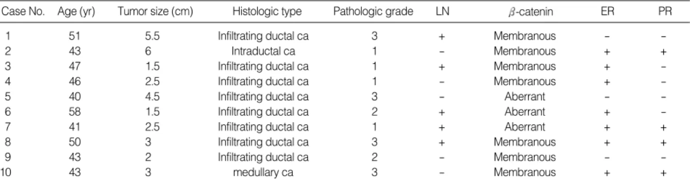
These clinical and histopathological features were summa- rized in Table 1.
Microarray analysis
Ten pairs of breast cancer and corresponding adjacent non- cancerous breast tissues were analyzed by MAGICTMmicroar- ray (Macrogen, Inc., Korea). This array contains 4,600 cDNAs, isolated from cDNA library of human breast cancer, and is composed of 4,370 known genes and 230 unknown genes (EST). For synthesizing double-stranded cDNA probe, label- ing kit provided by Macrogen was used. Microarray hybridiza- tion was performed according to manufacturer’s instructions.
Briefly, reverse transcription was performed on 30-50 g of total RNAs for 1 hr at 42℃with a T7-oligo (dT) 24 primer
and Superscript II reverse transcriptase (Life Technologies, Inc., Rockville, MD, U.S.A.). Second-strand cDNA synthe- sis was performed for 2 hr at 16℃with Escherichia coli DNA polymerase I, DNA ligase, and RNAse H (Life Technolo- gies, Inc., Rockville, MD, U.S.A.), followed by incubation in solulion containing 50 mmol/L NaOH/ 0.1 mmol/L EDTA for 10 min at 65℃, which caused degradation of the rRNA and tRNA. After phenolchloroform extraction in vitro, tran- scription was performed for 6 hr at 37℃using Cy5UTP and Cy3CTP, and a T7-Megascript kit (Ambion, Inc., Austin, TX, U.S.A.), and then fragmented for 35 min at 95℃. Hybri- dization (at 62℃for 16 hr) with the labeled cDNA in 6 mL of hybridization solution (6× SSC, 0.2% SDS, 5× Den- hardt solution, 1 mg/mL salmon sperm DNA) was followed by stringent washing (0.05× SSC, 0.2% SDS, at 58℃for 1 hr).
Data analysis
Images were analyzed by using the ImaGeneTManalysis software (BioDiscovery, Inc., Marina del Rey, CA, U.S.A.).
Spots showing no signal, or obvious defects, were excluded from the analysis. The local background was subtracted from the remaining spots and the ratios of net fluorescence from the Cy5-specific channel to the net fluorescence from the Cy3-specific channel were calculated for each spot. For this purpose, we determined transcript abundance using a custom algorithm, and the data set was trimmed of genes expressed at extremely low levels; that is, a gene was excluded if its 70 percentile value was less than 0.2 in 2 [log (tumor/normal)]
value. The results represented RNA expression in cancer relative to the corresponding noncancerous breast tissue.
A two-way pairwise average-linkage cluster analysis as an unsupervised clustering was applied. The cluster analysis was performed with Cluster 2.11, and the resulting expression map was visualized with TreeView 1.5. A set of microarray containing 4,600 cDNAs in duplicate was hybridized with a mixture of Cy3-labeled cDNA probes corresponding to can- cer tissues and Cy5-labeled cDNA probes corresponding to
Case No. Age (yr) Tumor size (cm) Histologic type Pathologic grade LN -catenin ER PR
1 51 5.5 Infiltrating ductal ca 3 + Membranous - -
2 43 6 Intraductal ca 1 - Membranous + +
3 47 1.5 Infiltrating ductal ca 1 + Membranous + -
4 46 2.5 Infiltrating ductal ca 1 - Membranous + -
5 40 4.5 Infiltrating ductal ca 3 - Aberrant - -
6 58 1.5 Infiltrating ductal ca 2 + Aberrant + -
7 41 2.5 Infiltrating ductal ca 1 + Aberrant + +
8 50 3 Infiltrating ductal ca 3 + Membranous + +
9 43 2 Infiltrating ductal ca 2 - Membranous - -
10 43 3 medullary ca 3 - Membranous + +
Table 1.Clinicopathologic summary of the breast cancer patients
Pathologic grade, histologic grade by Elston & Ellis method (35); LN, axillary lymph node metastasis; ER, estrogen receptor; PR, progesterone receptor;
ca, carcinoma.
noncancerous tissues. To identify genes with significantly dif- ferent expressions in breast cancer and noncancerous tissues, rational Cy3/Cy5 cut off values were determined. The differ- entially expressed genes, identified from cDNA microarray comparisons, were assigned to a modification of the NCBI Clusters of Orthologous Gene classification by searching the OMIM and PubMed databases (15). The genes were then assigned to 10 known functions (cell cycle regulator, transcrip- tion, oncogene/tumor suppressor gene, metabolism, cell struc- ture/motility, signal transduction, gene/protein expression, DNA repair, angiogenesis, and immunology) and a miscel- laneous category.
Semiquantitative RT-PCR
Single-stranded cDNA was synthesized with oligo (dT) primer in a 20- L reaction, from 5 g of total SuperScript Preamplification System for First Strand cDNA Synthesis System (Life Technologies, Inc., Rockville, MD, U.S.A.) and diluted up to final volume of 80 L. PCR was then performed with 1 L of cDNA, for 1 cycle of 94℃for 2 min, followed by 25 cycles of 94℃for 30 sec, 55℃for 30 sec, and 72℃for 1 min, using a gene-specific primer and Taq polymerase.
Primers were as follows; U76248 (Siah) sense, 5′-AAGC- TGTGATGTCCCATCTC-3′, antisense, 5′-AGTCTGTAG- GCAAAGTTCTC-3′, AF009674 (Axin) sense, 5′-TGAGA- ACTCCAGACCGTTGT-3′, antisense, 5′-TGCACATACC- TCTGCTTGGA-3′, D79995 sense, 5′-GGATGACGTGAT- ACTCAATG-3′, antisense, 5′-GGTAGAAAGGCTGGGC- TCTT-3′, X06825 (Tropomyosin 2 ) sense, 5′-CTGGTGA- TCCTGGAAGGAGA-3′, antisense, 5′-GCTTCTCCTCC- AACAGTTTG-3′.
Amplification of the correct target DNA was confirmed by mobility of gel electrophoresis and sequencing after sub- cloning into pGEM-T easy vector (Promega, Madison, WI, U.S.A.). -actin was used as an internal control to confirm equal amount between the templates.
Immunohistochemical stain
Paraffin-embedded 5 m-thick breast tumor sections were analyzed for -catenin expression by immunohistochemical staining. Immunohistochemical staining was performed using monoclonal mouse anti-(human Ig) antibody directed against -catenin (1:30, Zymed, CA, U.S.A.). Deparaffinization of all sections was performed through a series of xylene baths and rehydration was performed through graded alcohols. To enhance the immunoreactivity, microwave antigen retrieval at 750 W for 30 min in citrate buffer (pH 6.0) was performed.
After blocking the endogenous peroxidase activity with 5%
hydrogen peroxidase for 10 min, the primary antibody incu- bation for -catenin was performed at room temperature for 100 min. Detection of the immunoreactive staining was car- ried out by the avidin-biotin-peroxidase complex method
using Histostain-plus kit (Zymed, CA, U.S.A.). The antigen- antibody reaction was visualized using 3-amino-9-ethylcar- bazole as a chromogen. The Mayer’s hematoxylin counterstain was performed.
Immunohistochemical staining for -catenin expression was divided into two groups according to their immunohis- tochemical patterns by a pathologist: normal membranous pattern seen in non-neoplastic epithelium and aberrantly accumulated pattern showing nuclear and/or cytoplasmic staining with or without membranous staining.
RESULTS
Expression profiling revealed differentially expressed genes in breast cancer and noncancerous tissues
We categorized ten pairs of breast cancer by comparing the global gene expression patterns of cancer and noncancerous tissue after data trimming. Of the 4,600 genes, a total of 1,084 genes was left after data trimming. By unsupervised hierar- chical clustering based on 1,084 genes, the cancer and non- cancerous tissues were successfully distinguished (data not shown). However, this algorithm using most of the genes on the array would not classify samples among cancer tissues by subgroups associated with clinical and histopathological fea- tures including age, tumor size, status of axillary lymph nodes, histologic type of the tumor, pathologic grade, and hormone- receptor status.
When cut off values were set to 2.0, for the ratio between the cancerous and noncancerous tissues, 106 (of which 55 were known) and 49 (of which 28 were known) genes or cDNA were up- or down-regulated, respectively, in >60% of the breast cancer samples. These genes were classified in terms of 10 functions (cell cycle regulator, transcription, oncogene/
tumor suppressor gene, metabolism, cell structure/motility, signal transduction, gene/protein expression, DNA repair, angiogenesis, and immunology), as shown in Table 2. In the cancerous tissues, the genes related to cell cycle regulator, tran- scription, metabolism, cell structure/motility and signal trans- duction were mostly up-regulated. We identified consistent results of several genes that had some roles in cancer biology previously suggested by other workers, including pituitary tumor-transforming gene (PTTG) (16), histone acetyltrans- ferase (HAT) (17), c-src tyrosine kinase (CSK) (18), and insu- lin-like growth factor binding protein (7).
Validation of DNA microarray by semiquantitative RT-PCR
To verify the reproducibility of these gene lists, we perform- ed semiquantitative RT-PCR using the same RNA used in the microarray analysis. Ten pairs of the cancerous and corre- sponding noncancerous tissue samples were analyzed using
Function Accession No. Gene 2 log (T/N) Frequency
Cell cycle regulator D79995 KIAA0173 gene product -1.16 7
NM_012330 Histone acetyltransferase -1.3 6
NM_11722 Deoxynucleotidyltransferase -2.17 6
D14678 Kinesin-like 2 4.81 7
X66365 Cyclin-dependent kinase 6 2.74 7
U76248 Seven in absentia (Drosophila) homolog2 2.64 7
AF086947 Dynectin 1 2.76 6
U40705 Telomerin repeat binding factor 2.72 6
AB017430 Kinesin-like 4 2.67 6
NM_018667 Neutral sphingomyelinase II 2.32 6
AF095289 Pituitary tumor-transforming 3 1.8 6
X14850 H2A histone family, member X 1.48 6
Transcription NM_016331 Zinc finger protein ANC-2H01 2.24 9
U09412 Zinc finger protein 134 2.59 8
NM_013242 Similar to mouse Glt3 2.4 8
Oncogene/tumor suppressor gene AJ250014 KIAA0849 protein -1.6 7
K02276 Translocated t(8;14) c-myc oncogene -1.07 6
Y09160 Rho guanine nucleotide exchange factor 1 2.74 7
AF009674 Axin 1.53 7
Metabolism NM_015831 Acetylcholinesterase -1.48 6
Z25535 Nucleoporin 153 kDa -1.67 6
S69232 Electron-transferring-ubiquinone oxidoreductase -2.55 6
X59960 Sphingomyelin phosphodiesterase 1 3.26 7
M91211 Advanced glycosylation end product 2.92 7
X69433 Isocitrate dehydrogenase 2 2.21 7
AF069250 Acid-inducible phosphoprotein 2.33 6
AF064254 VLCS-H1 protein 11.5 6
Cell structure/Motility U61234 Tubulin-specific chaperone c -2.6 7
X06825 Tropomyosin 2 (beta) -1.18 6
M60922 Flotillin 2 -1.97 6
AB006780 Lectin 3.34 8
X14420 Collagen, type III, alpha 1 4.61 7
U80184 Flightless I (Drosophila) homolog 3.32 7
U33931 Erythrocyte membrane protein band 7.2 2.69 7
AF006087 Actin related protein 2/3 complex, subunit 4 1.93 6
Signal transduction L28824 c-src tyrosine kinase -1.43 7
AF078103 Develpomentally regulated GTP binding protein -1.81 7
AF263541 Dual-specificity tyrosine-(Y)-phophatase -2.46 7
M35878 Insulin-like growth factor binding protein -1.2 6
AF189009 Ubiquilin 2 -1.83 6
AF070599 Protein phosphatase 1 3.98 7
NM_004409 Dystrophia myotonica-protein kinase 3.28 7
U10099 POM-ZP3 mRNA 2.98 7
AF114012 Tumor necrosis factor (ligand) superfamily 2.61 7
U02680 Protein tyrosine kinase 9 3.03 6
AB014561 95kDa retinoblastoma protein binding protein 3.02 6
X78947 Connective tissue growth factor 1.37 6
Gene/protein expression U46571 Tetratricopeptide repeat domain 2 -2.06 8
NM_020365 Eukaryotic translation initiation factor -1.51 7
X70944 Splicing factor proline/glutamine rich -2.97 7
X76013 Glutamyl-tRNA synthetase -2.08 6
AB014546 Ring finger protein 8 4.34 7
NM_014018 Mitochondrial ribosomal protein 4.36 6
D13641 KIAA0016 3.76 6
DNA repair NM_016042 CGI-102 protein 3.56 8
M74905 N-methylpurine-DNA glycosylase 2.39 7
(Table 2, continued next) Table 2.Representative differential expression in breast cancer, defined a 2-fold or greater change relative to expression in noncancer- ous tissues, for various functional classes of genes
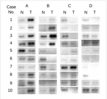
four randomly selected genes; U76248 (Siah), AF009674 (Axin), D79995, and X06825 (Tropomysin 2 ). Of these genes, two genes associated with -catenin regulation, Siah, the mammalian homologue of Drosophila seven in absentia, and Axin, were included. The results of the breast cancer genes found from the semiquantitative RT-PCR analyses were mostly consistent with those from the microarray studies (Fig. 1).
Genes associated with -catenin regulation
When we reviewed genes that were differentially expressed in cancer and noncancerous tissue (Table 2), we could readily identify genes associated with -catenin regulation. Three cancerous tissues (case 5-7) showing immunohistochemically aberrant accumulation of -catenin revealed down-regulation of Siah and Axin genes and up-regulation of Wnt and c-myc genes. Of the remaining seven cancerous tissues without aber- rant -catenin expression, four cases (case 1, 2, 8, 9) showed that the results of the expression of these genes were vice versa and remaining three cases (case 3, 4, 10) showed inconsistent findings (Fig. 2, Table 3).
DISCUSSION
We have globally analyzed gene expression of breast can- cer and noncancerous tissues to elucidate the characteristic changes associated with the carcinogenesis and progression in breast cancer. The cancer and noncancerous tissues were distinguished by gene expression profiling. In the cancer tis- sues, genes related to cell cycle, transcription, metabolism, cell structure/motility and signal transduction were mostly up-regulated in this study. We categorized ten pairs of breast cancer by comparing the global gene expression patterns of cancer and noncancerous tissue after data trimming. In this study, genes with expression ratios that varied at least 2-fold between the cancer and noncancerous tissues in 60% of the samples were selected. Although it resulted in the loss of
Function Accession No. Gene 2 log (T/N) Frequency
DNA repair J04973 Human cytochrome bc-1 complex core protein II 2.33 7
U12134 RAD52 (S. cerevisiae) homolog 2.4 6
Angiogenesis AF035121 Kinase insert domain receptor -2 6
U03644 CBF1 interacting corepressor 2.46 7
Immunology X14723 Clusterin -1.9 8
X53961 Lactotransferrin -2.19 7
AF110908 TNF receptor-associated factor 3 2.81 6
U12255 Fc fragment of IgG, receptor, transporter 2.3 6
X57809 Immunoglobulin lambda locus 1.73 6
J03909 Interferon, gamma-inducible protein 30 1.42 6
Table 2.(continued from the previous page)Representative differential expression in breast cancer, defined a 2-fold or greater change relative to expression in noncancerous tissues, for various functional classes of genes
2 log (T/N), 2 log (Tumor/Noncancerous tissue).
2 log (T/N) Case No.
Wnt-1 Siah Axin c-myc
1 -0.8 1.9 1.5 -1.5
2 -1.3 2.3 1.4 -1.5
3 F 3.5 0.6 -1.7
4 F 3.1 2.1 1.3
5 0.8 -0.6 -0.6 1.7
6 0.7 -0.8 -0.6 1.5
7 1.2 -1.4 -1.3 1.8
8 -1.8 4.1 2 -1.2
9 -1.9 2.1 1.6 -1.2
10 2 1.5 1.5 -2.5
Table 3.Differential expression pattern of genes associated with -catenin regulation according to the immunohistochemical findings for -catenin
2 log (T/N), 2 log (Tumor/Noncancerous tissue); F, failed.
Case A No.
1 2 3 4 5 6 7 8 9 10
N T
B
N T
C
N T
D
N T
Fig. 1.The results of semiquantitative RT-PCR for the 4 tested genes in the 10 breast cancer tissues and the noncancerous tissues, con- sisting with those from the microarrary studies. N, noncancerous tissue; T, tumor tissue; A, U76248 (Siah); B, AF009674 (Axin); C, D79995; D, X06825 (Tropomyosin 2 ).
some information, trimming in this manner decreased the possibility that the clustering algorithm would be strongly influenced by genes with little or no expression.
Most of the genes associated with the cell cycle were up- regulated, and examplified by the cyclin-dependent kinase 6 (CDK6) and PTTG. CDK6 is considered as a cdc-2 related kinase, which can play roles in the regulation of the mam- malian cell cycle. The cyclin D/CDK6 complex phosphory- lates the retinoblastoma protein. Under-phosphorylated pRb binds to the E2F family of transcription factors. Phosphory- lation of the pRb unshackles the E2F proteins, which in turn activates the transcription of several genes whose products are essential for progression through the S phase (19). Because the state of the pRb phosphorylation is crucial in determin- ing the cell cycle progression, the up-regulation of CDK6 in cancer tissues was not unexpected. PTTG has been identified as a key protein in mitotic regulation (20) and is highly ex- pressed in pituitary tumor (21) and other neoplasms (22, 23).
PTTG protein has been recognized as a mammalian securin protein that maintains binding of sister chromatids during mitosis. Overexpression of a nondegradable PTTG disrupts sister chromatid separation. PTTG overexpression results in genetic instability (24). Puri et al. (25) revealed a high level of expression of PTTG1 mRNA in both seminomatous and non-seminomatous testicular tumors, epithelial cell, sex-cord stromal cell, and germ cell tumors of the ovary, and in situ and invasive ductal carcinoma of the breast. In this study of breast cancer, PTTG was up-regulated in the cancerous tissue, which agrees with the results from a previous study (25) and suggests a certain role in tumorigenesis of breast cancer. On the other hand, the HAT directly links chromatin modifica-
tion to genetic activation and has also important activities in many cellular processes including cell cycle progression, transcription and DNA replication (17). Developmental aber- rations in mice and certain human cancers are associated with HAT mutations, further highlighting the important of these enzymes to normal cell growth and differentiation (26). In our study, this gene was down-regulated in cancer tissue which is consistent with the mechanism underlying tumor forma- tion of mutations of HAT genes involved with the regulation and control of the cell cycle.
The loss of tumor suppressor gene is a key event in many, possibly all, human tumors, as the physiological function of these genes are in the regulation of normal cell growth includ- ing the cell cycle and nuclear transcription. We have identi- fied a tumor suppressor gene that has previously been proposed by other research (18). The CSK has been shown to down- regulate the tyrosine kinase activity of the c-src oncoprotein through tyrosine phosphorylation of the c-src carboxyl termi- nus. The CSK gene could therefore potentially function as an antioncogene. Myoui et al. (27) examined the role of CSK in bone metastasis using an animal model with an inoculation of breast cancer cells and reported that CSK was an essential molecule for bone resorption by osteoclasts, which were cen- tral players in osteolytic bone metastases. They suggested the notion that CSK was a potential target molecule for design- ing novel therapeutic interventions, especially for bone metas- tases in breast cancer. In our study, CSK was down-regulated in cancer tissue of seven cases, which suggested that they need- ed to closer follow-up for bone metastasis. Identification of such a target may improve the efficiency of developing ther- apeutics for breast cancer.
Fig. 2.Immunohistochemical stain for -catenin in breast cancer. (A) Membranous pattern from case 1 (×200). (B) Aberrantly accumulat- ed pattern in the nucleus and cytoplasm from case 5 (×400).
A B
cDNA microarray provided a powerful alternative with an unprecedented view scope in monitoring of the gene expres- sion levels and led to discoveries of regulatory pathways in- volved in complicated biological processes. Differences in gene expression are likely to explain the phenotype variation. We used it to answer the specific question whether could be dis- tinguished by their gene expression profiles and whether these could be related to differences in biological effects. Lin et al.
(10) reported that high -catenin activity significantly corre- lated with poor prognosis of the patients and was a strong and independent prognostic factor in breast cancer. Lim et al. (28) reported that aberrant expression of -catenin was identified in 79% of breast cancer and there was correlation of aberrant expression of -catenin with lymph node metastasis, survival rate, and survival length in Korean patients with breast can- cer. -catenin is an executor of Wnt signaling. Components of Wnt signaling pathway include proto-oncogene products, such as -catenin, tumor suppressor proteins, such as APC, and Axin. It has been reported that Wnt genes are sometimes overexpressed in human breast cancer and there is a growing evidence that downstream components of the Wnt signaling pathway are activated in a significant proportion of breast tumors (29). Axin, a negative regulator of this pathway, pro- motes phoasphorylation of serine/threonine in exon 3 of - catenin by forming a complex with APC and GSK-3 . Phos- phorylated -catenin is quickly degraded via an ubiquitin- proteasome pathway in the cytoplasm. Upon Wnt signaling, phosphorylation of -catenin functions as a transcriptional regulator in the nucleus coupled with molecules of the Tcf/Lef family (13, 14). Siah is the mammalian homolog of Drosophila seven in absentia, required for formation of the R7 photore- ceptor cells during eye development (30). Siah-1 mediated downregulation of -catenin was induced by p53, providing a link between p53 and cell cycle regulation through a novel -catenin degradation pathway (31). Our study indicated that -catenin can be involved in breast cancer formation and/or progression and may serve as a target for breast cancer therapy, if we identified gene respect to regulatory pathways involved in -catenin expression. In this study, three cancer tissues show- ing immunohistochemically aberrant accumulation of -ca- tenin in the nucleus and/or cytoplasm revealed down-regula- tion of Siah and Axin genes and up-regulation of Wnt and c- myc genes. Of the remaining seven cancer tissues without aberrant -catenin expression, four cases showed that the results of these genes expression were vice versa. Although the remaining three cases showed inconsistent findings, our study revealed consistent findings of the mechanism of - catenin expression in tumorigenesis of breast cancer. However, because the cases studied had short follow-up periods, we can- not determine the relation between -catenin expression and prognostic significance.
We have analyzed whole cancer tissues in this study instead of focusing only on cancer cells to better describe the entire aspect of breast cancer, because breast cancer tissues as other
cancers generally contain multiple nonepithelial cell types such as fibroblast, smooth muscle cell, endothelial cell, infil- trating lymphocyte, and macrophage. Recent advances in cancer research have revealed the relevance of epithelial-stro- mal interaction including extracellular matrices, matrix met- alloproteinases, and angiogenic factors in cancer progression (32). Accordingly, targets of cancer therapeutics have been recently extended from molecules of cancer origin to molecules to stroma origin such as those related to angiogenesis and matrix remodeling (33, 34). As this study, it was based on whole tissue samples, the list of genes up-regulated in cancer tissues contained and may still contain many genes for stroma.
With further functional study, potentially therapeutic target molecules of stroma might be identified.
These results provide not only a new molecular basis for understanding biological properties of breast cancer, but also useful resources for future development of therapeutic targets for breast cancer. To investigate the molecular understanding of regulatory pathways involved in the complicated carcino- genic processes, especially associated with the expression of -catenin, a downstream effector of Wnt-mediated tumorige- nesis in breast cancer, was also suggested. The genes suggested by this analysis may serve as candidates for more detailed exam- ination by using a larger number of clinical samples and veri- fication at the protein level.
ACKNOWLEDGEMENT
This work was supported by the Korea Science and Engi- neering Foundation (KOSEF) through the MRCCMT at the Dong-A University.
REFERENCES
1. Shalon D, Smith SJ, Brown PO. A DNA microarray system for analyz- ing complex DNA samples using two-color fluorescent probe hybri- dization. Genome Res 1996; 6: 639-45.
2. Sorlie T, Perou CM, Tibshirani R, Aas T, Geisler S, Johnsen H, Hastie T, Eisen MB, van de Rijn M, Jeffrey SS, Thorsen T, Quist H, Matese JC, Brown PO, Botstein D, Eystein Lonning P, Borresen-Dale AL.
Gene expression patterns of breast carcinomas distinguish tumor subclasses with clinical implications. Proc Natl Acad Sci USA 2001;
98: 10869-74.
3. van’t Veer LJ, Dai H, van de Vijver MJ, He YD, Hart AAM, Mao M, Peterse HL, van der Kooy K, Marton MJ, Witteveen AT, Schreiber GJ, Kerkhoven RM, Roberts C, Linsley PS, Bernards R, Friend SH.
Gene expression profiling predicts clinical outcome of breast cancer.
Nature 2002; 415: 530-6.
4. Jenssen TK, Kuo WP, Stokke T, Hovig E. Associations between gene expressions in breast cancer and patient survival. Hum Genet 2002;
111: 411-20.
5. van de Vijver MJ, He YD, van’t Veer LJ, Dai H, Hart AAM, Voskuil
DW, Schreiber GJ, Peterse JL, Roberts C, Marton MJ, Parrish M, Atsma D, Witteveen A, Glas A, Delahaye L, van der Velde T, Barte- link H, Rodenhuis S, Rutgers ET, Friend SH, Bernards R. A gene- expression signature as a predictor of survival in breast cancer. N Engl J Med 2002; 347: 1999-2009.
6. Huang E, Cheng SH, Dressman H, Pittman J, Tsou MH, Horng CF, Bild A, Iversen ES, Liao M, Chen CM, West M, Nevins JR, Huang AT. Gene expression predictors of breast cancer outcomes. Lancet 2003; 361: 1590-6.
7. Gruvberger S, Ringner M, Chen Y, Panavally S, Saal LH, Borg A, Ferno M, Peterson C, Meltzer PS. Estrogen receptor status in breast cancer is associated with remarkably distinct gene expression patterns.
Cancer Res 2001; 61: 5979-84.
8. Kim HS, Jung JH, Park HY, Lee YH, Chung EJ, Kim MK, Kim JC.
Gene expression profile analysis of human breast cancer using cDNA microarrays. J Korean Breast Cancer Soc 2003; 6: 58-67.
9. Han W, Chung KW, Ahn SJ, Noh DY, Youn YK, Oh SK, Choe KJ.
Gene expression profiles of primary breast cancer tissue using cDNA microarray. J Korean Breast Cancer Soc 2002; 5: 284-90.
10. Lin SY, Xia W, Wang JC, Kwong KY, Spohn B, Wen Y, Pestell RG, Hung MC. -catenin, a novel prognostic marker for breast cancer:
its roles in cyclin D1 expression and cancer progression. Proc Natl Acad Sci USA 1999; 97: 4262-6.
11. Bienz M, Clevers H. Linking colorectal cancer to Wnt signaling. Cell 2000; 103: 311-20.
12. Peifer M, Polakis P. Wnt signaling in oncogenesis and embryogene- sis: a look outside the nucleus. Science 2000; 287: 1606-9.
13. Morin PJ, Sparks AB, Korinek V, Barker N, Clevers H, Vogelstein B, Kinzler KW. Activation of -catenin-Tcf signaling in colon can- cer by mutations in -catenin or APC. Science 1997; 275: 1787-90.
14. Rubinfeld B, Souza B, Albert I, Muller O, Chamberlain SH, Masiarz FR, Munemitsu S, Polakis P. Association of the APC gene product with
-catenin. Science 1993; 262: 1731-4.
15. Lam AK. Molecular biology of esophageal squamous cell carcino- ma. Crit Rev Oncol Hematol 2000; 33: 71-90.
16. McCabe C. Genetic targets for the treatment of pituitary adenomas:
focus on the pituitary tumor transforming gene. Curr Opin Pharma- col 2001; 1: 620-5.
17. Johnstone RW. Histone-deacetylase inhibitors: novel drugs for the treatment of cancer. Nat Rev Drug Discov 2002; 4: 287-99.
18. Armstrong E, Cannizzaro L, Bergman M, Huebner K, Alitalo K. The c-src tyrosine kinase (CSK) gene, a potential antioncogene, localizes to human chromosome region 15q23-25. Cytogenet Cell Genet 1992;
60: 119-20.
19. Sherr CJ. Cancer cell cycles. Science 1996; 274: 1672-7.
20. Zhang X, Horwitz GA, Prezant TR, Valentini A, Nakashima M, Bron- stein MD, Melmed S. Structure, expression, and function of human
pituitary tumor-transforming gene (PTTG). Mol Endocrinol 1999;
13: 156-66.
21. Zhang X, Horwitz GA, Heaney AP, Nakashima M, Prezant TR, Bron- stein MD, Melmed S. Pituitary tumor transforming gene (PTTG) expression in pituitary adenomas. J Clin Endocrinol Metab 1999;
84: 761-7.
22. Dominguez A, Ramos-Morales F, Romero F, Rios RM, Dreyfus F, Tortolero M, Pintor-Toro JA. hpttg, a human homologue of rat pttg, is overexpressed in hematopoietic neoplasms. Evidence for a transcrip- tional activation function of hPTTG. Oncogene 1998; 17: 2187-93.
23. Saez C, Japon MA, Ramos-Morales F, Romero F, Segura DI, Tortolero M, Pintor-Toro JA. hpttg is over-expressed in pituitary adenomas and other primary epithelial neoplasias. Oncogene 1999; 18: 5473-6.
24. Zou H, McGarry TJ, Bernal T, Kirschner MW. Identification of a vertebrate sister-chromatid separation inhibitor involved in transfor- mation and tumorigenesis. Science 1999; 285: 418-22.
25. Puri R, Tousson A, Chen L, Kakar SS. Molecular cloning of pituitary tumor transforming gene 1 from ovarian tumors and its expression in tumors. Cancer Lett 2001; 163: 131-9.
26. Marmorstein R, Roth SY. Histone acetyltransferases: function, struc- ture, and catalysis. Curr Opin Genet Dev 2001; 11: 155-61.
27. Myoui A, Nishimura R, Williams PJ, Hiraga T, Tamura D, Michigami T, Mundy GR, Yoneda T. C-SRC tyrosine kinase activity is associated with tumor colonization in bone and lung in an animal model of human breast cancer metastasis. Cancer Res 2003; 63: 5028-33.
28. Lim SC, Lee MS. Significance of E-cadherin/beta-catenin complex and cyclin D1 in breast cancer. Oncol Rep 2002; 9: 915-28.
29. Brown AM. Wnt signaling in breast cancer: have we come full circle?
Breast Cancer Res 2001; 3: 351-5.
30. Carthew RW, Rubin GM. Seven in absentia, a gene required for spec- ification R7 cell fate in the Drosophila eye. Cell 1990; 63: 561-77.
31. Liu J, Stevens J, Rote CA, Yost HJ, Hu Y, Neufeld KL, White RL, Matsunami N. Siah-1 mediates a novel -catenin degradation path- way linking p53 to the adenomatous polyposis coli protein. Mol Cell 2001; 7: 927-36.
32. Matrisian LM, Cunha GR, Mohla S. Epithelial-stromal interactions and tumor progression: meeting summary and future directions. Can- cer Res 2001; 61: 3844-6.
33. Curran S, Murray GI. Matrix metalloproteinase: molecular aspects of their roles in tumor invasion and metastasis. Eur J Cancer 2000;
36: 1621-30.
34. Deplanque G, Harris AL. Anti-angiogenic agents: clinical trial design and therapies in development. Eur J Cancer 2000; 36: 1713-24.
35. Elston CW, Ellis IO. Pathological prognostic factors in breast can- cer. I. The value of histological grade in breast cancer: experience from a large study with long-term follow-up. Histopathology 1991;
19: 403-10.