INTRODUCTION
We report a rare case of surgically confirmed total absence of the hemidiaphragm in a 70-yr-old woman with the clini- cal presentation of an acute mesenteroaxial gastric volvulus and diaphragmatic eventration. The acute gastric volvulus (AGV) and herniated abdominal organs resolved spontaneous- ly without reconstruction of the diaphragm.
CASE REPORT
A 70-yr-old woman, previously in good health was trans- ferred to our hospital with the history of left sided chest pain, non-bilious vomiting and shortness of breath for four days.
Six days before admission, the patient had a routine health check-up including chest radiography and upper gastroin- testinal study (UGIS) using a gastric bloating agent at a local clinic. According to the referring clinic, the chest radiogra- phy and UGIS were normal (Fig. 1A, 2A). However, after tak- ing the gastric bloating agent the patient developed increasing
epigastric pain and nausea.
On admission, the respiratory rate of the patient was 38 breaths/min and the oxygen saturation was 91% in room air.
Physical examination showed absent breath sounds and dull to percussion findings at the left lung base. The chest radio- graphy revealed gastric air-fluid levels and bowel loops in the left thoracic cavity. After the placement of a nasogastric tube, the chest radiography showed displacement of the tip of the tube to the dilated stomach located in the left thoracic cavity (Fig. 1B). An UGIS using gastrograffin was performed with- out a bloating agent (Fig. 2B). The UGIS showed no transit of the gastrograffin through the duodenum and a hook-like image was noted at the medial portion of the herniated stom- ach. A chest CT revealed absence of the left side of the dia- phragm (Fig. 3). The impression was that the patient had a mesenteroaxial gastric volvulus with a diaphragmatic hernia.
The patient underwent surgical exploration.
A left posterolateral thoracotomy was done at the sixth inter- costal space. We examined the surface of the entire lung field but could not find any diaphragmatic tissue. The abdominal organs (stomach, spleen, splenic flexure of colon) were present
517
Hye Young Sung1, Se Hyun Cho1, Sung Bo Sim2, Jin Il Kim1,
Dae Young Cheung1, Soo-Heon Park1, Joon-Yeol Han1, Se Min Lee1, Chee Ho Noh1, Yong-Bum Park1, Seung Eun Jung3, Seon Hui Lee2, and Kyu Yong Choi1
Division of Gastroenterology, Departments of Internal Medicine1, Thoracic Surgery2, and Radiology3, The Catholic University of Korea, Seoul, Korea
Address for correspondence Se Hyun Cho, M.D.
Division of Gastroenterology, Department of Internal Medicine, The Catholic University of Korea, St. Mary’s Hospital, 62 Yeouido-dong, Yeongdeungpo-gu, Seoul 150-713, Korea
Tel : +82.2-3779-1269, Fax : +82.2-780-3132 E-mail : [email protected] J Korean Med Sci 2009; 24: 517-9
ISSN 1011-8934
DOI: 10.3346/jkms.2009.24.3.517
Copyright � The Korean Academy of Medical Sciences
Congenital Hemidiaphragmatic Agenesis Presenting as Reversible Mesenteroaxial Gastric Volvulus and Diaphragmatic Hernia:
A Case Report
A 70-yr-old woman complained of left sided chest pain and non-bilious vomiting for four days after taking a gastric bloating agent for an upper gastrointestinal study. The chest radiography revealed gastric air-fluid levels and bowel loops in the left thoracic cavity. An emergency thoracotomy was performed. The abdominal organs (stom- ach, spleen, splenic flexure of the colon) were in the left thorax and the entire left hemidiaphragm was absent. There were no diaphragmatic remnants visible for re- construction of the left diaphragm. We provided warm saline irrigation and performed a left lower lobe adhesiotomy. Thirteen days after surgery, the chest radiography showed improvement in the herniation but mild haziness remained at the left lower lung field. Here we present the oldest case of congenital diaphragmatic agenesis presenting with transient gastric volvulus and diaphragmatic hernia.
Key Words : Congenital Hemidiaphragmatic Agenesis; Mesenteroaxial Gastric Volvulus; Hernia, Diaphrag- matic; Spontaneous Resolution
Received : 15 November 2007 Accepted : 23 February 2008
in the left thoracic cavity (Fig. 4). We attempted to rearrange the contents for devolvulation but could not achieve this due to the presence of severe pleural adhesions. Warm saline irri- gation and a left lower lobe adhesiotomy were performed.
There was no diaphragmatic tissue; we could not repair the diaphragm and return the abdominal organs, from the tho- rax, to the abdomen. The pleural cavity was closed in the usual manner. Conservative treatment was continued with noth- ing per oral and nasogastric drainage. Follow-up UGIS was performed 10 days after surgery (Fig. 2C); the abdominal organs remained in the left thoracic cavity but there was no disturbance of gastric passage. The patient improved by 12 days after surgery, on a soft diet. Thirteen days after surgery the chest radiography showed resolution of the herniation but mild haziness remained at the left lower lung field (Fig.
1C). The patient was discharged 20 days after admission.
During the 1 yr of follow-up, the patient continued to do well.
DISCUSSION
The diaphragm is derived from the septum transversum, pleuroperitoneal membrane and the thoracic wall, while the gastrohepatic ligament and dorsal mesentery are minimally involved. Agenesis of the hemidiaphragm involves non-devel- opment of all of the related components and is very rare (1).
Cunniff et al. (2) reported a 6% incidence of diaphragmatic agenesis in their review of 102 liveborn children with con- genital diaphragmatic defects. This anomaly is usually asso- ciated with pulmonary hypoplasia, resulting in progressive respiratory failure and death in the neonatal period. However, some infants survive with this condition. In a review of the literature, asymptomatic presentation of a complete diaphrag- matic agenesis has been previously reported (3, 4). In such rare cases, the gastrophrenic and gastrosplenic ligaments may have supported the intraabdominal organs so that they did
518 H.Y. Sung, S.H. Cho, S.B. Sim, et al.
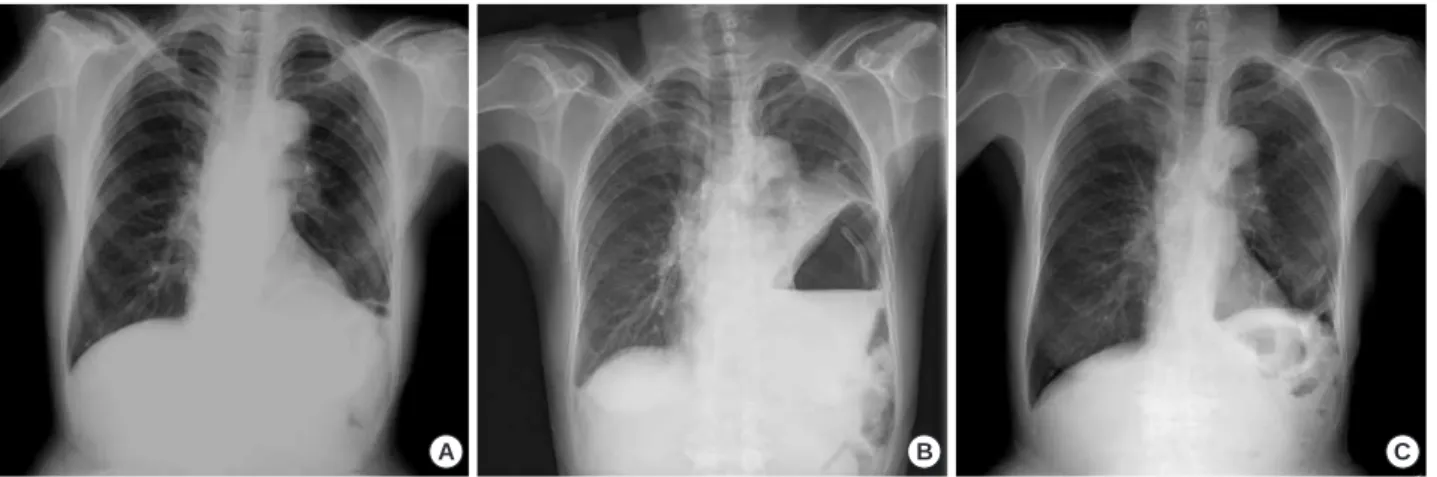
Fig. 1.Chest radiographies. (A) There was no abnormal finding reported on from the local clinic 4 days before admission. (B) On admis- sion, gastric air-fluid and bowel loops were observed in the left thoracic cavity and a coiled nasogastric tube was seen in the stomach. (C) The herniation improved but haziness was still seen at the left lower lobe after surgical intervention.
A B C
Fig. 2.Findings of upper gastrointestinal study (UGIS). (A) There was no disturbance of gastric passage at the local clinic 4 days before admission. (B) There were typical signs of a mesenteroaxial gastric volvulus; the stomach was in a vertical position with the pylorus higher than the cardia, and a double air-fluid level was seen; the antrum and pylorus were located inside the hernia with a hook like sign. (C) No passage disturbance of contrast media from the stomach into the duodenum; however, the stomach was still deviated into the thorax.
A B C
not protrude into the thorax. In addition, the presence of pleuroperitoneal adhesions may have played a role in prevent- ing herniation (5, 6). Loss of such physiologic balance may result from a sudden increase in the abdominal pressure.
Acute gastric volvulus and herniation of the abdominal organs into the thorax are associated with two factors: gastric distension and diaphragmatic eventration. It may be explained by an increased space around the stomach under a left diaph- ragmatic defect and by laxity of the gastrophrenic and gas- trosplenic ligaments. Conditions that increase the intra-abd- ominal pressure such as excessive coughing or pregnancy may increase the intra-gastric pressure, leading to strangulation of the stomach with a diaphragmatic defect (5, 7). Singh et al. (7) reported a diaphragmatic agenesis in an adult similar to our case; a 22-yr-old primigravida (gestation 38 weeks) presented with an acute gastric volvulus and diaphragmatic agenesis was confirmed surgically. The elevated abdominal pressure, by the pregnancy, likely played an important role in the pathogenesis of the AGV.
In our case, the patient lived for 70 yr without any medical problems even though she had only a right hemidiaphragm.
Just before this event, the patient had an UGIS for routine evaluation at a local clinic. The gastric bloating agent that was used for the UGIS might have led to the gastric distention and pressure overload. Therefore, the pressure imbalance may
have caused the gastric volvulus and abdominal organ her- niation into the thorax. On surgical exploration, we confirmed the absence of the left diaphragm. However, we could not reconstruct it. Spontaneous resolution of the gastric volvulus and herniation of abdominal organs was observed, as the abdominal pressure decreased.
REFERENCES
1. Langman J. Medical embryology, 4th ed. Baltimore: Williams and Willkins 1981; 282-317.
2. Cunniff C, Jones KL, Jones MC. Patterns of malformation in children with congenital diaphragmatic defects. J Pediatr 1990; 116: 258-61.
3. Tzelepis GE, Ettensohn DB, Shapiro B, McCool FD. Unilateral absen- ce of the diaphragm in an asymptomatic adult. Chest 1988; 94: 1301-3.
4. Wakai A, Winter DC, O’Sullivan GC. Unilateral diaphragmatic age- nesis precluding laparoscopic cholecystectomy. JSLS 2000; 4: 259-61.
5. Okoye BO, Bailey DM, Cusick EL, Spicer RD. Prophylactic gastro- pexy in the asplenia syndrome. Pediatr Surg Int 1997; 12: 28-9.
6. Qazi A, Awadalla S. Wandering spleen: a rare cause of mesenteroax- ial gastric volvulus. Pediatr Surg Int 2004; 20: 878-80.
7. Singh G, Bose SM. Agenesis of hemidiaphragm in adults. Aust NZJ Surg 1993; 63: 327-8.
Congenital Hemidiaphragmatic Agenesis Presenting as Gastric Volvulus and Diaphragmatic Hernia 519
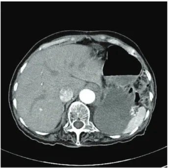
Fig. 3.CT finding at the level of the lower thorax demonstrating nor- mal right (arrow) and absent left hemidiaphragm and loops of bo- wel in left thorax.
Fig. 4.Intraoperative findings. There was no diaphragmatic tissue.
The abdominal organs (stomach, spleen, and splenic flexure of colon) were present in the left thoracic cavity.
Spleen
Spleen Left lung