Copyright © 2013 The Korean Society for Bone and Mineral Research
This is an Open Access article distributed under the terms of the Creative Commons Attribution Non-Commercial Li- cense (http://creativecommons.org/licenses/by-nc/3.0/) which permits unrestricted non-commercial use, distribu- tion, and reproduction in any medium, provided the original work is properly cited.
pISSN 2287-6375 eISSN 2287-7029
Bone Morphogenetic Protein-2 Desensitizes
MC3T3-E1 Osteoblastic Cells to Estrogen Through Transcriptional Downregulation of Estrogen
Receptor 1
Osamu Ishibashi
Department of Life and Environmental Sciences, Osaka Prefecture University, Sakai, Japan
Background: Estrogens exert preferable effects on bone metabolism through two es- trogen receptors (ERs), ER1 and ER2, which activate the transcription of a set of genes as ligand-dependent transcription factors. Thus, growth factors and hormones which mod- ulate ER expression in the bone, if any, may possibly modulate the effect of estrogens on bone metabolism. However, research as to which of these molecules regulate the ex- pression of ERs in osteoblasts has not been well documented. Methods: A reporter as- say system developed in this study was used to explore molecules that modulate ER1 expression in MC3T3-E1 osteoblastic cells. Gene expression was analyzed by reverse transcription-polymerase chain reaction. Results: A pilot study using the reporter sys- tem revealed that bone morphogenetic protein (BMP)-2 negatively regulated ER1, but not ER2, expression in MC3T3-E1 cells. Consistently, estradiol-induced reporter activity via an estrogen responsive element was strongly suppressed in MC3T3-E1 cells pretreat- ed with BMP-2. Conclusions: BMP-2 desensitizes osteoblastic cells to estrogen through downregulation of ER1 expression.
Key Words: Bone morphogenetic proteins, Osteoblasts, Receptors estrogen
INTRODUCTION
Estrogen plays an important role in various tissues, including the bone, brain and heart. In the bone of women and female animals, estrogen exerts a prefera- ble effect in its turnover.[1,2] Conversely, estrogen deficiency in postmenopausal women engenders accelerated bone resorption thereby leading to osteoporosis.
[3] The actions of estrogen are known to be mediated by two isoforms of nuclear receptors, estrogen receptor (ER)1 and ER2 (also designated as ERα and ERβ, re- spectively), which also function as ligand-dependent transcriptional factors. In the bone, both osteoblasts (bone-forming cells) and osteoclasts (bone-resorbing cells) have been demonstrated to express ERs,[4] indicating that estrogen directly acts on bone turnover. The two ER isoforms exhibit different properties in terms of ligand-specificity, tissue distribution and downstream intracellular signaling.[5-7]
These differences were reflected by the distinct phenotypes of mice, which were Corresponding author
Osamu Ishibashi
Department of Life and Environmental Sciences, Osaka Prefecture University, 1-1 Gakuen-cho, Nakaku, Sakai, Osaka 599-8531, Japan
Tel: +81-72-254-9474 Fax: +81-72-254-9921
E-mail: [email protected] Received: August 30, 2013
Revised: October 16, 2013 Accepted: October 16, 2013
No potential conflict of interest relevant to this article was reported.
This work was supported by a Grant-in-Aid for Young Scientists (B) from the Ministry of Education, Culture, Sports, Science and Technology (MEXT), and by grants from Mochida Memorial Foundation for Medical and Pharmaceutical Research and Astellas Foundation for Research on Medicinal Resources.
Original Article
deficient in either or both of ER1 and ER2.[8-10] To date, the interplay between estrogen and bone-related growth factors, e.g., transforming growth factor (TGF)-β[11-13]
and bone morphogenetic proteins (BMPs),[14] in osteo- blastic cells has been demonstrated. Further, the signaling cross-talk between estrogen and interleukin-6 has also been reported.[15] However, research on how the expres- sion of ERs in osteoblasts are regulated by bone-related growth factors or hormones has not been well documented.
In order to search for bioactive molecules that regulate the expression of ERs in osteoblasts at the transcriptional level, this study developed MC3T3-E1 osteoblastic cell lines which are stably transfected with a reporter construct that has the luciferase gene under the control of a mouse ER1 gene promoter. By conducting a pilot study using this re- porter system, the research also identified BMP-2 as a possi- ble suppressor of ER1 expression. Through a reverse tran- scription-polymerase chain reaction (RT-PCR), the study also verified that mRNA expression of ER1, but not that of ER2, in MC3T3-E1 was significantly downregulated by BMP-2. Fur- ther, the research also demonstrated that the responsive- ness of MC3T3-E1 cells to estradiol (E2) was pronouncedly suppressed by the pretreatment with BMP-2. These results indicate that BMP-2 is a possible negative regulator of ER1 gene expression in osteoblastic cells.
METHODS 1. Reagents
Recombinant human BMP-2 (rhBMP-2) and 1alpha,25- dihydroxy-vitamin D3 (1,25-[OH]2D3) were kindly provided by Astellas Pharma Co. Ltd (Tokyo, Japan). E2 and recombi- nant mouse epidermal growth factor (rmEGF) were pur- chased from Sigma-Aldrich Co. (St. Louis, MO). Phorbol- 12-myristate-13-acetate (PMA) and dibutyryl cyclic ade- nosine monophosphate (dbcAMP) were purchased from Wako (Osaka, Japan).
2. Cell culture
Mouse calvaria-derived MC3T3-E1 osteoblastic cells were purchased from Riken Bioresource Center (Tsukuba, Ibaraki, Japan). The cells were maintained in α-minimum essential medium (Invitrogen, Carlsbad, CA, USA) contain- ing 10% fetal bovine serum at 37°C in an atmosphere of 5% CO2/95% air.
3. Construction of reporter plasmids
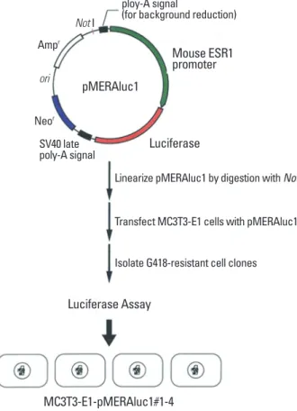
A mouse genomic DNA fragment encompassing the ER1 promoter region of 2.8 kbp upstream of its transcription initiation site was obtained by screening of a mouse ge- nomic DNA library using the 32P-labeled 5’-untranslated region of a mouse ER1 cDNA as a probe. The fragment was then subcloned into a pGVB2 vector (Nippongene, Tokyo, Japan) at its multicloning site. A fragment encompassing a neomycin-resistance gene derived from pSV2neo was then inserted into the vector in order to construct a final plas- mid designated pMERAluc1 (Fig. 1).
4. Stable transfection of MC3T3-E1 cells with the reporter construct under control of ER1 gene promoter
To establish a reporter system for identifying bioactive molecules in order to modulate ER1 gene expression, MC3T3-E1 cells were transfected with pMERAluc1. MC3T3- E1 cells were transfected with linearized pMERAluc1 using FuGENE 6 transfection reagent (Roche Diagnostics, Basel, Switzerland) and cultured in the presence of 500 µg/mL
Mouse ESR1 promoter ploy-A signal
(for background reduction)
Linearize pMERAluc1 by digestion with NotI Luciferase
SV40 late poly-A signal
Not I
pMERAluc1 Neor
Ampr ori
Transfect MC3T3-E1 cells with pMERAluc1 Isolate G418-resistant cell clones Luciferase Assay
MC3T3-E1-pMERAluc1#1-4
Fig. 1. Construction of the reporter plasmids used in this study.
G418 (Invitrogen) for 2 weeks. G418-resistant cells which formed colonies were separately trypsinized using metal cloning rings and transferred to wells of 24-well culture plates to further culture in the presence of 500 µg/mL G418. When reaching semi-confluence, these cells were expanded in a larger culture apparatus to allow propaga- tion; finally, 4 clones of these cells (designated MC3T3-E1- MERAluc1#1-4) could be successfully isolated as stably transformed cell lines.
5. Luciferase reporter assay
ER1 promoter-driven transcription of the luciferase gene in MC3T3-E1 cells was evaluated by luciferase reporter as- say using Picagene Luminescence Kit (Wako). The cells were lysed in LCβ lysis buffer (Wako), and luciferase activi- ties in the cell lysates were measured using a TD-20/20 lu- minometer (Promega, Madison, WI, USA), as previously de- scribed.[16]
6. Transient transfection
To validate the results obtained using the stable clones MC3T3-E1-MERAluc1#1-4, MC3T3-E1 cells were also trans- fected transiently with pMERAluc1. Transfection of the cells were performed using a FuGene6 transfection re- agent, as previously described.[7] Twenty-four hours after the transfection, rhBMP-2 was added at a final concentra- tion of 250 ng/mL. After 24-hour exposure with rhBMP-2, the cells were harvested and lysed in LCβ lysis buffer. The measurement of luciferase activity in the lysates were per- formed as described above.
7. RT-PCR
Cellular total RNAs were extracted using TRIzol reagent (Invitrogen, Carlsbad, CA, USA), according to the manufac-
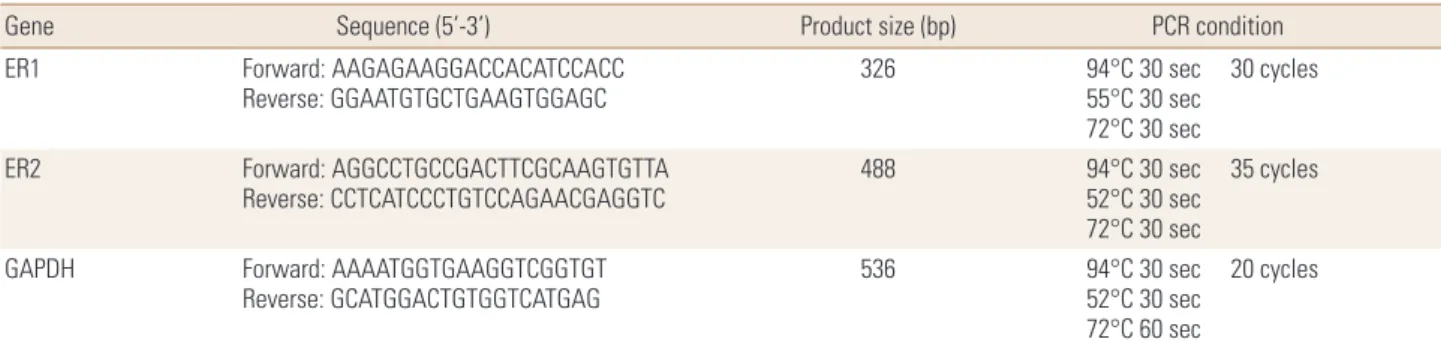
turer’s protocol. RT-PCR for mouse ER1, ER2 and glyceral- dehyde-3-phosphate dehydrogenase gene (GAPDH) was performed as described earlier [16]. Primers used in this study are listed in Table 1.
8. Statistical analysis
Statistical analysis was performed using an unpaired student’s t test. The results are shown as the means±SD.
RESULTS
1. Establishment of MC3T3-E1 cell lines stably harboring ER1 gene promoter-driven luciferase reporter
By conducting the procedures described in the Materials and Methods section, four discrete MC3T3-E1 cell clones stably harboring the luciferase reporter gene downstream of ER1 gene promoter, which were designated as MC3T3- E1-MERAluc1#1-4 (Fig. 1), were finally isolated.
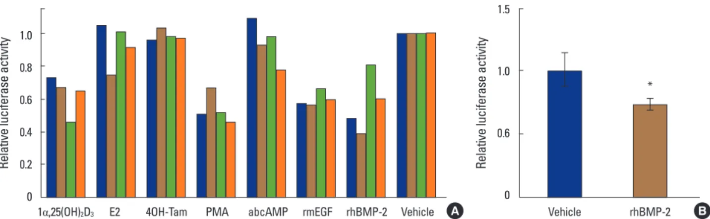
2. Identification of factors regulating ER1 mRNA expression in MC3T3-E1 cells
Relative luciferase activities in MC3T3-E1-MERAluc1 cells treated with several bioactive molecules, such as 1,25- (OH)2D3, E2, 4-hydroxy-tamoxifen (4-OH-Tam), PMA, db- cAMP, rmEGF and rhBMP-2, were measured. Of these mol- ecules, 1,25-(OH)2D3, PMA, mEGF and rhBMP-2 decreased the reporter activity driven by the ER1 promoter in all the stable cell clones (Fig. 2A). By contrast, E2, 4OH-Tam and dbcAMP had no effect on the promoter activity (Fig. 2A). In this study, the BMP-2-elicited downregulation of ER1 pro- moter activity was focused, because the functional inter- play between BMPs and estrogens in osteoblasts has not been much addressed so far, although both are well known
Table 1. Primers used in this study
Gene Sequence (5’-3’) Product size (bp) PCR condition
ER1 Forward: AAGAGAAGGACCACATCCACC
Reverse: GGAATGTGCTGAAGTGGAGC 326 94°C 30 sec
55°C 30 sec 72°C 30 sec
30 cycles
ER2 Forward: AGGCCTGCCGACTTCGCAAGTGTTA
Reverse: CCTCATCCCTGTCCAGAACGAGGTC 488 94°C 30 sec
52°C 30 sec 72°C 30 sec
35 cycles
GAPDH Forward: AAAATGGTGAAGGTCGGTGT
Reverse: GCATGGACTGTGGTCATGAG 536 94°C 30 sec
52°C 30 sec 72°C 60 sec
20 cycles
PCR, polymerase chain reaction; ER, estrogen receptor; GAPDH, glyceraldehyde-3-phosphate dehydrogenase gene.
to play important roles in osteoblast function.
To verify the above result, the effect of rhBMP-2 on ER1 gene promoter activity was examined using MC3T3-E1 cells transiently transfected with pMERAluc1. As expected, rhBMP-2 significantly suppressed the ER1 gene promoter- driven luciferase activity in MC3T3-E1 in this experiment (Fig. 2B).
3. BMP-2 suppresses ER1 mRNA expression in MC3T3-E1 cells
The study then examined whether the ER1 gene promot- er activity suppressed by BMP-2 was reflected by the altera- tion in ER1 mRNA expression in BMP-2-treated MC3T3-E1 cells. RT-PCR analyses revealed that ER1 mRNA expression in the cells was suppressed starting from 6 hours after the BMP-2 treatment (Fig. 3A). Conversely, rhBMP-2 did not change the mRNA expression of ER2 (Fig. 3B).
1.0
0.8
0.6
0.4
0.2
0
Relative luciferase activity Relative luciferase activity
1α,25(OH)2D3 E2 4OH-Tam PMA abcAMP rmEGF rhBMP-2 Vehicle Vehicle rhBMP-2 1.5
1.0
0.6
0
Fig. 2. Bone morphogenetic protein-2 downregulates a reporter activity driven by mouse estrogen receptor1 gene promoter in MC3T3-E1 cells. (A) MC3T3-E1-MERAluc1 cells (clone#1-4) were treated with 1alpha,25-dihydroxy-vitamin D3 (1,25-[OH]2D3; 10 nM), estradiol (E2; 100 nM), 4-hydroxy- tamoxifen (4-OH-Tam; 100 nM), phorbol-12-myristate-13-acetate (PMA; 10 nM), dibutyryl cyclic adenosine monophosphate (dbcAMP; 1 mM), re- combinant mouse epidermal growth factor (rmEGF; 100 ng/mL) and recombinant human bone morphogenetic protein-2 (rhBMP-2; 250 ng/mL). After a 48-hour incubation, these cells were harvested and lysed then, luciferase activities in the lysates were measured. Luciferase activities in the ve- hicle-treated cells were set at 1. (B) MC3T3-E1 cells were transiently transfected with pMERAluc1 (0.5 µg) along with pSV-β-gal (0.3 µg) for nor- malization of transfection efficiency, incubated for 24 hours and exposed by rhBMP-2 (250 ng/mL) or vehicle. Forty-eight hours later, the cells were harvested and lysed in LCβ lysis buffer. Luciferase activities were measured as described in the Materials and Methods section. The results are in- dicated as relative values when the normalized luciferase activity of the vehicle-treated cells is set at 1. *P<0.05.
*
A B
Fig. 3. Bone morphogenetic protein-2 downregulates expression of estrogen receptor (ER)1 mRNA, but not that of ER2, in MC3T3-E1 in a time-de- pendent manner. Total RNAs were isolated from MC3T3-E1 cells treated with bone morphogenetic protein (BMP)-2 or vehicle for the indicated pe- riods, and estrogen receptor (ER)1 expression was examined by reverse transcription-polymerase chain reaction (RT-PCR) analysis. ER1 mRNA ex- pression decreased from 6 to 24 hours after BMP-2 exposure (A). On the other hand, ER1 mRNA expression did not largely change over the period in the vehicle-treated MC3T3-E1 cells (B). Glyceraldehyde-3-phosphate dehydrogenase gene (GAPDH) expression was also analyzed as an internal control.
0 1 3 6 12 24 ER1
GAPDH
Culture time (hr)
0 1 3 6 12 24
ER2
GAPDH
Culture time (hr)
0 1 3 6 12 24
0 1 3 6 12 24
A B
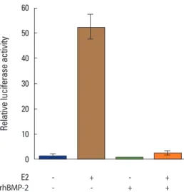
4. BMP-2 suppresses E2-induced ERE-mediated reporter activation in MC3T3-E1 cells
Next, the study observed whether the BMP-2-elicited downregulation of ER1 mRNA expression leads to reduced responsiveness of MC3T3-E1 cells to E2 by luciferase assay.
As conveyed in Fig. 4, the E2-induced luciferase activity driven by the ERE-containing promoter was markedly sup- pressed when the cells were pretreated with BMP-2, indicat- ing that BMP-2 desensitizes MC3T3-E1 osteoblastic cells to estrogen through the transcriptional downregulation of ER1.
DISCUSSION
In the present study, a reporter system was established in order to identify the molecules that potentially regulate ER1 gene expression in mouse osteoblastic MC3T3-E1 cells.
7 molecules were evaluated in a pilot study. Further, it was demonstrated that rhBMP-2 among the molecules suppre- ssed ER1 gene expression, leading to a restrained respon- siveness of the cells to E2.
So far, a number of bioactive molecules, besides estro-
gens, including hormones, growth factors, cytokines and chemical compounds, have been shown to play a role in the regulation of osteoblast functions. More importantly, estrogens and these molecules have been reported to act on osteoblasts, not only independently, but also interac- tively.[11-15,17] However, information regarding bioactive molecules that modulate ER mRNA expression in estrogen- responsive cells remains limited. Of these molecules, EGF, 1,25-(OH)2D3 and its agonistic derivatives have been previ- ously demonstrated to decrease the ER1 expression in MCF- 7, an ER-positive human breast tumor-derived cell line.
[18,19] Correspondingly, in this study, these two molecules decreased ER1 promoter activities in MC3T3-E1-MER Aluc1 clones (Fig. 2), suggesting that the suppression of ER1 ex- pression by these molecules could be a general event for many cell types. Conversely, estrogens have been reported to increase ER1 mRNA levels in MCF-7[20,21] as well as in the endothelium.[22,23] Against our expectations in the present study, however, the ER1 gene promoter activity was not affected by E2 and 4-OH-Tam, a tissue-selective agonist of ERs (Fig. 2). Considering that there have been no report so far that describes the presence of estrogen re- sponsive elements (EREs) in the ER1 gene promoter, the discrepancy may be explained by assuming that the ER1 mRNA level may be post-transcriptionally regulated.
Very recently, Matsumoto et al.[24] have reported that estrogen exerts a stimulatory effect on osteoblast differen- tiation through the activation of signaling mediated by BMP-4, which is structurally similar to BMP-2. Correspond- ing to the results of this study, the authors also indicated that ER1 expression was significantly suppressed by BMP-4 treatment, suggesting the presence of a negative feedback loop for osteoblast differentiation.
Overall, the results presented herein indicate that the prefer- able effect of estrogens on bone metabolism may be abrogat- ed by BMP treatment. Although it currently remains unknown whether our findings are true in vivo, this study will serve as a warning for the clinical use of BMPs. Further studies are cur- rently ongoing in order to elucidate a mechanistic insight of our findings, and the results will be reported elsewhere.
ACKNOWLEDGEMENTS
The author thanks Dr. Hiroyuki Kawashima, a sometime professor at Niigata University Graduate School of Medical
60
50
40
30
20
10
0
E2 - + - +
rhBMP-2 - - + +
Relative luciferase activity
Fig. 4. Bone morphogenetic protein-2 suppresses estradiol (E2)-induced estrogen responsive element (ERE)-mediated reporter activation in MC3T3-E1 cells. MC3T3-E1 cells transfected with pERE-G-luc, a reporter plasmid harboring the firefly luciferase gene under the control of a β- globin gene promoter, were pretreated with recombinant human bone morphogenetic protein-2 (rhBMP-2; 250 ng/mL) for 48 hours. These cells were then treated with estradiol (E2; 100 nM) for 24 hours, and the in- duced luciferase activities were evaluated. pCMVsport-β-gal, a reporter plasmid designed to express a β-galactosidase gene constitutively, was simultaneously transfected for normalization. The normalized values of the luciferase activity for the non-treated MC3T3-E1 cells are set at 1. Data are presented as the mean±SD of the three experiments. **P<0.01.
and Dental Sciences, for his helpful guidance and encour- agement. This work was supported by Grant-in-Aid for Young Scientists (B) from the Ministry of Education, Culture, Sports, Science and Technology (MEXT) (No. 13771092) and by grants from Mochida Memorial Foundation for Medical and Pharmaceutical Research and Astellas Foundation for Research on Medicinal Resources.
REFERENCES
1. Turner RT, Riggs BL, Spelsberg TC. Skeletal effects of estro- gen. Endocr Rev 1994;15:275-300.
2. Khosla S, Oursler MJ, Monroe DG. Estrogen and the skele- ton. Trends Endocrinol Metab 2012;23:576-81.
3. Venken K, Callewaert F, Boonen S, et al. Sex hormones, their receptors and bone health. Osteoporos Int 2008;19:1517-25.
4. Bland R. Steroid hormone receptor expression and action in bone. Clin Sci (Lond) 2000;98:217-40.
5. Nilsson S, Gustafsson JÅ. Estrogen receptors: therapies targeted to receptor subtypes. Clin Pharmacol Ther 2011;
89:44-55.
6. Heldring N, Pike A, Andersson S, et al. Estrogen receptors:
how do they signal and what are their targets. Physiol Rev 2007;87:905-31.
7. Ishibashi O, Yamagishi T, Hanada K, et al. Tamoxifen ago- nism and estrogen antagonism of c-fos gene promoter activity through non-consensus-responsive elements in MC3T3-E1 osteoblasts. Biochem Biophys Res Commun 2001;289:705-11.
8. Hewitt SC, Korach KS. Oestrogen receptor knockout mice:
roles for oestrogen receptors alpha and beta in reproduc- tive tissues. Reproduction 2003;125:143-9.
9. Emmen JM, Korach KS. Estrogen receptor knockout mice:
phenotypes in the female reproductive tract. Gynecol En- docrinol 2003;17:169-76.
10. McCauley LK, Tözüm TF, Rosol TJ. Estrogen receptors in skeletal metabolism: lessons from genetically modified models of receptor function. Crit Rev Eukaryot Gene Expr 2002;12:89-100.
11. Hawse JR, Subramaniam M, Ingle JN, et al. Estrogen-TGF- beta cross-talk in bone and other cell types: role of TIEG, Runx2, and other transcription factors. J Cell Biochem 2008;
103:383-92.
12. Matsuda T, Yamamoto T, Muraguchi A, et al. Cross-talk be-
tween transforming growth factor-beta and estrogen re- ceptor signaling through Smad3. J Biol Chem 2001;276:
42908-14.
13. Wu L, Wu Y, Gathings B, et al. Smad4 as a transcription co- repressor for estrogen receptor alpha. J Biol Chem 2003;
278:15192-200.
14. Yamamoto T, Saatcioglu F, Matsuda T. Cross-talk between bone morphogenic proteins and estrogen receptor sig- naling. Endocrinology 2002;143:2635-42.
15. Yamamoto T, Matsuda T, Junicho A, et al. Cross-talk be- tween signal transducer and activator of transcription 3 and estrogen receptor signaling. FEBS Lett 2000;486:143-8.
16. Ishibashi O, Kawashima H. Cloning and characterization of the functional promoter of mouse estrogen receptor beta gene. Biochim Biophys Acta 2001;1519:223-9.
17. Kato S, Masuhiro Y, Watanabe M, et al. Molecular mecha- nism of a cross-talk between oestrogen and growth factor signalling pathways. Genes Cells 2000;5:593-601.
18. Stoica A, Saceda M, Fakhro A, et al. Regulation of estrogen receptor-alpha gene expression by 1, 25-dihydroxyvita- min D in MCF-7 cells. J Cell Biochem 1999;75:640-51.
19. Swami S, Krishnan AV, Feldman D. 1alpha,25-Dihydroxyvi- tamin D3 down-regulates estrogen receptor abundance and suppresses estrogen actions in MCF-7 human breast cancer cells. Clin Cancer Res 2000;6:3371-9.
20. Lonard DM, Nawaz Z, Smith CL, et al. The 26S proteasome is required for estrogen receptor-alpha and coactivator turnover and for efficient estrogen receptor-alpha trans- activation. Mol Cell 2000;5:939-48.
21. Reid G, Hübner MR, Métivier R, et al. Cyclic, proteasome- mediated turnover of unliganded and liganded ERalpha on responsive promoters is an integral feature of estrogen signaling. Mol Cell 2003;11:695-707.
22. Ihionkhan CE, Chambliss KL, Gibson LL, et al. Estrogen causes dynamic alterations in endothelial estrogen recep- tor expression. Circ Res 2002;91:814-20.
23. Tschugguel W, Dietrich W, Zhegu Z, et al. Differential regu- lation of proteasome-dependent estrogen receptor alpha and beta turnover in cultured human uterine artery endo- thelial cells. J Clin Endocrinol Metab 2003;88:2281-7.
24. Matsumoto Y, Otsuka F, Takano-Narazaki M, et al. Estrogen facilitates osteoblast differentiation by upregulating bone morphogenetic protein-4 signaling. Steroids 2013;78:513- 20.