77
INTRODUCTION
Over the last two decades, the field of esthetic restorations has substantially expanded to satisfy patients’demands.
Traditional treatment approaches (full coverage crown) would involve the removal of large amounts of sound tooth substances, which has adverse effects on pulp, gingiva, and crown bio- mechanics.1Therefore, it is important to preserve much tooth structure in place. One of the most conservative treatment modal- ities is porcelain laminate veneers (PLVs). Since their intro- duction by Pincus in 1930, PLVs have been a popular dental treatment modality. The clinical survival rate of PLVs is high. Observation periods of PLVs reported in the literature range from 18 months to 15 years.2-4One study reported that 98.4%
of 186 PLVs placed over a 5-year period was rated as successful.2 Another study showed that the estimated survival probabili- ty of 182 PLVs over a period of 12 years is 94.4%.5PLVs are indicated not only for treating tooth discoloration, but also for restoring fractured, worn dentition and malformed teeth.
Swift et al. reported that PLVs provide suitable esthetics and reliable functional strength.6With the conservative approach, esthetic and functional results may be achieved. To date, following the study of Magne and colleagues who demonstrated the efficacy of bonding porcelain to enamel (as strong as natural dentition), the use of PLVs may be expanded to more
challenging cases.7 CASE REPORT
In this case, a 25-year-old male patient lost his maxillary left central incisor long before. From 2001, he had been treated with pre- and post-operative orthodontic therapy and gnatho- surgery to improve his facial appearance. In this process, the maxillary left lateral incisor was moved on the position of maxillary left central incisor and the maxillary left canine was moved on the position of maxillary left lateral incisor. The patient wanted to improve esthetics of anterior dentition. First, the space between anterior dentitions was analyzed. Analysis showed asym- metry in anterior dentitions because of the absence of the max- illary left central incisor and size difference between maxillary central and lateral incisors. Analysis also showed lingual inclination of the left lateral incisor. The maxillary left later- al incisor was more yellowish than the maxillary right central incisor and incisal edge of the maxillary right central incisor was concave due to attrition (Fig. 1).
1. Treatment planning
Porcelain laminate veneer was planned to improve esthetics of the anterior dentitions through transforming the shape of the
DOI:10.4047/jap.2010.2.3.77
Esthetic improvement in the patient with one missing maxillary central incisor restored with
porcelain laminate veneers
Dong-Jin Park, DDS, Jae-Ho Yang, DDS, PhD, Jai-Bong Lee, DDS, PhD, Sung-Hun Kim, DDS, PhD, Jung-Suk Han*, DDS, PhD Department of Prosthodontics and Dental research Institute, School of Dentistry, Seoul National University, Seoul, Korea
This article describes esthetic improvement in a patient with a missing maxillary left central incisor. Space analysis of the anterior dentition showed that minor tooth rearrangement was needed. Optimal space distribution for restorations was attained by orthodontic treatment. Through trans- forming tooth shape with porcelain laminate veneers, the maxillary left lateral incisor was transformed into central incisor and the maxillary left canine into a lateral incisor. The maxillary right central incisor was also restored for esthetic improvement. In a case of changing a tooth shape with porcelain laminate veneers, pre-treatment evaluation, space analysis and diagnostic wax-up are important factors.[J Adv Prosthodont 2010;2:77-80]
Corresponding author: Jung-Suk Han
Department of Prosthodontics and Dental research Institute, School of Dentistry, Seoul National University, 275-1, Yeongeon-dong, Jongno-gu, Seoul, 110-768, Korea Tel. 82 2 2072 2661: e-mail, [email protected]
Received August 5, 2010 / Last Revison September 8, 2010 / Accepted September 12, 2010
CASE REPORT J Adv Prosthodont 2010;2:77-80
KEY WORDS. Porcelain laminate veneers, Missing central incisor, Orthodontic treatment
ⓒ 2010 The Korean Academy of Prosthodontics
This is an Open Access article distributed under the terms of the Creative Commons Attribution Non-Commercial License (http://creativecommons.org/licenses/by- nc/3.0) which permits unrestricted non-commercial use, distribution, and reproduction in any medium, provided the original work is properly cited.
78 J Adv Prosthodont 2010;2:77-80
teeth with porcelain laminate veneer. The treatment plan was to transform the maxillary left lateral incisor into a central incisor and the maxillary left canine into a lateral incisor. The restora- tion of the maxillary right central incisor was also included into the plan for esthetic improvement. The maxillary central incisors are mesio-distally larger than the lateral incisors for about 2 mm, and it was necessary to do minor tooth rearrange- ment by orthodontic treatment. After the tooth rearrange- ment, the space of the maxillary left lateral incisor became equal to that of the maxillary right central incisor. In addition, cross-bite of the maxillary left lateral incisor was resolved (Fig. 2).
2. Diagnostic wax-up
Diagnostic wax-up was performed including the maxillary right central incisor, the maxillary left lateral incisor and maxillary left canine on the study cast (Fig. 3). Mockup is crit- ical for fabrication of the provisional restorations, enabling the
patient and clinician to evaluate esthetics, and to preview the anticipated result.8
3. Intraoral preparations
After duplicating diagnostic wax-up cast, putty index (Exafine putty type, GC, Tokyo, Japan) was made for tooth prepa- rations. Selective preparations were performed with the guide of putty index using tapered diamond burs (Shofu, Kyoto, Japan).
The preparation margins were placed at the equi-gingival level for esthetics. Preparation margins and surfaces were fin- ished with a low speed white stone bur (Shofu, Kyoto, Japan) under water spray (Fig. 4, 5).
4. Provisional restorations
Provisional restorations were fabricated by Luxatemp� (DMG, Hamburg, Germany) using putty attained from the diag- nostic wax-up cast. After 20 seconds of spot etching (Scotchbond etchant, 3M ESPE, MN, USA) for 20 seconds at the labial sur- faces, provisional restorations were set using flowable light- cured composite resin (AELITEFLOTM, Bisco, IL, USA).
5. Impressions
A week later, provisional restorations were removed and abut- ments were cleaned with a rubber cup. Before the impression was taken, retraction cord (UltraPak #00; Ultradent, UT, USA) was packed in the gingival sulcus to exposure the preparation margin. The final impression was taken with polyvinylsiloxane impression materials (Exafine putty type;
GC Corporation, Tokyo, Japan / Honigum Light; DMG, Hamburg, Germany) light body polyvinylsiloxane (Honigum Light, DMG, Hamburg, Germany) using 1-step impression technique.
Esthetic improvement in the patient with one missing maxillary central incisor restored with porcelain laminate veneers Park DJ et al.
Fig. 1. Preoperative view.
Fig. 2. Minor tooth rearrangement was completed. The space of maxillary left lateral incisor became equal to that of maxillary right central incisor.
Fig. 3. Diagnostic wax-up. The maxillary left lateral incisor and maxillary left canine were transformed into a central incisor and lateral incisor.
79 J Adv Prosthodont 2010;2:77-80
6. Placement and evaluation
Porcelain laminate veneers were fabricated with IPS e.max Press system (Ivoclar-vivadent, Schaan, Liechtenstein). After IPS e.max Press copings were made, they were layered with glass ceramics (IPS e.max Ceram Powder; Ivoclar-vivadent, Schaan, Liechtenstein) and then finishing and glazing were done (Fig. 6). IPS e.max Press has high flexural strength (400 MPa) and esthetics.9Also, the advantage of this method is need- less of a refractory cast. Porcelain laminate veneers were cemented with the light-cured resin cement (Variolink II;
Ivoclar-vivadent, Schaan, Liechtenstein). After a month from the delivery, as shown in Fig. 7 and 8, the gingiva was healthy and the interproximal area was fully filled with inter- dental papilla. Patient was satisfied with the harmonious shape and contour of the restorations which also showed good function.
Esthetic improvement in the patient with one missing maxillary central incisor restored with porcelain laminate veneers Park DJ et al.
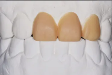
Fig. 6. Porcelain laminate veneers were checked on the master cast for esthetics and adaptation.
Fig. 7. Frontal view of cemented restorations (1 month after delivery).
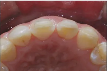
Fig. 8. Occlusal view of cemented restorations (1 month after delivery).
Fig. 5. Occlusal view of the tooth preparations.
Fig. 4. Frontal view of the tooth preparations.
80 J Adv Prosthodont 2010;2:77-80
CONCLUSION
This clinical report shows that porcelain laminate veneers can meet esthetic and functional desires of the patient with miss- ing maxillary central incisor. Porcelain laminate veneer is a great choice to change tooth shape because of relatively minor tooth reduction, short treatment time and acceptable esthetics.
In a case of changing a tooth shape with porcelain laminate veneers, pre-treatment evaluation, space analysis and diagnostic wax-up are important factors.
REFERENCES
1. Claman L, Alfaro MA, Mercado A. An interdisciplinary approach for improved esthetic results in the anterior maxilla. J Prosthet Dent 2003;89:1-5.
2. Aristidis GA, Dimitra B. Five-year clinical performance of porcelain laminate veneers. Quintessence Int 2002;33:185-9.
3. Peumans M, De Munck J, Fieuws S, Lambrechts P, Vanherle G, Van Meerbeek B. A prospective ten-year clinical trial of porce- lain veneers. J Adhes Dent 2004;6:65-76.
4. Wiedhahn K, Kerschbaum T, Fasbinder DF. Clinical long- term results with 617 Cerec veneers: a nine-year report. Int J Comput Dent 2005;8:233-46.
5. Fradeani M, Redemagni M, Corrado M. Porcelain laminate veneers: 6- to 12-year clinical evaluation--a retrospective study.
Int J Periodontics Restorative Dent 2005;25:9-17.
6. Swift EJ Jr, Friedman MJ. Critical appraisal: porcelain veneer outcomes, part II. J Esthet Restor Dent 2006;18:110-3.
7. Magne P, Magne M, Belser UC. Adhesive restorations, centric relation, and the Dahl principle: minimally invasive approach- es to localized anterior tooth erosion. Eur J Esthet Dent 2007;2:260-73.
8. Griffin JD Jr. Correction of congenitally missing lateral incisors with porcelain veneers. Pract Proced Aesthet Dent 2006;18:475- 80.
9. Albakry M, Guazzato M, Swain MV. Biaxial flexural strength, elastic moduli, and x-ray diffraction characterization of three press- able all-ceramic materials. J Prosthet Dent 2003;89:374-80.
Esthetic improvement in the patient with one missing maxillary central incisor restored with porcelain laminate veneers Park DJ et al.