Primary Extracranial Fibrous Meningioma of the Maxillary Sinus: A Case Report and
Literature Review
상악동에 발생한 일차성 섬유질형 수막종:
증례 보고 및 문헌 고찰
Hyunwoo Cho, MD , Sanghyeon Kim, MD* , Myongjin Kang, MD , DongWon Kim, MD
Department of Radiology, Dong-A University College of Medicine, Busan, Korea
Meningioma is a common neoplasm of the central nervous system; however, primary extracra- nial meningioma of the paranasal sinus, especially the maxillary sinus, is rare. We report a case of primary extracranial meningioma (fibrous type) of the maxillary sinus and present a litera- ture review of the imaging features that correlate with fibrous meningioma.
Index terms Fibrous Meningioma; Maxillary Sinus; Computed Tomography, X-Ray;
Magnetic Resonance Imging; Angiography
INTRODUCTION
Meningioma is the most frequent primary non-glial tumor of the central nervous sys- tem, comprising approximately 13–26% of all primary intracranial neoplasms (1, 2).
Rarely, meningioma develops extracranially, without association with the central ner- vous system, through transformation of embryonic arachnoid cell rests (1). The most common histopathological subtype of meningiomas is meningothelial meningioma, occurring in approximately 60% of cases; however, all other histopathology subtypes have been reported to occur in primary extracranial meningiomas (2). The fibrous me- ningioma of the maxillary sinus is extremely rare, with only a few case reports describ- ing the imaging features (3-5). Here, we present a case of primary fibrous meningioma of the maxillary sinus with computed tomography (CT), magnetic resonance imaging (MRI), and angiography findings.
Received April 14, 2020 Revised May 15, 2020 Accepted May 31, 2020
*Corresponding author Sanghyeon Kim, MD Department of Radiology, Dong-A University College of Medicine,
37 Nakdong-daero 550beon-gil, Saha-gu, Busan 49315, Korea.
Tel 82-51-240-5367 Fax 82-51-253-4931 E-mail [email protected] This is an Open Access article distributed under the terms of the Creative Commons Attribu- tion Non-Commercial License (https://creativecommons.org/
licenses/by-nc/4.0) which permits unrestricted non-commercial use, distribution, and reproduc- tion in any medium, provided the original work is properly cited.
ORCID iDs Hyunwoo Cho https://
orcid.org/0000-0002-1377-3905 Sanghyeon Kim
https://
orcid.org/0000-0002-0731-3806 Myongjin Kang
https://
orcid.org/0000-0002-2447-9314 DongWon Kim
https://
orcid.org/0000-0003-0207-9587
CASE REPORT
A 38-year-old man presented to our hospital with a one-year history of nasal obstruction. No other nasal symptoms such as epistaxis, rhinorrhea, and smell disorder were noted. The pa- tient did not have any other illness or trauma history. Nasal endoscopic examination revealed a grayish, firm, non-bleeding mass that appeared to be located in the right maxillary sinus.
MRI examination revealed a solid mass measuring 4.22 cm × 4.91 cm × 4.62 cm in the right maxillary sinus. The mass appeared slightly hypointense on the T1-weighted image (Fig. 1A) and moderate hypointense on the T2-weighted image (Fig. 1B). Although the con- trast-enhanced T1-weighted image showed heterogeneous enhancement of the mass (Fig.
1C), the 5-minute delayed image showed homogeneous enhancement. Diffusion-weighted imaging demonstrated no restricted diffusion (Fig. 1D). The mean apparent diffusion coeffi- cient value was 1.27 × 10⁻³ mm2s-1.
CT scan was performed to evaluate bony involvement and the surrounding bony anatomy.
An unenhanced CT scan showed an expansile hyperdense mass (Fig. 1E) with multiple small calcifications in the right maxillary sinus. The anterior wall of the maxillary sinus was erod- ed by the expanding mass.
Histological examination of the endoscopic biopsy specimen confirmed fibrous meningio- ma, characterized by fibroblast-like spindle cells embedded in a collagen and reticulin fiber- rich matrix. Mitosis, anaplastic change, and necrosis were not observed.
The patient underwent preoperative endovascular embolization. Pre-embolization selec- tive external carotid artery angiogram revealed tumor blushing (Fig. 1F), with dysplastic dila- tation of tumor vessels originating from the right maxillary artery. Tumor blushing persisted in the delayed phase, and no demonstrable arteriovenous shunt was noted. The mass was embolized using polyvinyl alcohol particles, and the tumor was endoscopically excised from the sinus 3 days postembolization.
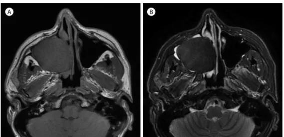
Fig. 1. A 38-year-old man with primary fibrous meningioma of the maxillary sinus.
A. The axial T1-weighted image shows a slightly hypointense mass measuring 4.22 cm × 4.91 cm × 4.62 cm in the right maxillary sinus.
B. T2-weighted image shows a moderate hypointense mass.
A B
DISCUSSION
Primary extradural meningiomas in the head account for 0.8–1.8% of all meningiomas (6).
A review by Mattox et al. (2) revealed that only 5 of 163 reported cases were of primary me- ningioma of the maxillary sinus. Similar to intracranial meningiomas, extracranial meningio- mas may exhibit a variety of different histological patterns. The most common histopathologi- cal subtype of primary extracranial meningioma of the head is meningothelial meningioma (53.4%). Other common subtypes are transitional (12.3%), psammomatous (11.7%), and fi- Fig. 1. A 38-year-old man with primary fibrous meningioma of the maxillary sinus.
C. The axial contrast-enhanced liver acquisition with volume acceleration image (left) shows a heterogeneous enhancement of the mass, and the 5-minute delayed contrast-enhanced T1-weighted image (right) shows a homogeneous enhancement of the mass.
D. The diffusion-weighted image at a b-value of 1000 sec/mm2 (left) demonstrates no diffusion restriction, and the apparent diffusion coeffi- cient value (right) is 1.27 × 10⁻³ mm2/s-1.
E. The axial unenhanced CT image shows an expansile hyperdense mass with multiple small calcifications in the right maxillary sinus. The anterior wall of the right maxillary sinus is eroded by the mass (arrow).
F. The early arterial (left) and late phases (right) of the right external carotid artery angiogram show tumor blushing with dysplastic vessels originating from the right maxillary artery and persistent staining. No significant arteriovenous shunt is observed.
C
E
D
F
brous (6.7%) meningiomas (6). Thus, primary, fibrous meningioma of the maxillary sinus is extremely rare, with only 3 reported cases using CT or MRI findings (Table 1) (3-5).
MRI is the modality of choice for evaluating meningiomas because it provides superior tis- sue differentiation. The typical MRI features of meningiomas are as follows: isointensity to slight hypointensity relative to gray matter on the T1-weighted sequence; isointensity to slight hyperintensity relative to gray matter on the T2 sequence; and avid, homogeneous en- hancement after contrast agent administration. The signal intensity of meningiomas on T2- weighted images is closely related to the histological subtype, for example fibrous meningio- mas show prominent hypointensity due to the presence of the fibrous component (4). CT is useful for detecting calcifications and demonstrating the effects of tumor on the adjacent bone for differential diagnosis. Calcification is observed in up to 25% of meningiomas, which is associated with slow growth and lower grade (7). Hyperostosis of the adjacent bone is asso- ciated with benign meningioma, and osseous destruction indicates atypical or malignant meningioma (8). However, our case and 3 previous cases of benign meningioma demonstrat- ed maxillary sinus wall erosion or destruction without clear hyperostosis. A relatively thin bony wall of the maxillary sinus and pressure may have contributed to the erosion or de- struction of the bone rather than to hyperostosis. In addition, Thomson et al. reported that a paranasal meningioma was observed along with adjacent bone erosion in many cases (1).
In accordance with previous studies, the imaging findings of our case suggest a fibrous meningioma. However, as meningioma of the maxillary sinus is rare, differential diagnoses should also include T2-hypointense lesions such as fungal sinusitis, solitary fibrous tumor, ossifying fibroma, or fibrous dysplasia. The majority of sinus fungus balls show marked hy- pointensity surrounded by hyperintense mucosal walls on T2-weighted images. Fungus balls can be also suggested by the presence of hyperintense portions within the mass on T1- weighted images, regardless of calcification on the CT scan. Furthermore, the bony margins of the involved sinus are usually intact. Sinonasal solitary fibrous tumors usually demon- strate bone remodeling, thinning, or local absorption on CT scans, and isointensity or hy- pointensity on T2-weighted images. However, contrary to our case, the lesion tends to be more heterogeneous in signal intensity on T2-weighted images and demonstrates a marked enhancement on contrast-enhanced T1-weighted images. Ossifying fibroma usually exhibits hypointensity in the peripheral ossified areas of the lesion and hyperintense central cystic
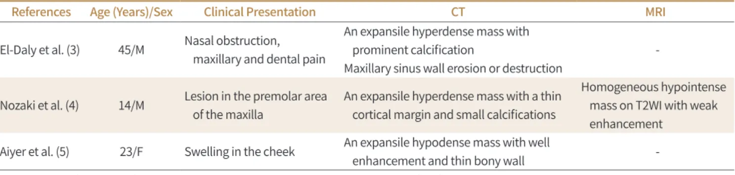
Table 1. Summary of the Clinical Presentations and Imaging Findings of Previously Reported Cases of the Maxillary Sinus Fibrous Meningioma
References Age (Years)/Sex Clinical Presentation CT MRI
El-Daly et al. (3) 45/M Nasal obstruction, maxillary and dental pain
An expansile hyperdense mass with prominent calcification
Maxillary sinus wall erosion or destruction
-
Nozaki et al. (4) 14/M Lesion in the premolar area of the maxilla
An expansile hyperdense mass with a thin cortical margin and small calcifications
Homogeneous hypointense mass on T2WI with weak enhancement
Aiyer et al. (5) 23/F Swelling in the cheek An expansile hypodense mass with well
enhancement and thin bony wall -
CT = computed tomography, MRI = magnetic resonance imaging, T2WI = T2-weighted imaging
area on T2-weighted images. Depending on the degree of calcification, it may present as a diffuse and homogeneous T2-hypointense mass, but can be more locally aggressive than in our case (9). Fibrous dysplasia of the sinonasal cavity must also be included in the differential diagnosis. However, this usually demonstrates heterogeneity and hypointensity on T2- weighted images and a characteristic ground-glass appearance on CT scans.
Treatment of choice for primary benign, extracranial meningioma is gross total resection.
Preoperative embolization is effective for reducing intraoperative blood loss and facilitating surgical resection by tumor softening. Meningioma generally appears with a “sunburst” or
“spokewheel” pattern on angiography and enhances early during the arterial phase and re- mains well opacified after the venous phase. Arteriovenous shunting suggests malignancy (10). In our case, pre-embolization angiography showed delayed tumor blushing without ar- teriovenous shunt, probably resulting from the fibrotic component of the tumor.
In conclusion, we presented a case of primary benign, fibrous meningioma of the maxil- lary sinus. Although primary extracranial meningioma (fibrous subtype) is extremely rare with no typical imaging findings, knowledge of atypical imaging characteristics will assist the differential diagnosis.
Author Contributions
Conceptualization, K.S.; investigation, all authors; supervision, K.S.; visualization, C.H.; writing—
original draft, C.H., K.S.; and writing—review & editing, K.M., K.D.
Conflicts of Interest
The authors have no potential conflicts of interest to disclose.
REFERENCES
1. Thompson LD, Gyure KA. Extracranial sinonasal tract meningiomas: a clinicopathologic study of 30 cases with a review of the literature. Am J Surg Pathol 2000;24:640-650
2. Mattox A, Hughes B, Oleson J, Reardon D, McLendon R, Adamson C. Treatment recommendations for pri- mary extradural meningiomas. Cancer 2011;117:24-38
3. El-Daly A, Pitman KT, Ferguson BJ, Snyderman CH. Primary extracranial meningioma of the maxillary an- trum. Skull Base Surg 1997;7:211-215
4. Nozaki S, Yamazaki M, Koyama T, Kubota Y, Kitahara H, Yoshizawa K, et al. Primary extracranial meningio- ma of the maxillary sinus presenting as buccal swelling. Asian J Oral Maxillofac Surg 2011;23:134-137 5. Aiyer RG, Prashanth V, Ambani K, Bhat VS, Soni GB. Primary extracranial meningioma of paranasal sinuses.
Indian J Otolaryngol Head Neck Surg 2013;65:384-387
6. Liu Y, Wang H, Shao H, Wang C. Primary extradural meningiomas in head: a report of 19 cases and review of literature. Int J Clin Exp Pathol 2015;8:5624-5632
7. Zeng L, Liang P, Jiao J, Chen J, Lei T. Will an asymptomatic meningioma grow or not grow? A meta-analy- sis. J Neurol Surg A Cent Eur Neurosurg 2015;76:341-347
8. Saloner D, Uzelac A, Hetts S, Martin A, Dillon W. Modern meningioma imaging techniques. J Neurooncol 2010;99:333-340
9. Salina ACI, Souza PMM, Gadelha CMDC, Aguiar LB, Castro JDV, Barreto ARF. Ossifying fibroma: an uncom- mon differential diagnosis for T2-hypointense sinonasal masses. Radiol Case Rep 2017;12:313-317 10. Wilson CB. Meningiomas: genetics, malignancy, and the role of radiation in induction and treatment. The
Richard C. Schneider lecture. J Neurosurg 1994;81:666-675
상악동에 발생한 일차성 섬유질형 수막종:
증례 보고 및 문헌 고찰
조현우 · 김상현* · 강명진 · 김동원
수막종은 중추신경계에서 흔한 종양이지만, 부비동, 특히 상악동에 위치한 일차성 두개외 수 막종은 매우 드물다. 본 연구에서는 상악동에서 발생한 일차성 섬유질형 수막종의 증례를 보 고하고, 문헌의 섬유질형 수막종의 영상 소견과 함께 고찰하고자 한다.
동아대학교 의과대학 영상의학교실