Epidemiological Characteristics of the First Water-Borne Outbreak of Cryptosporidiosis in Seoul, Korea
The first case of human cryptosporidiosis was reported in Korea in 1995; however, an outbreak of Cryptosporidium has not been reported in Korea until now. This paper describes the first outbreak of cryptosporidiosis in Korea. On May 24, 2012, a local public health center filed a report on 126 residents with gastrointestinal symptoms in an old apartment complex in Seoul. Epidemiological investigations were implemented on 125 of the 126 patients. The patients were reported continuously over a period of 22 days.
Diarrhea was the most common clinical symptom, and lasted for 5 days on average. The tap water was the only common exposure of the patients. During the environmental investigation it was discovered that the water and septic tanks were situated closely and that the waste water pipes were corroded where they passed over the water pipes.
Cryptosporidium parvum was detected in 3 of the 7 stool specimens by PCR-RFLP. A number of Cryptosporidium oocysts were also detected in the water specimens from the water tank. In conclusion, Cryptosporidium parvum was the key causal pathogen of this outbreak. It is presumed that the tap water was contaminated by a sewage leak from the aged pipelines.
Key Words: Cryptosporidium parvum; Cryptosporidiosis; Food and Waterborne Disease Outbreak; Epidemiological Investigation; Fecal Contamination
Shinje Moon,1 Wooseok Kwak,1 Sangwon Lee,1 Won Kim,2 Jaeyeon Oh,3 and Seung-Ki Youn1
1Division of Epidemic Intelligence Service, Korea Centers for Disease Control and Prevention, Cheongwon; 2Division of Health Policy, Seoul Metropolitan Government, Seoul; 3Healthcare Department of Dongdaemun-gu Public Health Center, Seoul, Korea
Received: 7 August 2012 Accepted: 14 May 2013 Address for Correspondence:
Seung-Ki Youn, PhD
Division of Epidemic Intelligence Service, Korea Centers for Disease Control and Prevention, 187 Osongsaengmyeong 2-ro, Cheongwon 363-951, Korea
Tel: +82.43-719-7190, Fax: +82.43-719-7219 E-mail: [email protected]
http://dx.doi.org/10.3346/jkms.2013.28.7.983 • J Korean Med Sci 2013; 28: 983-989
INTRODUCTION
Cryptosporidium is a minute coccidian parasite with worldwide distribution. Two species of Cryptosporidium, C. hominis and C.
parvum, are the major micro-organisms of human infection (1, 2). Recently, the outbreaks of cryptosporidiosis have increased due to increase in recreational water use (3-5). The first human infection of Cryptosporidium was reported in Korea in 1995 (6) and since then, epidemiologic studies were carried out on a di- verse range of regions (7-11). Although a positive rate of Crypto- sporidium oocysts in human less than 3% was reported in most of the regions, some regions reported a very high positive rate of more than 40%. The livestock in such regions also displayed high level of positive rates. Therefore, it is possible to presume that zoonotic transmission is the most important cause of Cryp- tosporidium infection in Korea. However, in spite of such high level of positive rates in specific regions, an outbreak of Crypto- sporidium has not been reported in Korea until now. This study is reporting the first outbreak of Cryptosporidiosis in Korea. This outbreak could be deemed to be highly significant in that it has displayed unique epidemiologic characteristics irrelevant to the usage of recreational water or the zoonotic transmission.
MATERIALS AND METHODS Study setting
This study is a case series study. On May 24, 2012, a local public health center filed a report on 126 residents with diarrhea in an old apartment complex in Seoul, Korea. An epidemiological in- vestigation was immediately undertaken by a Seoul Metropoli- tan Epidemic Intelligence Service (EIS) officer in Seoul along with infectious disease inspectors from the local Public Health Center. Korea Centers for Disease Control and Prevention (KCDC) was consulted on this epidemiological investigation. Clinical specimens including stool specimens and rectal smear samples were taken for microbiological examination during the epide- miological investigation. Water specimens from the water tank in the apartment complex and the adjacent building were col- lected by the Waterworks Research Institute of the Seoul Met- ropolitan Government.
Case definitions
The case was defined as the residents of the apartment complex, who had frequently suffered from watery diarrhea more than twice, or two of the following symptoms, namely, watery diar- rhea, abdomen pain, fever and vomiting in May 2012.
Data collection and pathogen detection
Data were collected by using the structured case report form during the epidemiologic investigation. The case report form included the following variables: the demographic features such as sex, age and address, the common exposures, clinical features and date of onset.
The clinical specimens were primarily examined by the Seoul Research Institute of Public Health and Environment for the bacteriological and virological examination as the Epidemio- logic Investigation Guideline for Food and Waterborne Diseases from KCDC (12). The stool specimens were subsequently exam- ined by the Korea National Institute of Health for parasitologi- cal examination. The protozoa detection was undertaken by polymerase chain reaction (PCR) and polymerase chain reac- tion–restriction fragment length polymorphism (PCR-RFLP) (13). PCR was performed in 20 μL reactions containing 4 μL of DNA template, 10 pM of each primer, 1.25 U of Ex Taq DNA poly- merase (iNtRON Biotechnology, Seongnam, Korea), 1.5 mM MgCl2, and 0.2 mM of each dNTP. The primers that were used were CP2-415F (upstream), 5´-CCCACGCGAAGTTGAAGTA- AC-3´ and CP2-415-R (downstream), 5´-CTTAGGTTGCTTGC- TTGGAGTTGG-3´. The amplified size was 415 bp fragment.
The PCR conditions used for first-step PCR were 1 cycle of 94oC for 5 min, 35 cycles of 94oC for 30 sec, 53oC for 30 sec, and 72oC for 90 sec, and final extension of 1 cycle of 72oC for 10 min. The nested-PCR was performed using 1 cycle of 94oC for 5 min, 35 cycles of 94oC for 30 sec, 55oC for 30 sec, and 72oC for 90 sec, and final extension of 1 cycle of 72oC for 10 min. For the nested-PCR, the primers were 5´-CAACCAGAAGTTGAGGTT-3´ (upstream) and 5´-CTAGTATGCTTCAGACCATGA-3´ (downstream). The nested PCR amplified 171 bp fragment. Each PCR product ob- tained by nested-PCR analysis was purified with a QIAquick Gel Extraction kit (QIAGEN Inc., Valencia, CA, USA) and was digested with restriction enzymes of BsiEI to identification of Cryptosporidium hominis and C. parvum (13). The water speci- mens were examined by the Waterworks Research Institute of Seoul Metropolitan Government. The analyzed pathogens are listed on Table 1.
Ethics statements
The study protocol was approved by the institutional review board of Korea National Institute of Health (IRB No. 2013- 01C0N-02-P). The informed consent was waived by the board.
RESULTS
The apartment complex, consisting of three buildings, was built in 1977. A total of 228 households (564 people) were residing in the apartment complex at the time of the incident. Total num- ber of patients reported was 126 during the period of epidemio- logic investigations with prevalence rate of 22.3% (126/564). Of the 126 patients, epidemiologic investigations were implement- ed on 125 patients.
Demographic data
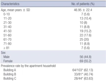
There were more females (55.2%) than the males (44.8%) and the mean age was 46.95 yr. The prevalence rates in each apart- ment were as follows: 62.1% per one household for building-A, 40.7% for building-B and 63.6% for building-C (Table 2). The
Table 1. Pathogens examined in the rectal smear samples, stool specimens and water specimens
Specimens Number of samples Microorganisms
Rectal smear samples 29 Bacteria (10): Salmonella spp., Shigella spp., Staphyloccocus aureus, Vibrio spp., Listeria monocytogenes, Yersinia enterocolitica, Bacillus cereus, Escherichia coli, Clostridium perfringens, Campylobacter jejuni Virus (5): Rotavirus, Norovirus, Adenovirus, Astrovirus, Sapovirus
Stool specimens 7
Bacteria (10): Salmonella spp., Shigella spp., Staphyloccocus aureus, Vibrio spp., Listeria monocytogenes, Yersinia enterocolitica, Bacillus cereus, Escherichia coli, Clostridium perfringens, Campylobacter jejuni Virus (5): Rotavirus, Norovirus, Adenovirus, Astrovirus, Sapovirus
Protozoa (3): Cryptosporidium spp., Giardia lamblia, Entamoeba histolytica
Water specimens 1 L for viruses
2 L for bacteria
3 L for protozoa
Virus: Enterovirus (by TCVA), Norovirus (by PCR)
Bacteria: Total coliforms, General bacteria, Psychrophilic bacteria, Fecal Streptococci, Sulfite reducing spore forming anaerobes, Salmonella spp., Shigella spp., Escherichia coli, Enterococcus spp., Pseudomonas aeruginosa
Protozoa: Cryptosporidium spp., Giardia lamblia
Table 2. Demographic characteristics of the patients in the outbreak (n = 125)
Characteristics No. of patients (%)
Age, mean years ± SD 0-10
11-20 21-30 31-40 41-50 51-60 61-70 71-80 ≥ 81
46.95 ± 22.4 7 (5.6) 13 (10.4) 10 (8) 11 (8.8) 19 (15.2) 22 (17.6) 25 (20) 11 (8.8)
7 (5.6) Sex
Male
Female 56 (44.8)
69 (55.2) Prevalence rate by the apartment household
Building A Building B Building C
64/103* (62.13) 33/81* (40.74) 28/44* (63.63) Data are No. (%) of patients, unless otherwise indicated. *No. of patients/Household in a building.
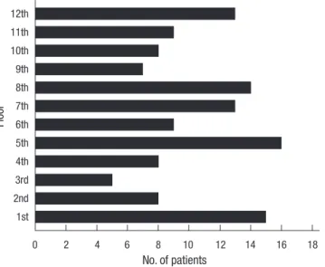
patients were reported throughout the entire floors of the apart- ment (Fig. 1).
Outbreak pattern
The patient was first found on May 3 and the last patient on May 28. The peak time of the incidence was May 18 (Fig. 2).
Clinical manifestations
Of the 125 patients, 121 patients suffered from diarrhea, while 73 patients suffered from abdominal pain, 34 patients suffered from fever and 26 patients suffered from vomiting. On average diarrhea was continued for five days (Fig. 3).
Epidemiologic investigations
As the results of epidemiologic investigations, it was found that there were no abnormalities prior to the outbreak through which all the patients could be commonly exposed to the pathogen.
The tap water was the only common source of exposure of the
patients to the pathogen. Accordingly, investigation was con- ducted on the usage of tap water among the patients. All the patients used tap water for domestic purposes including cook- ing. A total of 116 out of 125 patients used tap water for drinking while the remaining 9 did not. Of the 116 patients who used tap water for drinking, 50 used water purifier, 60 boiled the tap wa- ter, and 6 boiled the water from the water purifier. There was no patient who drank tap water without any kind of treatments.
Environmental investigations
The water tank that stored the tap water was buried underground in the vacant lot at the center of the apartment complex. Septic tanks were situated within the distance of 3meters in its sur- rounding. The external wall of the underground water tank was constructed with concrete and had no crack. The upper portion of the underground water tank was covered with manhole. Struc- tural abnormality of the pipeline was observed during the inves- tigation of the pipeline structure. The wastepipes for the septic tank crossed over the water pipes for the water tank. These pipes were aged and worn out (Fig. 4).
Floor
No. of patients
0 2 4 6 8 10 12 14 16 18
12th 11th 10th 9th 8th 7th 6th 5th 4th 3rd 2nd 1st
Fig. 1. Patient distribution by building floor into the apartment complex.
No. of patients
Diarrhea Abdominal pain Fever Nausea Vomiting 140
120
100
80
60
40
20
0
Fig. 2. Clinical presentations in the outbreak.
Fig. 3. Patients with gastrointestinal symptoms by onset, May 4-May 27, 2012.
No. of patients
20 18 16 14 12 10 8 6 4 2 0
Date of onset
05-0405-0505-0605-0705-0805-0905-1005-1105-1205-1305-1405-1505-1605-1705-1805-1905-2005-2105-2205-2305-2405-2505-2605-2705-28
Laboratory results
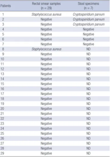
During the epidemiological investigation, 20 rectal smear sam- ples and six stool specimens were collected from the residents suffering from severe diarrhea. Additionally, nine rectal smear
samples and one stool specimen were taken from the residents who started diarrhea after the investigation. Staphylococcus au- reus was detected in two specimens among 29 rectal smear sam- ples and seven stool specimens, and C. parvum had been de- tected in 3 of the 7 stool specimens by PCR-RFLP (Table 3 and Fig. 5). Also, a number of bacteria and C. parvum oocysts were detected in the tap water of underground water tank. On the other hand, there was no other pathogen detected in the tap water of the nearby buildings (Table 4).
Public health measures
Due to the suspicion of tap water as the source of infection, the supply of tap water was cut immediately. During this period of stoppage in the supply of tap water, the pipeline structure was corrected and the water tank and pipelines were disinfected.
Cutting off of the tap water supply was maintained until no Cryp- tosporidium oocysts were detected through continuous water test, and it took approximately 4 weeks before the Cryptosporid- ium oocysts were found no more. There was no report of new patient since the cutting off of the tap water supply. Although there was a report of patients with recurring symptoms during the same period, all the patients recovered without complica- Table 3. Pathogens detected in the rectal smear samples and stool specimens
Patients Rectal smear samples
(n = 29) Stool specimens
(n = 7) 1 Staphylococcus aureus Cryptosporidium parvum
2 Negative Cryptosporidium parvum
3 Negative Cryptosporidium parvum
4 Negative Negative
5 Negative Negative
6 Negative Negative
7 Negative Negative
8 Staphylococcus aureus ND
9 Negative ND
10 Negative ND
11 Negative ND
12 Negative ND
13 Negative ND
14 Negative ND
15 Negative ND
16 Negative ND
17 Negative ND
18 Negative ND
19 Negative ND
20 Negative ND
21 Negative ND
22 Negative ND
23 Negative ND
24 Negative ND
25 Negative ND
26 Negative ND
27 Negative ND
28 Negative ND
29 Negative ND
ND, Not done.
Table 4. Pathogens detected in the water specimens
Pathogens Underground
water tank The adjacent building Bacteria
General bacteria Psychrophilic bacteria Total coliforms Escherichia coli Fecal Streptococci
Sulfite reducing spore forming anaerobes Salmonella spp.
Shigella spp.
Enterococci
Pseudomonas aeruginosa
360 CFU/mL 1,000 CFU/mL
Positive Positive Positive Negative Negative Positive Positive Positive
Negative Negative Negative Negative Negative Negative Negative Negative Negative Negative Virus
Enterovirus
Norovirus Negative
Negative Negative Negative Parasite
Cryptosporidium spp.
Giardia lamblia TNTC
Negative Negative Negative TNTC, Too numerous to count.
Fig. 4. The corroded water pipes in the apartment complex, Seoul, Korea.
M
171 bp
1 2
Fig. 5. PCR-RFLP pattern of amplified Cryptosporidium parvum. The amplified DNA by PCR was digested with BsiEI. Lane 1, Marker; Lane 2, Control; Lane 3, Patient stool sample.
tions. The occurrence of patients was monitored for 5 weeks since the resuming of the tap water supply, and there was nei- ther report of new patients nor patients with recurring symp- toms.
DISCUSSION
Cryptosporidium sp. is diagnosed by identifying organism in intestinal biopsy material, and by microscopically detecting oo- cysts following modified acid fast stains or fluorescent stains in stool specimens (14-16). Cryptosporidium can also be detected by enzyme immunoassay or PCR (17-20). PCR could be an ex- cellent diagnostic method for Cryptosporidium sp. because PCR does not need highly trained expert and reduces the diagnostic labor time. Moreover, PCR-RFLP could differentiate the two species of Cryptosporidium, such as C. parvum and C. hominis.
In this study, C. parvum was detected by PCR-RFLP. Eight patients who recovered at the time of investigation refused the stool examination, and 22 among 29 patients, who had clinical symptoms during epidemiologic investigation only consented to taking of rectal smear samples, and denied taking of stool specimens. C. parvum was detected in 3 of the 7 stool specimens in this study. In addition, a number of C. parvum oocysts were detected in the tap water from the water tank. In the guidelines for confirmation of food-borne disease outbreak (21, 22), Cryp- tosporidium spp. can be confirmed as the causal pathogen for the outbreak if Cryptosporidium was detected in stool speci- mens or in small bowel biopsy of two or more ill persons, or if pathogen is detected in the epidemiologically implicated food.
Although small number of stool specimens was examined, C.
parvum could be detected in more than two patients and in the tap water, which was epidemiologically implicated. Therefore, it provided sufficient ground to decide C. parvum as the cause of the outbreak.
However, there is a limitation in concluding that all 126 re- ported patients have been infected by C. parvum because a number of different bacteria were detected in the tank water specimens. These bacteria implied stool contamination of the tap water. However, there was no bacterium or virus that iden- tified in the stool specimens and rectal smear samples except Staphylococcus aureus. S. aureus was detected in two rectal smear samples. It was presumed that possibility of S. aureus be- ing the major pathogen for this outbreak was low because the clinical symptoms in the patients differed from those caused by S. aureus. Based on the laboratory results, it was concluded that C. parvum was the key causal pathogen of this outbreak.
The numbers of water borne and food borne disease out- breaks in Korea were 355 in 2008, 227 in 2009, 254 in 2010, 236 in 2011 and 288 in 2012 respectively. Majority of outbreaks were due to infection at ordinary restaurants. In the large-scale out- breaks with more than 100 people being affected, the main cause
was consumption of contaminated food at group canteen, in- cluding schools. These outbreaks lasted less than 2 weeks. As the results of internal data review on the water borne and food borne disease outbreaks that reported to the Korea Centers for Disease Control and Prevention over the last 5 yr, there has not been any outbreak of waterborne disease like this case with large number of patients with more than 100 patients being affected over a period of more than 3 weeks.
Although there has not been any domestic report of the out- break of cryptosporidiosis until now, there have continually been literary reports on investigations of positive rates of Cryp- tosporidium oocysts in human in different regions. Low posi- tive rate of 0.5% was reported in Seoul (8), 1.1% in Chungju (9), 1.9% in Cheorwon (7), 2.2% for Chuncheon (8), 0.4% in Haman- gun, and 1.1% for Euryeong-gun (9). However, high positive rate of 10.5% was observed for Jeollanam-do, and while low positive rate of 3.7% was observed for urban regions such as Mokpo and Yeosu. Hwasun-gun displayed high positive rate of 40% (8), which continued to be maintained at the high rate during the investigations conducted afterwards (10). In addition, low posi- tive rate of 1.5% was observed in the island regions of Jeollanam- do (11). Such epidemiologic characteristics of Jeollanam-do were presumed to be the results of the zoonotic transmission because the high rates of Cryptosporidium infection were re- ported in the livestock in the regions with high positive rates.
However, this study was different from the previous ones be- cause the present outbreak was irrelevant with the zoonotic transmission.
The symptoms of cryptosporidiosis develop after an incuba- tion period about 1 week (23). Watery diarrhea is the main symp- tom with possible accompaniment by abdominal pain, nausea, vomiting, anorexia, fever and reduction in body weight. In gen- eral, the symptoms last approximately 5-10 days in immuno- competent individuals (24). Approximately 39% of the patients display biphasic patterns in which symptoms recur within sev- eral days to several weeks after the symptoms disappear (24- 26). This outbreak also displayed the majority of patients having symptoms of watery diarrhea accompanied by abdominal pain, fever and vomiting. Median duration of illness was about five days. In addition, the patients with recurrence of symptoms were reported in this outbreak. The clinical characteristics of patients in this outbreak coincided with the clinical symptoms of cryptosporidiosis.
The investigation on the usage of tap water was conducted to deduce the transmission route of Cryptosporidium. In spite of the investigation, the transmission route was not exactly identi- fied in this study. However, it was still possible that some of the residents might be exposed to sufficient quantity of Cryptospo- ridium oocysts that could cause infection through the usage of tap water for cooking and washing. The reason is that Crypto- sporidium spp. could cause infection with minute quantity in
the range of 10-30 oocysts (27).
In order to find the cause of contamination of the tap water, water specimens from the adjacent buildings were taken for an investigation. These buildings use water that has not passed through the underground water tank, even though the same water supply network is used. No pathogen was detected in the water specimen in these buildings. This indicates that there was no contamination of the tap water prior to being stored in the underground water tank. With the old pipelines of the water tank and the septic tanks and structural abnormalities of pipe- line network in the apartment complex, it is presumed that contaminated water was mixed with the water in the under- ground water tank by leakage of sewage in wastepipe.
There are several limitations in this study. Firstly, the epide- miological investigation to the patients without symptoms was not made. Secondly, the stool examinations from the patients who were already recovered were not conducted. Thirdly, the leakage of pipelines of the underground water tank and septic tank was not confirmed by means of dye tests. Fourthly, the cause of C. parvum contamination of the sewage was not clari- fied. For this, demographic data including occupation and un- derlying disease from all residents in the apartment complex should be collected. Because a number of the residents decline to participate in the epidemiological investigation, further eval- uation could not be conducted. However, the previous study about the prevalence of Cryptosporidium spp. in the Han-river which passed through Seoul showed that domestic wastewater from the urban region could be a source of Cryptosporidium spp. contamination (28).
In conclusion, the outbreak was due to C. parvum in the con- taminated water of underground water tank. It is presumed that the water in the underground water tank was contaminated by sewage in the septic tank through the old pipelines. This report is significant in that it is the first outbreak of cryptosporidiosis in Korea, and occurred in the urban area without the risk fac- tors like recreational water use or livestock infection. Since de- tection of Cryptosporidium spp. is not a test generally performed in the outbreak of waterborne disease, it can be easily overlooked unless careful attention is paid. If the causal pathogen in the outbreak that occurs in urban regions is not clear, then, it could be helpful to implement tests for the presence of Cryptosporidi- um species.
ACKNOWLEDGEMENTS
We express our gratitude to our colleagues in the Seoul RIHE, the Waterworks Research Institute of Seoul Metropolitan Gov- ernment and Department of Malaria and Parasite Disease in the National Institute of Health for the laboratory examination to detect the pathogen. Also, we thank Dr. Jae-Ran Yu for pro- viding the control DNA of C. parvum.
DISCLOSURE
All authors have no conflicts of interest to disclose.
REFERENCES
1. Chalmers RM, Davies AP. Minireview: clinical cryptosporidiosis. Exp Parasitol 2010; 124: 138-46.
2. Cama VA, Bern C, Roberts J, Cabrera L, Sterling CR, Ortega Y, Gilman RH, Xiao L. Cryptosporidium species and subtypes and clinical manifes- tations in children, Peru. Emerg Infect Dis 2008; 14: 1567-74.
3. Yoder JS, Harral C, Beach MJ; Centers for Disease Control and Preven- tion (CDC). Cryptosporidiosis surveillance - United States, 2006-2008.
MMWR Surveill Summ 2010; 59: 1-14.
4. Takagi M, Toriumi H, Endo T, Yamamoto N, Kuroki T. An outbreak of cryptosporidiosis associated with swimming pools. Kansenshogaku Zasshi 2008; 82: 14-9.
5. Baldursson S, Karanis P. Waterborne transmission of protozoan para- sites: review of worldwide outbreaks - an update 2004-2010. Water Res 2011; 45: 6603-14.
6. Kang YK, Lee HK, Kim SW, Chi JG. Cryptosporidiosis in a leukemia child with severe diarrhea. Seoul J Med 1995; 36: 29-34.
7. Seo M, Huh S, Chai JY, Yu JR. An epidemiological survey on Cryptospo- ridium parvum infection of inhabitants in Chorwon-gun, Kangwon-do.
Korean J Parasitol 2001; 39: 201-3.
8. Chai JY, Lee SH, Guk SM, Lee SH. An epidemiological survey of Crypto- sporidium parvum infection in randomly selected inhabitants of Seoul and Chollanam-do. Korean J Parasitol 1996; 34: 113-9.
9. Yu JR, Lee JK, Seo M, Kim SI, Sohn WM, Huh S, Choi HY, Kim TS. Prev- alence of cryptosporidiosis among the villagers and domestic animals in several rural areas of Korea. Korean J Parasitol 2004; 42: 1-6.
10. Park JH, Guk SM, Han ET, Shin EH, Kim JL, Chil JY. Genotype analysis of Cryptosporidium spp. prevalent in a rural village in Hwasun-gun, Re- public of Korea. Korean J Parasitol 2006; 44: 27-33.
11. Park JH, Kim HJ, Guk SM, Shin EH, Kim JL, Rim HJ, Lee SH, Chai JY. A survey of cryptosporidiosis among 2,541 residents of 25 coastal islands in Jeollanam-do (Province), Republic of Korea. Korean J Parasitol 2006;
44: 367-72.
12. Korea Centers for Disease Control and Prevention. Epidemiologic inves- tigation guideline for food and waterborne diseases 2012. Cheongwon:
Division of Epidemic Intelligence Service, Korea Centers for Disease Con- trol and Prevention, 2012.
13. Lee SH, Joung M, Yoon S, Choi K, Park WY, Yu JR. Multiplex PCR detec- tion of waterborne intestinal protozoa; Microsporidia, Cyclospora and Cryptosporidium. Korean J Parasitol 2010; 48: 297-301.
14. Ramirez NE, Ward LA, Sreevatsan S. A review of the biology and epide- miology of cryptosporidiosis in humans and animals. Microbes Infect 2004; 6: 773-85.
15. Garcia LS, Bruckner DA, Brewer TC, Shimizu RY. Techniques for the re- covery and identification of Cryptosporidium oocysts from stool speci- mens. J Clin Microbiol 1983; 18: 185-90.
16. Nielsen CK, Ward LA. Enhanced detection of Cryptosporidium parvum in the acid-fast stain. J Vet Diagn Invest 1999; 11: 567-9.
17. Chan R, Chen J, York MK, Setijono N, Kaplan RL, Graham F, Tanowitz
HB. Evaluation of a combination rapid immunoassay for detection of Giardia and Cryptosporidium antigens. J Clin Microbiol 2000; 38: 393-4.
18. Sulaiman IM, Xiao L, Lal AA. Evaluation of Cryptosporidium pavum genotyping techniques. Appl Environ Microbiol 1999; 65: 4431-5.
19. Sharp SE, Suarez CA, Duran Y, Poppiti RJ. Evaluation of the Triage Micro Parasite Panel for detection of Giardia lamblia, Entamoeba histolytica/
Entamoeba dispar, and Cryptosporidium parvum in patient stool speci- mens. J Clin Microbiol 2001; 39: 332-4.
20. Elwin K, Chalmers RM, Roberts R, Guy EC, Casemore DP. Modification of a rapid method for the identification of gene-specific polymorphisms in Cryptosporidium parvum and its application to clinical and epide- miological investigations. Appl Environ Microbiol 2001; 67: 5581-4.
21. Korea Centers for Disease Control and Prevention. Epidemiologic in- vestigation guideline for food and waterborne diseases 2013 revised ed.
Cheongwon: Division of Epidemic Intelligence Service, Korea Centers for Disease Control and Prevention, 2013.
22. Lynch M, Painter J, Woodruff R, Braden C; Centers for Disease Control and Prevention. Surveillance for foodborne-disease outbreaks: United States, 1998-2002. MMWR Surveill Summ 2006; 55: 1-42.
23. Huang DB, White AC. An updated review on Cryptosporidium and Giar- dia. Gastroenterol Clin North Am 2006; 35: 291-314.
24. Mac Kenzie WR, Hoxie NJ, Proctor ME, Gradus MS, Blair KA, Peterson DE, Kazmierczak JJ, Addiss DG, Fox KR, Rose JB, et al. A massive out- break in Milwaukee of cryptosporidium infection transmitted through the public water supply. N Engl J Med 1994; 331: 161-7.
25. MacKenzie WR, Schell WL, Blair KA, Addiss DG, Peterson DE, Hoxie NJ, Kazmierczak JJ, Davis JP. Massive outbreak of waterborne cryptospo- ridium infection in Milwaukee, Wisconsin: recurrence of illness and risk of secondary transmission. Clin Infect Dis 1995; 21: 57-62.
26. Newman RD, Sears CL, Moore SR, Nataro JP, Wuhib T, Agnew DA, Guer- rant RL, Lima AA. Longitudinal study of Cryptosporidium infection in children in northeastern Brazil. J infect Dis 1999; 180: 167-75.
27. DuPont HL, Chappell CL, Sterling CR, Okhuysen PC, Rose JB, Jakubows- ki W. The infectivity of Cryptosporidium parvum in healthy volunteers.
N Engl J Med 1995; 332: 855-9.
28. Lee MY, Cho EJ, Lee JH, Han SH, Park YS. A survey of Cryptosporidium oocysts in water supplies during a 10-year period (2000-2009) in Seoul.
Korean J Parasitol 2010; 48: 219-24.