Neurologic Manifestations of Enterovirus 71 Infection in Korea
Enterovirus 71 frequently involves the central nervous system and may present with a variety of neurologic manifestations. Here, we aimed to describe the clinical features, magnetic resonance imaging (MRI) findings, and cerebrospinal fluid (CSF) profiles of patients presenting with neurologic complications of enterovirus 71 infection. We retrospectively reviewed the records of 31 pediatric patients hospitalized with acute neurologic manifestations accompanied by confirmed enterovirus 71 infection at Ulsan University Hospital between 2010 and 2014. The patients’ mean age was 2.9 ± 5.5 years (range, 18 days to 12 years), and 80.6% of patients were less than 4 years old. Based on their clinical features, the patients were classified into 4 clinical groups: brainstem encephalitis (n = 21), meningitis (n = 7), encephalitis (n = 2), and acute flaccid paralysis (n = 1). The common neurologic symptoms included myoclonus (58.1%), lethargy (54.8%), irritability (54.8%), vomiting (48.4%), ataxia (38.7%), and tremor (35.5%).
Twenty-five patients underwent an MRI scan; of these, 14 (56.0%) revealed the characteristic increased T2 signal intensity in the posterior region of the brainstem and bilateral cerebellar dentate nuclei. Twenty-six of 30 patients (86.7%) showed CSF pleocytosis. Thirty patients (96.8%) recovered completely without any neurologic deficits;
one patient (3.2%) died due to pulmonary hemorrhage and shock. In the present study, brainstem encephalitis was the most common neurologic manifestation of enterovirus 71 infection. The characteristic clinical symptoms such as myoclonus, ataxia, and tremor in conjunction with CSF pleocytosis and brainstem lesions on MR images are pathognomonic for diagnosis of neurologic involvement by enterovirus 71 infection.
Keywords: Enterovirus A, Human; Neurologic Manifestations; Brain Stem; Magnetic Resonance Imaging; Cerebrospinal Fluid; Child; Korea
Kyung Yeon Lee, Myoung Sook Lee, and Dong Bin Kim
Department of Pediatrics, Ulsan University Hospital, University of Ulsan College of Medicine, Ulsan, Korea
Received: 24 August 2015 Accepted: 18 December 2015 Address for Correspondence:
Kyung Yeon Lee, MD
Department of Pediatrics, Ulsan University Hospital, 877 Bangeojinsunhwando-ro, Dong-gu, Ulsan 44033, Korea E-mail: [email protected]
http://dx.doi.org/10.3346/jkms.2016.31.4.561 • J Korean Med Sci 2016; 31: 561-567
INTRODUCTION
Enterovirus 71 is one of the main causative pathogens of hand, foot and mouth disease. Compared to other enterovirus sero- types, enterovirus 71 more frequently involves the central ner- vous system (CNS), and induces diverse neurologic features such as brainstem encephalitis, aseptic meningitis, and acute flaccid paralysis (1,2). Since enterovirus 71 was first described in 1974, large-scale epidemics of enterovirus 71 infection have been reported in Asia-Pacific regions including Malaysia, Tai- wan, and China and have caused many fatalities (3-6). Addi- tional epidemics of hand, foot and mouth disease caused by enterovirus 71 that included fatal cases occurred in Korea be- tween 2009 and 2013 (7-9).
Enterovirus 71 infection predominantly involves the brain- stem. Most of the fatalities caused by enterovirus 71 infection result from autonomic dysfunction such as pulmonary hemor- rhage in patients with brainstem encephalitis (1,2). Moreover, deaths usually occur within 6-24 hours after admission because the disease rapidly progresses to the critical stage (2,10). Hence,
early diagnosis of enterovirus 71 infection with neurologic in- volvement and prompt, appropriate management is important.
To aid in the early detection of neurologic involvement in en- terovirus 71 infection, we aimed to describe the diverse clinical features, magnetic resonance imaging (MRI) findings and cere- brospinal fluid (CSF) profiles of patients with neurologic com- plications associated with enterovirus 71 infection that were en- countered during a 5-year period in a single Korean institute.
MATERIALS AND METHODS Patients
We retrospectively reviewed the medical records of 31 children hospitalized with acute neurologic manifestations accompa- nied by confirmed enterovirus 71 infection at Ulsan University Hospital between January 2010 and December 2014. All patients included in this study were healthy prior to infection without neurologic problems. Of the 31 patients included in this study, 17 patients were included in our previous case series study and case report (11,12).
At the time of admission and during the hospitalization peri- od, all patients were queried about, and observed for, the pres- ence of myoclonus, tremor, ataxia, fever, vomiting, headache, seizure, irritability, lethargy, and decreased consciousness. Based on their clinical features, patients were classified into four clini- cal groups: 1) brainstem encephalitis characterized by myoclo- nus, tremor, ataxia, nystagmus, oculomotor palsies, bulbar pal- sy, and autonomic dysfunctions such as pulmonary edema, in various combinations, with or without neuroimaging evidence;
2) aseptic meningitis characterized by fever, headache, vomit- ing, and neck stiffness, without the other neurologic manifesta- tions; 3) encephalitis characterized by decreased consciousness, seizure, and fever without myoclonus, tremor, ataxia, and auto- nomic dysfunction; and 4) acute flaccid paralysis characterized by acute onset motor weakness of the extremities.
Neurological and virological diagnostic tests
MRI was performed on either a 3.0 Tesla system (Intera Achie- va; Philips, Best, The Netherlands) or a 1.5 Tesla system (Achie- va; Philips). The protocol included T1-weighted images, T2-wei- ghted images, fluid-attenuated inversion recovery (FLAIR) im- ages, and contrast-enhanced T1-weighted images. Virus exami- nations were conducted with stool and CSF collected from the patients using real-time reverse transcription polymerase chain reaction (RT-PCR) at the Division of Enteric and Hepatitis Vi- ruses, Korea Centers for Disease Control and Prevention. For genome detection and genotyping of the enterovirus, RT-PCR using Taq-Man technology was attempted on all stool and CSF samples obtained from patients. For genotyping, semi-nested RT-PCR was used to amplify part of the VP1 gene of the entero- virus based on the United States Centers for Disease Control and Prevention protocol for the detection of enterovirus by the use of confirmed enterovirus-positive specimens and real-time PCR. CSF analysis was performed to determine the white blood cell count, red blood cell count, levels of protein and glucose, and bacterial cultures.
Ethics statement
This study was approved by the institutional review board of Ulsan University Hospital (IRB No. 2015-06-044). Informed con- sent was waived by the board because this was a retrospective study.
RESULTS
Patient demographics and seasonal distribution
Eighteen patients were male and 13 patients were female. The age distribution is shown in Table 1. The mean age of the patients was 2.9 ± 5.5 years (range, 18 days to 12 years), and 80.6% of the patients were less than 4 years old. The majority of patients were 1 to 2 years old, accounting for 32.3% of the patients. The num- ber of cases appearing per year was as follows: 8 cases appeared in 2010, 8 cases in 2012, 12 cases in 2013, and 3 cases in 2014.
No cases appeared in 2011. As for the seasonal distributions, 10 cases (32.2%) developed in the spring (March-May), 18 cases (58.1%) in the summer (June-August), 2 cases (6.5%) in the au- tumn (September-November), and 1 case (3.2%) in the winter (December-February) (Fig. 1). The peak months for developing the disease were July (n = 11, 35.5%), May (n = 7, 22.6%), and June (n = 5, 16.1%).
Virological analyses
Stool samples obtained from 30 patients and CSF samples from 15 patients were examined for enterovirus 71. All 30 stool sam- ples were enterovirus 71-positive. Four of the CSF samples (26.7%) were enterovirus 71-positive.
Clinical features
All of the patients except one (96.8%) had a fever above 38°C, and 17 patients (54.8%) had body temperatures above 39°C. Twen- ty-five patients (80.6%) had a characteristic hand, foot and mouth disease rash before or during the neurologic symptoms. Neuro- logic symptoms or signs appeared within 4 days after the onset of skin lesions in the 25 patients that experienced the rash.
Fig. 1. Seasonal distribution of neurologic complications by enterovirus 71 infection.
Nuber of patients
Month
1 2 3 4 5 6 7 8 9 10 11 12 12
10
8
6
4
2
0 Table 1. Age distribution and clinical diagnosis according to patients’ age
Age (yr) Brainstem encephalitis
n = 21 (%)
Aseptic meningitis n = 7 (%)
Encephalitis n = 2 (%)
Acute flaccid paralysis n = 1 (%)
Total n = 31 (%)
< 2 9 (42.8) 4 (57.1) 1 (50.0) 1 (100.0) 15 (48.4)
2-4 8 (38.1) 2 (28.6) 0 (0.0) 0 (0.0) 10 (32.3)
4-6 1 (4.8) 0 (0.0) 0 (0.0) 0 (0.0) 1 (3.2)
> 6 3 (14.3) 1 (14.3) 1 (50.0) 0 (0.0) 5 (16.1)
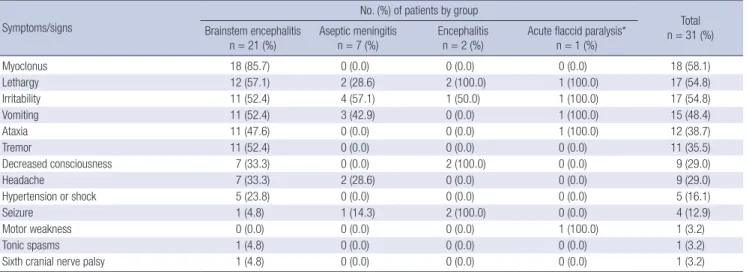
As mentioned above, the patients were classified into 4 clini- cal groups based on their clinical features: brainstem encepha- litis (n = 21), meningitis (n = 7), encephalitis (n = 2), and acute flaccid paralysis (n = 1). The neurologic features presented by the patients in each of the 4 clinical groups are summarized in Table 2.
One patient who was classified as being in the acute flaccid paralysis group showed acute motor weakness of the right low- er extremity in conjunction with clinical features of brainstem encephalitis such as ataxia. The most common neurologic fea- tures presented by the patients included myoclonus (n = 18, 58.1%), lethargy (n = 17, 54.8%), and irritability (n = 17, 54.8%).
In the 21 patients with brainstem encephalitis, myoclonus (n = 18, 85.7%), ataxia (n = 11, 52.4%), and tremor (n = 11, 52.4%) occurred more frequently than they did in the patients of the other 3 groups due to the definitions for disease classification.
Despite the low occurrence rate, some patients with brainstem encephalitis showed hypertension or hypotension (n = 5, 16.1%), tonic spasms (n = 1, 3.2%), and sixth cranial nerve palsy (n = 1, 3.2%). Three infants under 5 months of age who were classified into the aseptic meningitis group showed non-specific neuro- logic manifestations, such as lethargy and irritability along with
fever, yet they revealed CSF pleocytosis and were enterovirus 71-positive (CSF and stool samples in two patients; stool sam- ple in one patient). Seizures occurred in 4 patients (12.9%); of these, 2 patients were included in the encephalitis group, while the other 2 patients were included in the aseptic meningitis and brainstem encephalitis groups, respectively. The two patients included in the aseptic meningitis and brainstem encephalitis groups were 17 months and 19 months of age, respectively, and showed typical features of febrile seizures, which were general- ized seizures lasting 2-3 minutes accompanied by high fever.
MRI findings
MRI was performed on 25 patients. The locations of the lesions in the MR images for all 4 clinical groups are summarized in Table 3. Fourteen (56.0%) of the 25 patients revealed increased signal intensity in the posterior region of the brainstem (the pons in 12 patients, and the pons and midbrain in 2 patients) and bi- lateral cerebellar dentate nuclei on T2-weighted and FLAIR im- ages (Fig. 2A and B). Two patients (8.0%), who both had seizures, revealed leptomeningeal enhancement of the cerebral hemi- spheres without brain parenchymal lesions (Fig. 2C). Eight pa- tients (32.0%) showed no apparent abnormalities. None of the
Table 2. Neurologic features of the patients in the four clinical groups
Symptoms/signs
No. (%) of patients by group
Total n = 31 (%) Brainstem encephalitis
n = 21 (%) Aseptic meningitis
n = 7 (%) Encephalitis
n = 2 (%) Acute flaccid paralysis*
n = 1 (%)
Myoclonus 18 (85.7) 0 (0.0) 0 (0.0) 0 (0.0) 18 (58.1)
Lethargy 12 (57.1) 2 (28.6) 2 (100.0) 1 (100.0) 17 (54.8)
Irritability 11 (52.4) 4 (57.1) 1 (50.0) 1 (100.0) 17 (54.8)
Vomiting 11 (52.4) 3 (42.9) 0 (0.0) 1 (100.0) 15 (48.4)
Ataxia 11 (47.6) 0 (0.0) 0 (0.0) 1 (100.0) 12 (38.7)
Tremor 11 (52.4) 0 (0.0) 0 (0.0) 0 (0.0) 11 (35.5)
Decreased consciousness 7 (33.3) 0 (0.0) 2 (100.0) 0 (0.0) 9 (29.0)
Headache 7 (33.3) 2 (28.6) 0 (0.0) 0 (0.0) 9 (29.0)
Hypertension or shock 5 (23.8) 0 (0.0) 0 (0.0) 0 (0.0) 5 (16.1)
Seizure 1 (4.8) 1 (14.3) 2 (100.0) 0 (0.0) 4 (12.9)
Motor weakness 0 (0.0) 0 (0.0) 0 (0.0) 1 (100.0) 1 (3.2)
Tonic spasms 1 (4.8) 0 (0.0) 0 (0.0) 0 (0.0) 1 (3.2)
Sixth cranial nerve palsy 1 (4.8) 0 (0.0) 0 (0.0) 0 (0.0) 1 (3.2)
*The patient who was classified into the acute flaccid paralysis group also showed clinical features of brainstem encephalitis.
Table 3. Locations of lesions in magnetic resonance images in the patients of the four clinical groups
Image findings
No. (%) of patients by group Brainstem encephalitis
(n = 21) Aseptic meningitis
(n = 7) Encephalitis
(n = 2) Acute flaccid paralysis*
(n = 1)
Posterior part of brainstem† 12 (57.1) 1 (14.3) 1 (50.0) 1 (100.0)
Cerebellar dentate nuclei 12 (57.1) 0 (0.0) 1 (50.0) 1 (100.0)
Ventral nerve roots of spinal cord 0 (0.0) 0 (0.0) 0 (0.0) 1 (100.0)
Leptomeningeal enhancement 1 (4.8) 0 (0.0) 1 (50.0) 0 (0.0)
No lesion 7 (33.3) 1 (14.3) 0 (0.0) 0 (0.0)
MRI not performed 1 (4.8) 5 (71.4) 0 (0.0) 0 (100.0)
*The patient who was classified into the acute flaccid paralysis group showed simultaneous lesions in the brainstem and spinal cord on MRI; †Two of 14 patients who showed brainstem lesion in MR images had lesions in the pons and midbrain, while the other 12 patients had lesion only in the pons.
patients had abnormalities in brain regions outside the brain- stem and cerebellum. Twelve (57.1%) of the 21 patients present- ing with clinical brainstem encephalitis revealed the character- istic lesions in the dorsal brainstem and bilateral cerebellar den- tate nuclei on brain MR images. One additional patient (4.8%) showed leptomeningeal enhancement of the cerebral hemisph- eres and the remaining 7 patients (33.3%) had no abnormalities.
MRI could not be performed in 1 patient with clinical brainstem encephalitis (4.8%) due to the patient’s very critical condition including pulmonary hemorrhage and acute heart failure. In the 2 patients with clinical encephalitis, one showed leptomen- ingeal enhancement of the cerebrum without focal brain lesions, while the other patient, who presented with decreased conscious- ness, lethargy, irritability, and seizure, had lesions in the dorsal brainstem and bilateral cerebellar dentate nuclei. One patient with the clinical features of acute flaccid paralysis and brainstem encephalitis revealed contrast-enhancement of the bilateral ventral nerve roots at the lumbar spine level on spine MR imag- es, as well as abnormalities in the dorsal pons and cerebellar dentate nuclei on brain MR images. One patient with clinical aseptic meningitis displayed a small lesion in the left dorsal pons.
Ten (71.4%) of the 14 patients who displayed lesions in the bi- lateral dentate nuclei of the cerebellum on brain MR images exhibited ataxia. The remaining 4 patients (28.6%) did not show ataxia. Two (18.2%) of the 11 patients with ataxia revealed no
cerebellar lesions on brain MR images. Three (20%) of 15 pa- tients with brainstem and cerebellar lesions in MR images and 2 (25%) of 8 patients with no abnormality in MR images were managed in the intensive care unit. In 7 of the 14 patients with brainstem and cerebellar lesions on brain MR images, follow- up brain MRI was performed between 7 days and 1 month after the first images were obtained. All of the follow-up MR images revealed apparent improvement compared to the initial images.
CSF findings
Routine CSF analysis was performed in 30 patients. CSF exami- nation was not conducted in one patient because the patient’s parents refused the examination. The CSF findings of the pa- tients in each of the 4 clinical groups are summarized in Table 4.
Twenty-six patients (86.7%) showed CSF pleocytosis (range, 28 to 900 cells/mm3), whereas four patients (13.3%) revealed no pleocytosis (range, 0 or 1 cells/mm3). Sixteen patients (53.3%) revealed mild pleocytosis (10-100 cells/mm3). The mean pro- tein level was 48.4 mg/dL (range, 10.9 to 130.9 mg/dL), and 11 patients (36.7%) showed increased protein levels (> 50 mg/dL).
The mean glucose level was 66.7 mg/dL (range, 46 to 149 mg/dL).
Treatment and outcomes
We considered administration of intravenous immunoglobulin to patients with suspected enterovirus 71 infection who showed Table 4. Cerebrospinal fluid findings in the patients of the four clinical groups
Findings
No. (%) of patients by group Brainstem encephalitis
(n = 21) Aseptic meningitis
(n = 6) Encephalitis
(n = 2) Acute flaccid paralysis (n = 1)
Mean WBC count (range, cells/mm3) 129.8 (1-900) 181.3 (0-480) 275.0 (0-550) 20
Mean protein level (range, mg/dL) 48.1 (20.7-95.4) 55.8 (10.9-130.4) 32.5 (19-45.9) 42.8
Mean glucose level (range, mg/dL) 68.4 (52-149) 61.5 (52-75) 71.0 (64-78) 54
WBC, white blood cell.
Fig. 2. Magnetic resonance imaging findings in the brain. Note the high signal intensity lesion in the posterior portion of the pons and bilateral dentate nuclei of the cerebellum on the axial T2-weighted images in (A) and (B) (arrows). Note the leptomeningeal enhancement of both cerebral hemispheres on contrast-enhanced T1-weighted images in (C) (arrowheads).
A B C
1) apparent abnormal neurological signs such as decreased con- sciousness, acute motor weakness, ataxia, and daytime myoclo- nus; 2) lesions of brainstem, cerebellum, and spinal cord in MR images; 3) CSF pleocytosis; or 4) prolonged high fever (> 38.5°C for more than 2 days after hospitalization) despite symptomatic treatment. However, despite satisfying the aforementioned con- ditions, intravenous immunoglobulin administration was not considered in cases that showed 1) rapid improvement of ab- normal neurological sign and symptoms and fever in response to symptomatic treatment after admission and 2) relatively good general condition with signs of aseptic meningitis such as head- ache, vomiting, and neck stiffness without other neurologic man- ifestations. Milrinone was administered when patients had hy- pertension as well as hypotension.
Intravenous immunoglobulin was administered in 17 pati- ents. The other 14 patients were managed symptomatically. Five patients (16.1%) with hypertension or acute heart failure were managed with intravenous milrinone. In all 4 groups, the num- ber of patients treated with intravenous immunoglobulin was as follows: 14 (66.7%) of 21 patients with clinical brainstem en- cephalitis, 1 (14.3%) of 7 patients with aseptic meningitis, 1 (50%) of 2 patients with encephalitis, and 1 patient (100%) with acute flaccid paralysis. Milrinone was administered to 5 (23.8%) of 21 patients with brainstem encephalitis. The mean time from ad- mission to disappearance of fever was 2.5 days (range, 1-8 days) and 2.3 days (range, 1-5 days) in patients treated with intrave- nous immunoglobulin and those managed symptomatically, respectively. The mean duration of hospitalization was 9 days (range, 5-17 days) and 5.6 days (range, 3-10 days) for these 2 groups, respectively. The mean time from administration of in- travenous immunoglobulin to disappearance of fever was 1.4 days (range, 1-2 days). Twenty-seven patients (96.8%) recovered completely without neurologic deficits, whereas one patient (3.2%) with brainstem encephalitis died due to pulmonary hem- orrhage, acute heart failure and shock within 15 hours after ad- mission to the emergency department, despite aggressive man- agement including intravenous immunoglobulin, milrinone and empirical antimicrobial therapy.
DISCUSSION
In the present study, the clinical features we noted in patients with neurologic complications of enterovirus 71 infection were mostly concordant with the findings of previous studies (1,2,13- 15). The majority of patients with neurologic complications re- lated to enterovirus 71 were under the age of 4 years. The peak seasons of the disease were late spring to summer. The patients mostly had a high fever above 38°C and the characteristic skin rash of hand, foot and mouth disease before or during the neu- rologic manifestations. The characteristic clinical features of en- terovirus 71 brainstem encephalitis such as myoclonus, ataxia,
and tremor also often appeared, as reported in previous studies.
Brainstem encephalitis is the most critical presentation of CNS involvement in enterovirus 71 infection because it can cause pulmonary hemorrhage/edema, which may lead to death (10, 14). Although several hypotheses, such as the presence of re- ceptors for enterovirus 71 in the brainstem that induce tissue tropism, have been proposed, the pathophysiology is currently unknown (2). In this study, brainstem encephalitis was the most common neurologic manifestation of enterovirus 71 infection.
Enterovirus 71 brainstem encephalitis has been defined as an illness characterized by myoclonus, ataxia, nystagmus, oculo- motor palsies, and bulbar palsy, in various combinations, with or without confirmation by neuroimaging in conjunction with virus isolation from at least one site (throat swab, stool, CSF, or others) (15,16). Whereas many studies have documented that brainstem encephalitis is the most common neurologic com- plication of enterovirus 71 infection, as described in this study (2,3,15), other studies have reported that aseptic meningitis is the most common complication (13,17). These differences may result from the level of detail acquired when obtaining a patient’s medical history or from the level of observation regarding the presence of clinical symptoms such as myoclonus and tremor, since enterovirus 71 brainstem encephalitis is diagnosed based on clinical features even without neuroimaging, as mentioned above. Myoclonus in patients with enterovirus 71 brainstem encephalitis occurs during wakefulness and deep sleep, as well as light sleep, and may appear frequently, unlike sleep myoclo- nus (13,18). Myoclonus is not only the most common symptom, but is also associated with disease severity and a higher inci- dence of neurologic sequelae in children with enterovirus 71 infection (12,14,16). The brainstem is known as one of the ori- gin sites for myoclonus. Brainstem myoclonus typically pres- ents as generalized myoclonus, and particularly affects the up- per body in response to sudden unexpected stimuli (19). It of- ten begins in the sternocleidomastoid and trapezius muscles, and propagates rostrally to the brainstem-innervated cranial muscles and down the spinal cord (20). Nevertheless, many phy- sicians and parents often overlook it as a non-specific symptom of febrile illnesses in children with hand, foot and mouth disease by enterovirus 71 infection, which may possibly lead to misdi- agnosis of aseptic meningitis instead of brainstem encephalitis.
Hence, all patients with suspected enterovirus 71 infection should be queried or carefully observed for all clinical symptoms such as myoclonus and ataxia for early detection of CNS involvement, since significant differences in the outcome may exist between patients with brainstem encephalitis and aseptic meningitis.
The characteristic MRI findings of enterovirus 71 brainstem encephalitis are high signal intensities on T2-weighted and FL- AIR images in the posterior portion of the pons, medulla oblon- gata, midbrain, and bilateral dentate nuclei of the cerebellum, although the MR images may be normal (1,21). In children with
acute flaccid paralysis, MR images typically show high signal le- sions in the anterior horn cells of the spinal cord on T2-weight- ed images, and contrast-enhanced ventral root on T1-weighted images (22,23). Although the clinical features are positively cor- related with MRI findings (21,22,24), they are not necessarily consistent with MRI findings (11). The present study also showed the characteristic clinical features of enterovirus 71 CNS involve- ment together with the typical MRI findings in many patients.
However, MRI findings were not necessarily correlated with the neurologic symptoms and signs.
Although isolation of enterovirus 71 from the CSF is confir- matory of CNS infection by enterovirus 71, unlike other entero- viruses, the detection rate of enterovirus 71 in the CSF of patients with neurologic disease is very low (0%-10%) (1). Enterovirus 71 is isolated mainly from throat or stool specimens, and occa- sionally from skin vesicle fluid (2). Nevertheless, CSF examina- tion is essential in patients who have suspected CNS involve- ment associated with enterovirus 71 infection, since most pa- tients with neurologic complications revealed CSF pleocytosis (1). Mild lymphocytic pleocytosis of 10-100 cells per μL is typi- cal, but occasionally pleocytosis is not observed. This study also showed a high rate (86.7%) of CSF pleocytosis, despite a low vi- ral detection rate from the CSF.
Antiviral therapy is currently ineffective for treating enterovi- rus 71 infection (25). In patients with severe neurologic compli- cations of enterovirus 71 infection, such as brainstem encepha- litis, intravenous immunoglobulin and milrinone treatment are recommended (2). Intravenous immunoglobulin therapy pro- vides neutralization antibodies, decreases sympathetic outflow and attenuates cytokine production. Milrinone is a drug for con- gestive heart failure, and has both inotropic and vasodilator properties (26). A recent prospective, open-label, randomized controlled study showed that patients who were treated with intravenous milrinone (0.5 μg/kg/min) and intravenous immu- noglobulin had a lower 1-week mortality rate compared to pa- tients who were treated with dopamine or dobutamine and in- travenous immunoglobulin (18.2% vs. 57.9%) (27).
This study showed low mortality and morbidity rates com- pared to previous studies (1,2,4,5). CNS involvement in entero- virus 71 infection accompanied with cardiopulmonary failure is associated with a high mortality rate during the acute stage of the infection (30%-40%) and a poor long-term prognosis (28- 30), whereas CNS involvement without cardiopulmonary fail- ure is associated with good short- and long-term outcomes (30).
Although the reasons for the low incidences of cardiopulmo- nary failure and fatality in this study are unclear, the size of the epidemic, differences in the enterovirus 71 subgenotypes dur- ing epidemics, absence of medulla oblongata lesion in MR im- ages, treatment with intravenous immunoglobulin and milri- none, environmental factors such as climate, and the ethnicity or genetic background of the patients may affect the mortality
and morbidity rates.
This study has several limitations. Since this was a retrospec- tive study performed at a single institute, ascertainment bias may have occurred. In addition, because this was not a case- control study, more severely affected patients were managed with intravenous immunoglobulin and milrinone, whereas rel- atively mildly affected patients were managed symptomatically.
Hence, we cannot say to what degree the intravenous immuno- globulin and milrinone treatments contributed to the low mor- tality and morbidity rates observed in this study, although these are known to be efficient treatments.
In conclusion, this study demonstrated that brainstem en- cephalitis was the most common neurologic manifestation of enterovirus 71 infection. Identification of the characteristic clin- ical symptoms such as myoclonus, ataxia, tremor, CSF pleocy- tosis, and brainstem lesions on MR images is pathognomonic for the early diagnosis of enterovirus 71 infection with neuro- logic involvement. Early diagnosis and management of entero- virus 71 infection with CNS involvement may reduce the mor- tality and morbidity rates of patients with the disease.
DISCLOSURE
The authors have no potential conflicts of interest to disclose.
AUTHOR CONTRIBUTION
Conception and design of the study: Lee KY. Acquisition and analysis of data: Lee KY, Lee MS, Kim DB. Interpretation of data:
Lee KY. Manuscript preparation: Lee KY. Critical review of the manuscript: Lee KY, Lee MS, Kim DB. Manuscript approval: all authors.
ORCID
Kyung Yeon Lee http://orcid.org/0000-0001-6821-1056 Myoung Sook Lee http://orcid.org/0000-0001-7313-5676 Dong Bin Kim http://orcid.org/0000-0002-7006-3934 REFERENCES
1. Ooi MH, Wong SC, Lewthwaite P, Cardosa MJ, Solomon T. Clinical fea- tures, diagnosis, and management of enterovirus 71. Lancet Neurol 2010;
9: 1097-105.
2. Wang SM, Liu CC. Enterovirus 71: epidemiology, pathogenesis and man- agement. Expert Rev Anti Infect Ther 2009; 7: 735-42.
3. Schmidt NJ, Lennette EH, Ho HH. An apparently new enterovirus isolat- ed from patients with disease of the central nervous system. J Infect Dis 1974; 129: 304-9.
4. Chan LG, Parashar UD, Lye MS, Ong FG, Zaki SR, Alexander JP, Ho KK, Han LL, Pallansch MA, Suleiman AB, et al. Deaths of children during an
outbreak of hand, foot, and mouth disease in sarawak, malaysia: clinical and pathological characteristics of the disease. Clin Infect Dis 2000; 31:
678-83.
5. Ho M, Chen ER, Hsu KH, Twu SJ, Chen KT, Tsai SF, Wang JR, Shih SR; Tai- wan Enterovirus Epidemic Working Group. An epidemic of enterovirus 71 infection in Taiwan. N Engl J Med 1999; 341: 929-35.
6. Yang F, Ren L, Xiong Z, Li J, Xiao Y, Zhao R, He Y, Bu G, Zhou S, Wang J, et al. Enterovirus 71 outbreak in the People’s Republic of China in 2008. J Clin Microbiol 2009; 47: 2351-2.
7. Ryu WS, Kang B, Hong J, Hwang S, Kim J, Cheon DS. Clinical and etiologi- cal characteristics of enterovirus 71-related diseases during a recent 2-year period in Korea. J Clin Microbiol 2010; 48: 2490-4.
8. Choi CS, Choi YJ, Choi UY, Han JW, Jeong DC, Kim HH, Kim JH, Kang JH.
Clinical manifestations of CNS infections caused by enterovirus type 71.
Korean J Pediatr 2011; 54: 11-6.
9. Kim SJ, Kim JH, Kang JH, Kim DS, Kim KH, Kim KH, Kim YH, Chung JY, Bin JH, Jung E, et al. Risk factors for neurologic complications of hand, foot and mouth disease in the Republic of Korea, 2009. J Korean Med Sci 2013; 28: 120-7.
10. Chang LY, Lin TY, Hsu KH, Huang YC, Lin KL, Hsueh C, Shih SR, Ning HC, Hwang MS, Wang HS, et al. Clinical features and risk factors of pul- monary oedema after enterovirus-71-related hand, foot, and mouth dis- ease. Lancet 1999; 354: 1682-6.
11. Lee KY, Lee YJ, Kim TH, Cheon DS, Nam SO. Clinico-radiological spec- trum in enterovirus 71 infection involving the central nervous system in children. J Clin Neurosci 2014; 21: 416-20.
12. Lee KY, Yeh HR. Continuous myocloni and tonic spasms in a 2-month- old infant with enterovirus 71 brain stem encephalitis. Neuropediatrics 2015; 46: 52-5.
13. Hu Y, Jiang L, Peng HL. Clinical analysis of 134 children with nervous sys- tem damage caused by enterovirus 71 infection. Pediatr Infect Dis J 2015;
34: 718-23.
14. Huang CC, Liu CC, Chang YC, Chen CY, Wang ST, Yeh TF. Neurologic com- plications in children with enterovirus 71 infection. N Engl J Med 1999;
341: 936-42.
15. Wang SM, Liu CC, Tseng HW, Wang JR, Huang CC, Chen YJ, Yang YJ, Lin SJ, Yeh TF. Clinical spectrum of enterovirus 71 infection in children in sou- thern Taiwan, with an emphasis on neurological complications. Clin In- fect Dis 1999; 29: 184-90.
16. Huang MC, Wang SM, Hsu YW, Lin HC, Chi CY, Liu CC. Long-term cog- nitive and motor deficits after enterovirus 71 brainstem encephalitis in children. Pediatrics 2006; 118: e1785-8.
17. Cho HK, Lee NY, Lee H, Kim HS, Seo JW, Hong YM, Lee SJ, Lee SW, Cheon DS, Hong JY, et al. Enterovirus 71-associated hand, foot and mouth dis- eases with neurologic symptoms, a university hospital experience in Ko- rea, 2009. Korean J Pediatr 2010; 53: 639-43.
18. Lu HK, Lin TY, Hsia SH, Chiu CH, Huang YC, Tsao KC, Chang LY. Prog- nostic implications of myoclonic jerk in children with enterovirus infec- tion. J Microbiol Immunol Infect 2004; 37: 82-7.
19. Caviness JN, Brown P. Myoclonus: current concepts and recent advances.
Lancet Neurol 2004; 3: 598-607.
20. Cassim F, Houdayer E. Neurophysiology of myoclonus. Neurophysiol Clin 2006; 36: 281-91.
21. Shen WC, Chiu HH, Chow KC, Tsai CH. MR imaging findings of enterovi- ral encephaloymelitis: an outbreak in Taiwan. AJNR Am J Neuroradiol 1999; 20: 1889-95.
22. Chen CY, Chang YC, Huang CC, Lui CC, Lee KW, Huang SC. Acute flac- cid paralysis in infants and young children with enterovirus 71 infection:
MR imaging findings and clinical correlates. AJNR Am J Neuroradiol 2001;
22: 200-5.
23. Shen WC, Tsai C, Chiu H, Chow K. MRI of Enterovirus 71 myelitis with monoplegia. Neuroradiology 2000; 42: 124-7.
24. Zeng H, Wen F, Gan Y, Huang W. MRI and associated clinical characteris- tics of EV71-induced brainstem encephalitis in children with hand-foot- mouth disease. Neuroradiology 2012; 54: 623-30.
25. Kung YA, Hung CT, Liu YC, Shih SR. Update on the development of en- terovirus 71 vaccines. Expert Opin Biol Ther 2014; 14: 1455-64.
26. Shipley JB, Tolman D, Hastillo A, Hess ML. Milrinone: basic and clinical pharmacology and acute and chronic management. Am J Med Sci 1996;
311: 286-91.
27. Chi CY, Khanh TH, Thoa PK, Tseng FC, Wang SM, Thinh Q, Lin CC, Wu HC, Wang JR, Hung NT, et al. Milrinone therapy for enterovirus 71-induced pulmonary edema and/or neurogenic shock in children: a randomized controlled trial. Crit Care Med 2013; 41: 1754-60.
28. Chang LY, Hsia SH, Wu CT, Huang YC, Lin KL, Fang TY, Lin TY. Outcome of enterovirus 71 infections with or without stage-based management:
1998 to 2002. Pediatr Infect Dis J 2004; 23: 327-32.
29. Wang SM, Lei HY, Huang MC, Wu JM, Chen CT, Wang JN, Wang JR, Liu CC. Therapeutic efficacy of milrinone in the management of enterovirus 71-induced pulmonary edema. Pediatr Pulmonol 2005; 39: 219-23.
30. Chang LY, Huang LM, Gau SS, Wu YY, Hsia SH, Fan TY, Lin KL, Huang YC, Lu CY, Lin TY. Neurodevelopment and cognition in children after en- terovirus 71 infection. N Engl J Med 2007; 356: 1226-34.