http://dx.doi.org/10.4174/astr.2016.90.1.16 Annals of Surgical Treatment and Research
Stereotactic vacuum-assisted breast biopsy under lateral decubitus position
Sang Hyup Lee, Youn Joo Jung, Hyuk Jae Jung, Jee Yeon Kim1, Ki Seok Choo2, Kyung Jin Nam2, Hyun Yul Kim
Department of Breast and Thyroid Surgery, Research Institute for Convergence of Biomedical Science and Technology,
1Pathology, and 2Radiology, Pusan National University Yangsan Hospital, Yangsan, Korea
INTRODUCTION
Breast cancer is increasing each year in the Korean popula
tion. Breast cancer is diagnosed by physical examination, mammography or ultrasound and confirmed by biopsy [1].
Patients with palpable breast mass or breast mass identified by ultrasound is not difficult to diagnose. However, nonpalpable breast lesions or microcalcification, which are identified only in mammography, are difficult to diagnose. Wire localization
biopsy using a hook wire has been widely used for those lesions [2]. In recent years, stereotactic vacuumassisted breast biopsy (VAB) was introduced for accurate and even minimally invasive methods [3,4].
Stereotactic VAB is usually performed in the prone position, which requires expensive and specially dedicated tables.
Stereotactic VAB in the upright position overcomes these disad
vantages, and the procedure is easier and faster. However, main
taining this position is difficult and the procedure is performed Purpose: Stereotactic vacuum-assisted breast biopsy (VAB) has been established as a standard method for histological diagnosis of microcalcification or nonpalpable breast lesions on mammography. Generally, the procedure has been done under the prone position or upright sitting position. We herein attempt to evaluate clinical utility of Stereotactic VAB under lateral decubitus position.
Methods: One hundred six women (mean age, 51.2 years) with mammographically detected microcalcification underwent lateral decubitus positioning VAB using the 8G probe. In all cases, we obtained mammography specimens for identification of microcalcification and postprocedure mammography. We reviewed mean procedure time, pieces of specimen, pathology and follow-up mammography.
Results: The procedure took approximately 20 minutes (range, 15–24 minutes). Average number of obtained specimens was 8.5 pieces (range, 6–12 pieces). Microcalcifications were confirmed in both specimen mammography and microscopic slides. Of 106 cases, 10 cases were diagnosed as ductal carcinoma in situ. Additional surgical management was performed. Atypical ductal hyperplasias were found in 8 cases, and fibrocystic changes in 88 cases.
Conclusion: Stereotactic VAB using the 8G probe under lateral decubitus position does not need a dedicated table, and is easier to maintain the position. Also, this procedure is accurate and safe. Thus, stereotactic VAB using the 8G probe under lateral decubitus position will be a useful method for diagnosis of microcalcification or nonpalpable breast lesions on mammography.
[Ann Surg Treat Res 2016;90(1):16-20]
Key Words: Microcalcification, Stereotactic techniques, Lateral decubitus position, Breast, Biopsy
Reviewed January February March April May June July August September October November December
Received June 29, 2015, Revised September 8, 2015, Accepted September 30, 2015
Corresponding Author: Hyun Yul Kim
Department of Breast and Thyroid Surgery, Research Institute for Convergence of Biomedical Science and Technology, Pusan National University Yangsan Hospital, 20 Geumo-ro, Mulgeum-eup, Yangsan 50612, Korea
Tel: +82-55-360-2124, Fax: +82-55-360-2154 E-mail: [email protected]
This article was presented at the 2014 European Society of Surgical Oncology.
Copyright ⓒ 2016, the Korean Surgical Society
cc Annals of Surgical Treatment and Research is an Open Access Journal. All articles are distributed under the terms of the Creative Commons Attribution Non- Commercial License (http://creativecommons.org/licenses/by-nc/4.0/) which permits unrestricted non-commercial use, distribution, and reproduction in any medium, provided the original work is properly cited.
facing the patient, which causes anxiety and vasovagal syncope.
Therefore, lateral decubitus positioning stereotactic VAB was introduced to compensate for these problems. We evaluated the usefulness of lateral decubitus positioning stereotactic VAB in this study.
METHODS
Between January 2009 and December 2014, we performed and evaluated stereotactic VAB under lateral decubitus position on 106 lesions that were clustered microcalcifications in mammography requiring biopsy in our Breast Center. Lateral decubitus positioning stereotactic VAB was performed on all of these patients using the 8G probe (Mammotome; Ethicon Endo
Surgery, Cincinnati, OH, USA). Specimen mammography was obtained to confirm microcalcification.
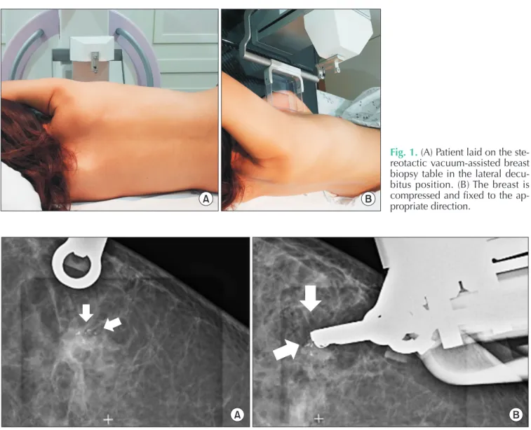
The patient lay on a stereotactic VAB table in the lateral
decubitus position with the affected breast side up (Fig. 1A). The breast was compressed and fixed to the appropriate direction (Fig. 1B). First, we checked the mammography. An additional 15o paired stereotactic mammography was obtained. Then the computer program calculated the threedimensional location, the horizontal (x) and vertical (y) distance and the depth (z) from the zero point of the stereotactic VAB device (Fig. 2).
The attached needle was moved to the skin incision site we measured. Local anesthesia was done through the breast parenchyma under aseptic conditions. After the skin incision within 4 mm, the 8G VAB probe was inserted through the incision site. Stereotactic 15o paired mammographies were checked to find the presence of dislocation due to lidocaine infection or needle insertion. We obtained a specimen after the needle location was confirmed by checking the image (Fig. 3).
One or two tissue specimens were collected from each of the six clockwise positions (2, 4, 6, 8, 10, and 12). If the calcification
A B
Fig. 1. (A) Patient laid on the ste
reotactic vacuumassisted breast biopsy table in the lateral decu
bitus position. (B) The breast is compressed and fixed to the ap
propriate direction.
A B
Fig. 2. (A) Mammography for confirmation of exact location of needle after introduction (arrows: microcalcification). (B)
is still identified on stereotactic mammography or was not identified in the specimen mammography, additional sampling was done. The needle was removed from the biopsy site when the procedure was successfully completed. The biopsy site was manually compressed. Skin closure with a piece of strip (Steri
Strip; 3M, St. Paul, MN, USA) and a compression bandage were used for hemostasis. These procedures were all performed by a single physician.
RESULTS
The average of patient age was 51.2 years (range, 35–76 years) and the average of procedure time was 20 minutes (range, 15–24 minutes). The average number of obtained specimens was 8.5 pieces (range, 6–12 pieces) (Table 1).
Mammography findings were distributed from category 3 to category 4 according to the breast imagingreporting and data system (BIRADS) classification. Micro calcification was observed in all specimen mammographies that underwent stereotactic VAB in 106 patients. The histological results for 10 patients (9.5%) were ductal carcinoma in situ (DCIS), 8 patients (7.5%) were focal atypical ductal hyperplasia (ADH), and 88 patients (83%) were benign (Table 2). There was neither vasovagal syncope event nor major complication. Minor hematoma was reported in 8 patients (7.5%).
Additional breast conserving surgery was performed for the 10 patients who were diagnosed with DCIS.
Additional wide excision was done for 2 of 8 patients diagnosed with ADH, due to diffusely scattered microcalcifica
tion. The final pathologic diagnosis was reported as ADH only.
After discussion with the patients, we decided close followup for 2 patients and another 6 patients diagnosed with ADH. Also, 88 patients diagnosed with fibrocystic change were followed up
6 months later by mammography without additional surgery.
They showed no interval change.
DISCUSSION
Traditionally, wire guided excisional biopsy is performed for nonpalpable breast lesion and mammographically detected microcalcification. However, this method is more invasive, leaves a larger scar, is more complicated, and more time
consuming than the percutaneous biopsy method. Recently, stereotactic VAB was introduced for breast lesions like these.
Many biopsy tools have been developed depending on the procedural approach and the position of the patient.
Stereotactic VAB can be classified as prone position [47], upright position [8,9], and lateral decubitus position method.
In the prone position, patients feel more safe. But a special table for the prone position is expensive and more space is required. It is difficult to perform a procedure at this table for patients who have small breasts such as Asian women. Also, it is impossible for lesions close to the chest wall.
The upright position method compensates for these disad
vantages. This method is more suitable for the average Asian woman who have small breast sizes. However, it is very difficult to fix the breast and maintain a stable position. Moreover, the procedure is performed in front of the patient; this causes Fig. 3. Specimen mammography shows a microcalcification
(arrow) two separate specimens.
Table 1. Clinical characteristics of stereotactic vacuum
assisted breast biopsy cases
Characteristic Value
Age (yr) 51.2 (35–76)
Procedure time (min) 20 (15–24)
No. of specimen 8.5 (6–12)
Complication after procedure
Yes (Hematoma) 8 (7.5)
No 98 (92.5)
Values are presented as mean (range) or number (%).
Table 2. Association of BIRADS category and pathologic results by stereotactic vacuumassisted biopsy
BIRADS category No.
(%) Pathology Pathologic
results 3 16 (15) Fibrocystic change, benign mass 14
Atypical ductal hyperplasia 2 4 90 (85) Fibrocystic change, benign mass 74 Atypical ductal hyperplasia 6
DCIS 6
DCIS → IDC 4
BIRADS, breast imagingreporting and data system; DCIS, ductal carcinoma in situ; IDC, invasive ductal carcinoma.
anxiety to the patient, back pain and vasovagal reaction.
To compensate for the disadvantages of the prone or upright
positioning for stereotactic VAB, a technique using an addon stereotactic unit with the patient in the decubitus position is used increasingly to avoid patient movement and syncope [10].
Recently, the decubitus table (DBI table, Medical Positioning Inc., Kansas City, MO, USA) has been developed and used with addon stereotactic units.
Laterally decubitus position was introduced to overcome the problems of these two methods. In particular, it is possible to obtain more specimens by using an 8G probe instead of an 11G probe, for more accurate and faster biopsy [11]. In our study, an expensive prone table was not necessary during stereotactic VAB and its lack did not cause vasovagal reaction.
In our center, stereotactic VAB has been used for biopsy of microcalcification since 2009. However, the procedure cost is still more expensive than surgical excision. When the cost was burdensome for the patient, surgical excision using a hook wire was done. Also, patients with breast parenchyma lesser than 3 mm, making the procedure impossible, had surgical excision.
According to BI-RADS, 90 patients (84.9%) were classified as category 4, which was the majority among the calcified lesions confirmed by biopsy. There were biopsy cases with category 3 patients, which were microcalcifications increasing during their followup or patients who were anxious about the microcalcification itself. In our clinic, there was no case diagnosed as malignancy in category 3 lesions, making the category an important factor to diagnosis before biopsy is done.
However, there were some cases diagnosed as malignancy. They were classified as category 3 in mammography, but category 4 in ultrasound showing a mass lesion. Therefore, BIRADS classi
fication is important in microcalcification lesions with no mass lesion shown in ultrasound.
In our study, there was no case diagnosed as malignancy in category 3, but there are some reports that the cancer rate of category 3 lesion is more than 20%. Therefore, biopsy is
needed even in category 3 lesions in clinically needed cases. For this reason, one radiologist and one breast surgeon judge the necessity of biopsy in these cases in our clinic. Biopsy is done when any clinician declares the necessity of biopsy.
Stereotactic VAB can focus precisely on clustered small lesions. Therefore, this procedure is a very excellent way to obtain a minimum of specimens quickly. However, it is possible to obtain only the specimen around the needle in distributed lesions. Also, VAB cannot function well in a patient whose breast parenchyma is thin. In recent years, there are a lot of solutions to these problems, but it is still difficult to per form a biopsy in lesions close to the chest wall or skin.
The complication rate of VAB is reported as approximately 1%–4%, but serious complications requiring treatment are rare [11,12]. In our study, there were only four cases of mild hematoma without any serious complication.
In conclusion, lateral decubitus position stereotactic VAB using 8G probe does not need a dedicated table, and is easier to maintain the position compared to upright stereotactic VAB.
It can also aid in obtaining a large size of specimen, reducing the number of trials and procedure time. This makes lateral decubitus positioning stereotactic VAB an accurate, safe and simple biopsy method; and also reduces the patient’s vasovagal reaction.
Our report has limitations such as the small number of cases with a shortterm follow up of four years. Longterm research of the false negative rate in stereotactic VAB is needed in category 4 patients who have been diagnosed with a benign lesion, because they can increase in size or possibly form a mass.
Thus, further improvements to the limitations our research can provide greater benefit to patients with microcalcification.
CONFLICTS OF INTEREST
No potential conflict of interest relevant to this article was reported.
1. Korean Central Cancer Registry. 2002 An
nual report of the Korean Central Cancer Registry. Gwacheon: Ministry of Health and Welfare; 2003.
2. Bae JW, Koo BW. Retrospective analysis of the needle localization biopsies for nonpalpable mammographic microcalcifi
cations of the breast. J Korean Surg Soc
1994;46:33541.
3. Kerlikowske K, Grady D, Rubin SM, Sandrock C, Ernster VL. Efficacy of screen
ing mammography. A metaanalysis.
JAMA 1995;273:14954.
4. Reynolds HE, Poon CM, Goulet RJ, Lazaridis CL. Biopsy of breast microcalcifi
cations using an 11gauge directional
vacuumassisted device. AJR Am J Roent
genol 1998;171:6113.
5. Meyer JE, Smith DN, DiPiro PJ, Denison CM, Frenna TH, Harvey SC, et al. Stereo
tactic breast biopsy of clustered microcal
cifications with a directional, vacuum
assisted device. Radiology 1997;204:5756.
6. Burak WE Jr, Owens KE, Tighe MB, Kemp
REFERENCES
L, Dinges SA, Hitchcock CL, et al. Vacuum
assisted stereotactic breast biopsy: histo
logic underestimation of malignant le
sions. Arch Surg 2000;135:7003.
7. Rotter K, Haentschel G, Koethe D, Goetz L, BornhofenPoschke A, Lebrecht A, et al. Evaluation of mammographic and cli
nical followup after 755 stereotactic va
cuumassisted breast biopsies. Am J Surg 2003;186:13442.
8. Walker TM. Impalpalbe breast lesions:
stereotactic core biopsy with `addon`
unit. The Breast 1997;6;12631.
9. Kirshenbaum KJ, Voruganti T, Overbeeke C, Kirshenbaum MD, Patel P, Kaplan G, et al. Stereotactic core needle biopsy of nonpalpable breast lesions using a con
ventional mammography unit with an addon device. AJR Am J Roentgenol 2003;
181:52731.
10. Welle GJ, Clark M, Loos S, Pauls D, Warden D, Sheffield M, et al. Stereotactic
breast biopsy: recumbent biopsy using addon upright equipment. AJR Am J Roentgenol 2000;175:5963.
11. Burbank F. Stereotactic breast biopsy:
comparison of 14 and 11gauge Mammo
tome probe performance and complica
tion rates. Am Surg 1997;63:98895.
12. Philpotts LE, Lee CH, Horvath LJ, Tocino I.
Canceled stereotactic coreneedle biopsy of the breast: analysis of 89 cases. Radiology 1997;205:4238.