2020 Korean Society for Surgery of the Hand, Ko- rean Society for Microsurgery, and Korean Society for Surgery of the Peripheral Nerve. All Rights re- served.
This is an open-access article distributed under the terms of the Creative Commons Attribution Non-Commercial license (http://creativecommons.
org/licenses/by-nc/4.0/), which permits unrestricted non-commercial use, distribution, and reproduction in any medium, provided the original work is prop- erly cited.
Calmette-Guérin (BCG) 골수염
김지영, 전지인, 장학
서울대학교 의과대학 서울대학교병원 성형외과
Bacille Calmette-Guérin Osteomyelitis of the Distal Radius in a Toddler
Ji-Young Kim, Ji-In Jeon, Hak Chang
Department of Plastic and Reconstructive Surgery, Seoul National University Hospital, Seoul National University College of Medicine, Seoul, Korea
BCG (Bacille Calmette-Guérin) vaccine has been administered safely to billions of peo- ple all over the world. The Tokyo-172 strain has reported to have a lower virulence and side effects than other strains. BCG osteomyelitis of distal radius is a very rare but se- rious complication due to generalized dissemination of BCG. We report a rare case of BCG osteomyelitis of the distal radius in a 21-month-old girl who had no underlying disorders. Although uncommon, BCG osteomyelitis should be considered a possible complication of BCG vaccination under certain clinical features for early diagnosis and proper treatment.
Keywords: BCG vaccine, Osteomyelitis, Toddler pISSN 2586-3290 · eISSN 2586-3533
Arch Hand Microsurg 2020;25(2):134-139 https://doi.org/10.12790/ahm.20.0017
Received: March 30, 2020 Revised: May 25, 2020 Accepted: May 25, 2020 Corresponding author:
Hak Chang
Department of Plastic and
Reconstructive Surgery, Seoul National University Hospital, Seoul National University College of Medicine, 101 Daehak-ro, Jongno-gu, Seoul 03080, Korea
Tel: +82-2-2072-3086 Fax: +82-2-3675-7792 E-mail: [email protected] ORCID:
https://orcid.org/0000-0002-1996-0680
Case Report
INTRODUCTION
In Korea, 95%–99% of children are vaccinated with BCG within 4 weeks after birth as a national policy for the management of tuberculosis. BCG was devel- oped as a live attenuated vaccine from the serial passage of Mycobacterium bovis [1]. Currently, three types of BCG strains (Moscow-368, Sofia SL222, and the To- kyo-172) are commonly used worldwide [2]. In Korea, the Pasteur 1173 P2, which had been used for more than 40 years, has been replaced by the Danish strain since 2007, and the Tokyo-172 was first introduced in Korea in 1993 and has been used steadily [3]. World Health Organization (WHO) recommends the intradermal methods with the merits of being able to administer accurate doses at a certain level. South Korea also recommends the intradermal method. Al- though the Korean national immunization program for BCG vaccination is based on the intradermal Danish strain, the percutaneous Tokyo-172 BCG vaccine is also used. The proportion of percutaneous Tokyo-172 BCG vaccination is esti- mated to be more than twice that of intradermal Danish BCG vaccination during the recent 10 years [4].
Among other BCG vaccines, Tokyo-172 strain has been reported to have a low- er virulence and side effects than other strains. But as a live bacterial vaccine, BCG causes dissemination beyond the vaccination site to various parts of the body. Osteomyelitis is a very rare but serious late complication of BCG-immuni-
zation in immunocompetent individuals from generalized dis- semination of BCG. Tokyo-172 strain is no exception. Here, we describe a rare case of BCG osteomyelitis of the distal radius in a 21-month-old girl who had no underlying immunodeficiency disorders.
CASE REPORT
A 21-month-old girl who was healthy and never had under- gone any disease presented symptoms of pseudoparalysis and tenderness with redness at her left wrist for a period of 1 month. There was no history of trauma, fever, or constitutional symptoms. According to the record of the Korea Centers for Disease Control and Prevention, she was vaccinated with To- kyo-172 BCG on the left upper arm at one month of age ad- ministered percutaneously using multipuncture method. At that time, as the doctor of the local clinic had an initial diagno- sis of a ruptured inclusion cyst, the wound was dressed into an arm splint. Although, 2 weeks of oral cefpodoxime (third-gen- eration cephalosporin antibiotics) therapy was maintained with a simple dressing, her wound was exacerbated and seemed to be cellulitis on her wrist (Fig. 1).
On admission, she had a normal body temperature of 36.6°C.
Blood workup was carried out and showed normal parameters ex- cept mild elevation of erythrocyte sedimentation rate (29 mm/hr).
A radiologic inspection was carried out and showed a distal pathologic geographic osteolytic lesion (Fig. 2). Magnetic reso- nance imaging (MRI) showed subacute osteomyelitis within the left distal radius and growth plate involvement with cortical dis- ruption. Also, an overlying infectious soft tissue enhancement with microabscess was noted (Fig. 3). These features suggested a low-virulent bacterial infection or mycobacterial infection.
Pus pocket was ruptured at dorsoradial aspect of the left wrist and debridement of the infected tissue was done through the opened pus pocket. After volar incision was made on the left wrist, a small hole was made at the radius volar side. Case- ous material was curettaged and aspirated through the hole. A
Fig. 1. Preoperative photograph on radial aspect of the left wrist.
About 2 x 1 cm sized ulcerative lesion involving subcutaneous layer.
Fig. 2. The wrist X-ray image taken at the time of suspicion of osteomyelitis shows a well-defined metaphyseal lytic lesion with geographic pattern. (A) Left image, anteroposterior view, (B) right image, lateral view.
Fig. 3. The wrist magnetic resonance image showing an aggressive lesion of the left distal radius combined with growth plate involvement and cortical disruption. (A) Left image, coronal view, (B) right image, axial view.
A
A
B
B
central physeal invasion was identified. All cultures including M. tuberculosis were negative but the Xpert MTB/RIF assay (Cepheid Inc., Sunnyvale, CA, USA; molecular identification of M. tuberculosis complex [MTBC] and resistance to rifampin [RIF]) identified M. tuberculosis with negative rifampin resis- tance. We simultaneously performed a BCG specific poly- merase chain reaction (BCG specific PCR) with the specimen obtained from an operating room for differential diagnosis of M. bovis-BCG or others. According to the result of Xpert MTB/
RIF assay, diagnosis of osteomyelitis of M. tuberculosis was made, and we started the treatment with a regimen of rifampin, isoniazid, and pyrazinamide. After 2 weeks later, multiplex PCR for BCG Tokyo-172 strain was positive following the BCG specific PCR report. The detection of BCG substrains using the multiplex PCR allowed us to differentiate BCG osteomyelitis from other mycobacterial infections. After the result, the stan- dard 12 months of anti-TB pharmacotherapy except pyrazin- amide was planned because M. bovis-BCG strain showed resis- tance to pyrazinamide. A 4-month follow-up X-ray showed a decreased extent of the osteolytic lesion and physeal growth (Fig. 4). At 8 months follow-up visits, she showed gradual im- provement and the pus no longer came out. She used her left forearm normally with a full range of movement.
DISCUSSION
The BCG vaccine is used to protect recipients against severe forms of tuberculosis like disseminated tuberculosis and ex-
trapulmonary tuberculosis. Serious adverse effects following BCG vaccination are rare. The most common adverse effects of BCG vaccination are local abscess. Osteitis or osteomyelitis is one of the rare consequences of BCG vaccination [5]. (Osteitis and osteomyelitis mean the infects of the bone and bone mar- row, respectively. However, medical literature usually does not clarify the distinction between osteitis and osteomyelitis [6].
Both conditions are referred to here as osteomyelitis.) The inci- dence of BCG osteomyelitis following the BCG vaccine in Ko- rea would be at least 4.08 cases per million during 2007-2017 [2]. Previously, the incidence of osteomyelitis following BCG Tokyo-172 vaccination was reported to be extremely low. The incidence of BCG osteomyelitis in Japan where Tokyo-172 strain is administered by percutaneous multiple puncture method was 0.01 per million in the 1980s, the lowest compared to other countries [7]. In Seoul National University Children's Hospital from January 2007 to March 2018, only 21 patients were diagnosed with BCG osteomyelitis [2].
With its rarity, its onset is slow and the symptoms are usually mild. These points make it difficult to diagnose BCG osteomy- elitis until it has well advanced. The most common symptoms of BCG osteomyelitis are mild pain and swelling of the overly- ing soft tissue, pseudoparalysis at the affected site, while fever was only accompanied in few [8]. The diagnosis is also easily confused with bacterial osteomyelitis because of clinical find- ings and isolated, well-bounded osteolytic lesions in radio- graphs. It is important to recognize the distinctive features of BCG osteomyelitis from bacterial osteomyelitis. Children with bacterial osteomyelitis commonly show high fever and bone pain of abrupt onset. Only a few BCG osteomyelitis accompany fever and bacterial osteomyelitis can develop at any age in chil- dren while BCG osteitis usually develops between 6 months and 5 years of age [9,10]. There is a long latent period between the BCG vaccination and the onset of symptoms of osteomyeli- tis because the incubation period of BCG osteomyelitis ranges from six to nine months after the vaccination, and in most cas- es, the metaphysis and epiphysis of long bones are affected. Ac- cording to a retrospective analysis of 222 cases, osteomyelitis was found in the femur (27%), tibia (19%), humerus (8%), and the sternum (15%) [10]. In our center, osteomyelitis at the dis- tal radius is the first case reported in toddler years (2–6 years old age). This also differs from disease caused by M. tuberculo- sis, which more commonly occurs in the spine and weight-bear- ing joints in older children and adults.
Since it is difficult to distinguish BCG strains from other M.
tuberculosis complexes by conventional culture or biochemical analysis, many previous reports of BCG osteomyelitis have Fig. 4. Four-month follow-up wrist X-ray with disappearance
of the lytic lesion. (A) Left image, anteroposterior view, (B) right image, lateral view.
A B
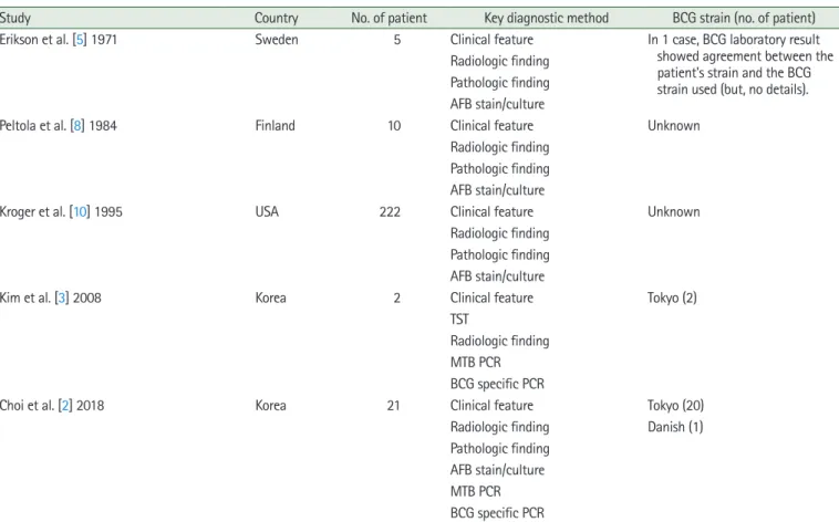
been clinically demonstrated without the characterization of isolates by laboratory methods (Table 1). In the absence of bac- terial evidence, the inclusion criteria for BCG osteomyelitis are typical histopathological findings, such as a type of epithelial cell granuloma with caseous necrosis, or typical radiological findings, such as a well-defined and eccentrically located de- struction in the metaphysis of a long bone, the history of BCG vaccination during the first year of life, and no evidence of pul- monary tuberculosis or a tuberculosis contact history [10].
Genetically diagnosed cases of BCG osteomyelitis were re- ported from Japan in 1997 and from Taiwan in 2004, but only two cases were distinguished from M. tuberculosis osteomyeli- tis, and it was not known whether the cause was due to M. bo- vis or a specific BCG substrain. In Korea, since the late 2000s,
there have been efforts to discriminate accurate BCG substrains using multiplex PCR technology, and the multiplex PCR meth- od has been utilized and proved to be a rapid and reliable method. BCG osteomyelitis is a rare complication, so many cli- nicians are unfamiliar with this diagnostic method. Compared
to the previous reports that focused on infection and technical aspects, we have described the process of diagnosing BCG os- teomyelitis with the molecular methods from the surgeon's point of view.
Most BCG osteomyelitis shows a favorable prognosis. It is known to have a good prognosis usually with surgical debride- ment and oral antituberculosis chemotherapy, and this is also the difference from M. tuberculosis infection [5]. In this case, after the anti-TB pharmacotherapy she was able to move his left wrist with a normal range of motion and activity, and the wound was recovered without additional surgical intervention.
Osteomyelitis induced by BCG vaccination is uncommon but should be considered a possible complication. Clinicians should consider BCG osteomyelitis based on clinical features when the BCG vaccinated child under 5 years old show osteo- myelitis, and select an appropriate diagnostic method for early diagnosis of BCG osteomyelitis and proper treatment.
Table 1. Diagnostic methods of BCG osteomyelitis and identification of BCG strain
Study Country No. of patient Key diagnostic method BCG strain (no. of patient) Erikson et al. [5] 1971 Sweden 5 Clinical feature In 1 case, BCG laboratory result
showed agreement between the patient's strain and the BCG strain used (but, no details).
Radiologic finding Pathologic finding AFB stain/culture
Peltola et al. [8] 1984 Finland 10 Clinical feature Unknown
Radiologic finding Pathologic finding AFB stain/culture
Kroger et al. [10] 1995 USA 222 Clinical feature Unknown
Radiologic finding Pathologic finding AFB stain/culture
Kim et al. [3] 2008 Korea 2 Clinical feature Tokyo (2)
TST
Radiologic finding MTB PCR BCG specific PCR
Choi et al. [2] 2018 Korea 21 Clinical feature Tokyo (20)
Radiologic finding Danish (1) Pathologic finding
AFB stain/culture MTB PCR BCG specific PCR
BCG, Bacille Calmette-Guérin; AFB, acid-fast bacillus; TST, tuberculin skin test; MTB, Mycobacterium tuberculosis; PCR, polymerase chain reaction.
CONFLICTS OF INTEREST
The authors have nothing to disclose.
REFERENCES
1. Khotaei GT, Sedighipour L, Fattahi F, Pourpak Z. Osteomyeli- tis as a late complication of Bacille Calmette-Guérin vaccina- tion. J Microbiol Immunol Infect. 2006;39:169-72.
2. Choi YY, Han MS, Lee HJ, et al. Mycobacterium bovis osteitis following immunization with Bacille Calmette-Guérin (BCG) in Korea. J Korean Med Sci. 2018;34:e3.
3. Kim SH, Kim SY, Eun BW, et al. BCG osteomyelitis caused by the BCG Tokyo strain and confirmed by molecular method.
Vaccine. 2008;26:4379-81.
4. Lee H, Dockrell HM, Kim DR, et al. The current status of BCG vaccination in young children in South Korea. Tuberc Respir Dis (Seoul). 2012;72:374-80.
5. Erikson U, Hjelmstedt A. Roentgenologic aspects of BCG os-
teomyelitis. Radiology. 1971;101:575-8.
6. Tiemann AH, Hofmann GO. Principles of the therapy of bone infections in adult extremities: are there any new devel- opments? Strategies Trauma Limb Reconstr. 2009;4:57-64.
7. Lotte A, Wasz-Hockert O, Poisson N, Dumitrescu N, Verron M, Couvet E. A bibliography of the complications of BCG vaccination. A comprehensive list of the world literature since the introduction of BCG up to July 1982, supplemented by over 100 personal communications. Adv Tuberc Res. 1984;21:
194-245.
8. Peltola H, Salmi I, Vahvanen V, Ahlqvist J. BCG vaccination as a cause of osteomyelitis and subcutaneous abscess. Arch Dis Child. 1984;59:157-61.
9. Vohra R, Kang HS, Dogra S, Saggar RR, Sharma R. Tubercu- lous osteomyelitis. J Bone Joint Surg Br. 1997;79:562-6.
10. Kroger L, Korppi M, Brander E, et al. Osteitis caused by bacille Calmette-Guérin vaccination: a retrospective analysis of 222 cases. J Infect Dis. 1995;172:574-6.
유아의 원위 요골부에 발생한 Bacille Calmette-Guérin (BCG) 골수염
김지영, 전지인, 장학
서울대학교 의과대학 서울대학교병원 성형외과
BCG 백신은 전세계적으로 널리 접종되고 있는 백신 중 하나로 그 중 Tokyo-172의 경우 비교적 안정하고 부작용이 드문 것으로 알려져 왔다. 원위 요골의 BCG골수염은 BCG 접종의 드문 합병증으로 BCG 균의 전신성 파종에 의해서 생긴다. BCG 접종에 의한 골수염은 드물지만 심각한 합병증을 유발할 수 있으며, 빠른 진단과 적절한 치료를 위해 감별진단으로 고려되어야 한다. 저자들은 생후 1개월에 BCG 접종을 받은 21개월된 여아에서 좌측 원위 요골부에 발생한 결핵성 골수염을 진단하였기에 보고하는 바이다.
색인단어: BCG 백신, 골수염, 유아
접수일 2020년 3월 30일 수정일 2020년 5월 25일 게재확정일 2020년 5월 25일 교신저자 장학
03080, 서울특별시 종로구 대학로 101, 서울대학교 의과대학 서울대학교병원 성형외과 TEL 02-2072-3086 FAX 02-3675-7792 E-mail [email protected] ORCID https://orcid.org/0000-0002-1996-0680