Hobnail hemangioma(HH) is a rare benign vascular endothelial neoplasm, but should be differentiated from other malignant vas- cular neoplasms such as patch stage Kaposi’s sarcoma or angiosarcoma. Clinically, it usually appears as a single patch or nodule. Micro- scopic characteristics are hobnail endothelial cells with prominent hyperchromatic nuclei and dissection of collagen pattern. We report a case of HH, which shows typical morpho- logic and histologic findings, but, has recur- rence unlike previous reports.
CASE REPORT
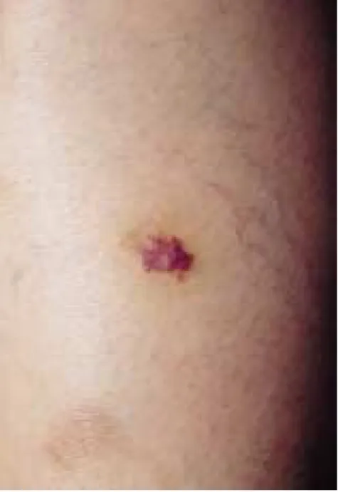
A 21-year-old woman was presented with a dusky-red patch on her left shin. She noticed the lesion five weeks ago. There was no
primitive lesion or trauma history. Examina- tion disclosed a well-demarcated but irregu- larly bordered patch, 1.2cm in diameter (Fig. 1). Histologic examination disclosed ectatic vascular channels with a single layer of plumped endothelial cells and occasional intraluminal papillary projections in the upper dermis (Fig. 2). Deeper in the dermis, the vascular channels were thinner and seemed to dissect the collagen bundles. Other findings included numerous extravasated erythrocytes and mild perivascular lymphocyte infiltrates and a few hemosiderin-laden macrophages.
Immunohistochemistry with monoclonal anti- bodies revealed a mild focal positive reaction with FactorVIII related antigen, and a nega- tive reaction with Ulex europaeus agglutinin I, but a strongly positive reaction with CD34 (Fig. 3). The patient underwent treatment with complete surgical excision with primary closure. Two years later, recur- rence was observed in the shin.
DISCUSSION
Hobnail hemangioma(HH) is a relatively rare benign acquired vascular tumor of en- dothelial origin. Typically, the tumor arises in young adults with an almost equal proportion
A Case of Hobnail Hemangioma
Shin Taek Oh, M.D., Seung Dong Lee, M.D., Sung Wook Kim, M.D.*, In Gang Jang, M.D.**, Baik Kee Cho, M.D.
Department of Dermatology, College of medicine, The Catholic University of Korea, Seoul, Korea, Cha
& Park Dermatologic Clinic*, Misarang Dermatologic Clinic**
Hobnail hemangioma(HH) is a benign acquired vascular tumor of endothelial origin which should be differentiated from other malignant vascular neoplasm such as Kaposi’s sarcoma or an- giosarcoma. We report a case of hobnail hemangioma in a 21-year-old woman who had a dusky-red patch on her left shin. Histologically, ectatic vascular channels with a single layer of plumped en- dothelial cells were seen and the vascular channels seemed to dissect the collagen bundles. She un- derwent treatment with surgical excision with primary closure.
(Ann Dermatol 14(1) 45-47, 2002).
Key Words : Hobnail hemangioma
Received April 20, 2001.
Accepted for publication August 22, 2001.
Reprint request to : Shin Taek Oh, Department of Der- matology, Taejon St. Mary’s Hospital, College of Med- icine, The Catholic University of Korea,
520-2, Daeheung-dong, Chung-ku, Taejon 301-012, Korea.
Tel. 042-220-9578, Fax. 042-253-5107
45
in men and women, but it may affect all age groups. It has a predilection for extremities, but can occur at any site of the body.
Clinically, it appears usually as a single, small, well-demarcated patch or nodule1.
Histologically, characteristic features are small size, hobnail endothelial nuclei, superfi- cial dilated blood vessels, progressive disap- pearance of the lesion into the reticular dermis and dissection of collagen1,2. Targetoid hemosiderotic hemangioma has in addition to the same histologic appearance as HH, a
marked deposition of hemosiderin. Clinically, the characteristic targetoid appearance is due to peripheral hemorrhage and the subse- quent deposition of hemosiderin3. But, Calonje proposed that HH is the more ap- propriate descriptive morphologic term re- gardless of the targetoid appearance or marked hemosiderin deposition2.
The main microscopic differential diagnosis is for the patch stage of Kaposi’s sarcoma (KS), which shares many similarities with HH. Factors favoring patch stage KS are the presence of plasma cells, spindle-shaped cells and apoptotic endothelial cells4. Other differential diagnoses include progressive lymphangioma, lymphangioma-like variants of KS, well-differentiated angiosarcoma (AS), and retiform hemangioendothelioma.
Progressive lymphangioma can be distin- guished by its large size and lack of superfi- cial “hobnailed” endothelial cells5,6. In lym- phangioma-like variants of KS, it occurs mainly in older persons and possesses the less extravasated erythrocytes7. Well-differentiated AS occurs predominantly in head and neck, and one can find less well differentiated areas with thorough examination8. Retiform he- mangioendothelioma has the same hobnail endothelial nuclei as the unique hobnail en- dothelial nuclei of HH, but has a retiform pattern of distribution2. HH can be ade- quately treated by simple surgical excision without tendency to recur1. In our case, the 46 ST Oh, et al.
Annals of Dermatology Vol. 14, No. 1, January 2002
Fig. 2. Ectatic vascular channels with a single layer of plumped endothelial cells in the superficial dermis (H & E, ×100).
Fig. 3. Positive reaction with CD34 monoclonal anti- body (×100).
Fig. 1. Dusky-red patch on the left shin.
lesion was treated under complete excision and primary closure. But, two years later, re- currence was observed in the vicinity of the original lesion.
HH is thought to be originated from vas- cular endothelium3. Factor VIII- related antigen has been reported as weakly positive;
Ulex europaeus agglutinin has been strongly positive9 or rarely positive1. CD34 is a very sensitive marker for the vascular en- dothelium, and showed variable reactivity for endothelial cells of HH1,3. In our case, immunohistochemistry revealed a mild focal positive reaction with Factor VIII related antigen, and a negative reaction with Ulex europaeus agglutinin I, but a strongly positive reaction with CD34. These results imply that the origin of HH is vascular endothelial cells, not lymphatic endothelaial cells.
REFERENCES
1. Guillou L, Calonje E, Speight P, Rosai J, Fletcher CDM. Hobnail hemangioma: a pseudomalignant vascular lesion with a reappraisal of targetoid he-
mosiderotic hemangioma. Am J Surg Pathol 1999;
23: 97-105.
2. Calonje E, Fletcher CDM, Wilson Jones E. Reti- form hemangioendothelioma: a distinctive form of low-grade angiosarcoma delineated in a series of 15 cases. Am J Surg Pathol 1994; 18: 115-125.
3. Santonja C, Torrelo A. Hobnail hemangioma. Der- matology 1995; 191: 154-15.
4. Chor PJ, Santa cruz DJ. Kaposi’s sarcoma: a clini- copathologic review and differential diagnosis. J Cutan Pathol 1991; 19: 6-20.
5. Lowe L. Targetoid hemosiderotic hemangioma:
self-assessment. J Cutan Pathol 1994; 21: 567-569.
6. Meunier L, Barneon G, Meynadier J. Acquired pro- gressive lymphangioma. Br J Dermatol 1994; 131:
706-708.
7. Gange RW, Wilson Jones E. Lymphangioma-like Kaposi’s sarcoma: a report of three cases. Br J Der- matol 1979; 100: 327-334.
8. Holden CA, Spittle MF. Angiosarcoma of the face and scalp, prognosis and treatment. Cancer 1987;
59: 1046-1057.
9. Santa Cruz DJ, Aronberg J. Targetoid hemosiderot- ic hemangioma. J Am Acad Dermatol 1988; 19:
550-558.
A Case of Hobnail Hemangioma 47