Letter to the Editor
258 Ann Dermatol
Received December 6, 2012, Revised March 23, 2013, Accepted for publication April 16, 2013
Corresponding author: Seung Phil Hong, Department of Dermatology, Dankook University Medical College, 119 Dandae-ro, Dongnam-gu, Cheonan 330-714, Korea. Tel: 82-41-550-6485, Fax: 82-41-552-7541, E-mail: [email protected]
This is an Open Access article distributed under the terms of the Creative Commons Attribution Non-Commercial License (http://
creativecommons.org/licenses/by-nc/3.0) which permits unrestricted non-commercial use, distribution, and reproduction in any medium, provided the original work is properly cited.
sity Medical School (BK+21) and Chonnam National University Hospital Biomedical Research Institute (CRE- 13118-7).
REFERENCES
1. Nishiyama H, Itoh K, Kaneko Y, Kishishita M, Yoshida O, Fujita J. A glycine-rich RNA-binding protein mediating cold- inducible suppression of mammalian cell growth. J Cell Biol 1997;137:899-908.
2. Yang C, Carrier F. The UV-inducible RNA-binding protein A18 (A18 hnRNP) plays a protective role in the genotoxic
stress response. J Biol Chem 2001;276:47277-47284.
3. Lleonart ME. A new generation of proto-oncogenes: cold- in- ducible RNA binding proteins. Biochim Biophys Acta 2010;
1805:43-52.
4. Hamid AA, Mandai M, Fujita J, Nanbu K, Kariya M, Kusakari T, et al. Expression of cold-inducible RNA-binding protein in the normal endometrium, endometrial hyperplasia, and en- dometrial carcinoma. Int J Gynecol Pathol 2003;22:240-247.
5. Artero-Castro A, Callejas FB, Castellvi J, Kondoh H, Carnero A, Fernández-Marcos PJ, et al. Cold-inducible RNA-binding protein bypasses replicative senescence in primary cells through extracellular signal-regulated kinase 1 and 2 activa- tion. Mol Cell Biol 2009;29:1855-1868.
http://dx.doi.org/10.5021/ad.2014.26.2.258
Pemphigus Vulgaris in Pregnancy Associated with Herpes Virus Type 1 Infection
Jiwon Gye, Chan Hee Nam, Ji Seok Kim, Jee Young Kim, Byung Cheol Park, Myung Hwa Kim, Seung Phil Hong
Department of Dermatology, Dankook University Hospital, Cheonan, Korea
Dear Editor:
Pemphigus vulgaris (PV) rarely occurs during pregnancy.
We report a case of PV associated with herpes simplex type 1 virus (HSV-1) which occurred in the third trimester of pregnancy.
A 32-year-old second gravida at 37 weeks’ gestation was admitted for multiple bullous skin lesions that persisted for over a month.
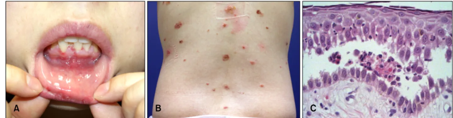
These vesicular and erosive lesions initiated from the periumbilical region and spread to the oral mucosa and the skin of the back (Fig. 1A, B). The diagnosis of PV was
confirmed by biopsy (Fig. 1C), and direct immufluore- scence detected anti-immunoglobulin G and C3 antibodies.
Anti-desmoglein 1 and anti-desmoglein 3 antibodies were elevated at 82.1 U/ml (normal, <14 U/ml) and 184.9 U/ml (normal, <7 U/ml) in peripheral blood. Tzanck smear and viral polymerase chain reaction (Seeplex STD B41 Detedtion; Seegene, Seoul, Korea) were done on the base of a vesicular lesion on the trunk. Tzanck smear was negative, but, viral polymerase chain reaction (PCR) was positive for HSV-1 (Fig. 2).
Prednisolone at a dose of 20 mg/d was initiated. Foll-
Letter to the Editor
Vol. 26 No. 2, 2014 259 Fig. 1. (A) Multiple erosions on oral mucosa and (B) scattered crusted vesicles and erosions on the back. (C) Suprabasal blisters with eosinophils and acantholytic cells in the epidermis (H&E, ×400).
Fig. 2. Polymerase chain reaction results show a positive band on 189 bp. M: 100 bp DNA ladder, Lane 1: positive result of the patient’s tissue for herpes simplex type 1 virus (189 bp), Lane 2:
normal control, Lane 3: positive control, Lane 4: sensitivity test, Lane 5: negative control.
owing PCR results, antiviral agent (valacyclovir, total 4,500 mg) was also given for 5 days. Lesions started to improve after starting the medication.
Two weeks after the diagnosis the patient was transferred to the labour unit for induced labour. The baby was in a good state. After giving birth, the dose of prednisolone was increased to 25 mg/d, and mycophenolate mofetil 1.0 g/d was added. After improvement in both vesicles and general health status, the patient was discharged. During follow-up, a subtle amount of skin lesion occurred which was controlled with topical steroid, oral mycofenolate mofetil 1.0 g/d, and intermittent oral steroid at a dose of 4 mg/d.
Though it is known that pemphigus develops as a result of the interaction between endogenous (genetic) factors and exogenous factors1, the primary stimulus for the autoimm- une response of PV is still unknown.
Pregnancy may aggravate certain autoimmune diseases, such as systemic lupus erythematosus, pemphigoid gesta- tionis, and PV2. The patient’s vesicular eruptions devel-
oped during the period of conception and were not controlled well at that time, so we supposed her condition was related to pregnancy.
Bullous diseases that are reported to be associated with pregnancy include pemphigoid gestationis pemphigus foli- aceus, and PV2,3. The most important diagnostic tool is histologic examination, and the two diseases mentioned above show subepidermal, subcorneal blisters. However PV, as seen in our patient, shows a suprabasal epidermal split with acantholysis and few eosinophils in the dermis, and perilesional skin shows intercelluar deposits of IgG and C3 in the epidermis on direct immunofluorescence4. A diagnosis of PV in this patient was made according to the histological features and clinical involvement of oral mucosa.
The possible role of viruses, especially HSV-1, has been proposed in the pathogenesis of PV. Positive viral PCR results suggest that viral factors, especially HSV and Ep- stein-Barr virus, are capable of inducing and/or exacer- bating pemphigus in a genetically susceptible host1. Infectious agents can stimulate the immune response in genetically susceptible individuals or in those with immu- ne deviating conditions such as pregnancy, leading to an increase in the production of cytokines. High levels of interferon-gamma induce the expression of human leuco- cyte antigen type 2 in the membranes of keratinocytes, making the structural site of PV antigen (epitope sprea- ding) immunologically active. Chronic, recurrent viral in- fections can also stimulate excessive production of inter- leukin (IL)-4 and IL-10, which result in a shift from TH1 to TH2 response, increasing antibody production. In addi- tion, they can also directly infect B and T lymphocytes, contributing to the production of autoreactive B lympho- cytes and autoimmune antibodies5. When infected, kera- tinocytes can pass through structural changes which favor the exposure of antigens1.
In summary, we report a case of a woman who showed
Letter to the Editor
260 Ann Dermatol
Received March 13, 2013, Revised April 11, 2013, Accepted for publication April 19, 2013
*These authors equally contributed as first authors.
Corresponding author: Ken Igawa, Department of Dermatology, Osaka University Graduate School of Medicine, 2-2 Yamadaoka, Suita-shi, Osaka, 565-0871, Japan. Tel: 81-6-6879-3031, Fax: 86-6879-3039, E-mail: [email protected]
This is an Open Access article distributed under the terms of the Creative Commons Attribution Non-Commercial License (http://
creativecommons.org/licenses/by-nc/3.0) which permits unrestricted non-commercial use, distribution, and reproduction in any medium, provided the original work is properly cited.
PV associated with HSV-1 in her second pregnancy, whi- ch was cured with steroids and antiviral agents.
REFERENCES
1. Brenner S, Sasson A, Sharon O. Pemphigus and infections.
Clin Dermatol 2002;20:114-118.
2. Fainaru O, Mashiach R, Kupferminc M, Shenhav M, Pauzner D, Lessing JB. Pemphigus vulgaris in pregnancy: a case rep- ort and review of literature. Hum Reprod 2000;15:1195- 1197.
3. Choi JH, Lim SW, Kim YJ, Bang JS, Lee JW, Suh MK, et al. A case of pemphigus foliaceus developed during pregnancy.
Korean J Dermatol 2001;39:1449-1451.
4. Elder DE, Elenitsas R, Johnson Jr BL, Murphy GF, Xu X. Le- ver's histopathology of the skin. 10th ed. Philadelphia:
Lippincott-Raven, 2009:247-261.
5. Brandão ML, Fernandes NC, Batista DP, Santos N. Refra- ctory pemphigus vulgaris associated with herpes infection:
case report and review. Rev Inst Med Trop Sao Paulo 2011;53:113-117.
http://dx.doi.org/10.5021/ad.2014.26.2.260
A Rare Case of Annular Pustular Psoriasis Associated with Pemphigus Foliaceus
Kenichi Kato
1,2,*, Takaaki Hanafusa
1,*, Ken Igawa
1, Motohiro Tatsumi
2, Yuji Takahashi
2, Takashi Yamanaka
2, Ichiro Katayama
11Department of Dermatology, Osaka University Graduate School of Medicine, Osaka,
2Department of Dermatology, Kansai Rosai Hospital, Hyogo, Japan
Dear Editor:
A 56-year-old Japanese woman suffered from multiple, scaly erythemas on the trunk for approximately 30 years.
She had been previously diagnosed with psoriasis vulgaris in the clinic due to the clinical appearance and histo- logical findings. She was also previously diagnosed with pemphigus foliaceus (PF) at our hospital 10 years ago, based on the clinical appearance and histological findings of a subcorneal blister, as well as on the direct immuno- fluorescence findings of superficial epidermal intercellular immunoglobulin (Ig) G deposition. During her clinical course, serum anti- desmoglein (Dsg)-1 IgG antibody le-
vels were elevated to 99 index points in 2010, whereas anti-Dsg-3 antibody levels remained at <5 index points.
Recently, she was treated for PF by administration of oral betamethasone (0.5 mg/d) and cyclosporine (100 mg/d).
She was admitted to our hospital because the annular erythemas with pustular margins on her trunk were exacerbated and accompanied by high fever (Fig. 1). No lesions resembling PF were seen. Laboratory findings were as follows (abnormal values are underlined): white blood cell count, 21.3×10³/μl (neutrophils: 72%); C-reactive protein: 16.4 mg/L. Anti-Dsg1 and anti-Dsg3 antibodies were within the normal range. Because of the clinical