ISSN 2234-3806 • eISSN 2234-3814
http://dx.doi.org/10.3343/alm.2013.33.4.288
Rapid Determination of Chimerism Status Using Dihydrorhodamine Assay in a Patient with X-linked Chronic Granulomatous Disease Following
Hematopoietic Stem Cell Transplantation
Hyun-Young Kim, M.D.1, Hee-Jin Kim, M.D.1, Chang-Seok Ki, M.D.1, Dae Won Kim, M.D.1, Keon Hee Yoo, M.D.2, and Eun-Suk Kang, M.D.1
Departments of Laboratory Medicine and Genetics1 and Pediatrics2, Samsung Medical Center, Sungkyunkwan University School of Medicine, Seoul, Korea
Chronic granulomatous disease (CGD) is a rare genetic disease, which is caused by de- fects in the NADPH oxidase complex (gp91phox, p22phox, p40phox, p47phox, and p67phox) of phagocytes. This defect results in impaired production of superoxide anions and other re- active oxygen species (ROS), which are necessary for killing bacterial and fungal microor- ganisms and leads to recurrent, life-threatening bacterial and fungal infections and granu- lomatous inflammation. The dihydrorhodamine (DHR) flow cytometry assay is a useful di- agnostic tool for CGD that can detect absent or reduced NADPH oxidase activity in stimu- lated phagocytes. We report a patient with X-linked CGD carrying a novel mutation of the CYBB gene whose chimerism status following hematopoietic stem cell transplantation (HSCT) has been rapidly determined using the DHR assay. The level of DHR activity corre- lates well with short tandem repeat PCR analysis. Considering the advantages of this sim- ple, rapid, and cost-effective procedure, serial measurement of DHR assay would facilitate the rapid determination of a patient’s engraftment status, as a supplementary monitoring tool of chimerism status following HSCT.
Key Words: Chronic granulomatous disease, Dihydrorhodamine assay, Chimerism
Received: November 9, 2012 Revision received: December 24, 2012 Accepted: February 26, 2013 Corresponding author: Eun-Suk Kang Department of Laboratory Medicine and Genetics, Samsung Medical Center, 81 Irwon-ro, Gangnam-gu, Seoul 135-710, Korea Tel: +82-2-3410-2703 Fax: +82-2-3410-2719 E-mail: [email protected]
© The Korean Society for Laboratory Medicine.
This is an Open Access article distributed under the terms of the Creative Commons Attribution Non-Commercial License (http://creativecom- mons.org/licenses/by-nc/3.0) which permits unrestricted non-commercial use, distribution, and reproduction in any medium, provided the original work is properly cited.
INTRODUCTION
Chronic granulomatous disease (CGD) is a rare genetic disease with an incidence of approximately 1 in 200,000 [1]. CGD is caused by a defect in the NADPH oxidase complex of phago- cytes, which results in impaired production of superoxide an- ions and other reactive oxygen species (ROS), which are neces- sary for killing bacterial and fungal microorganisms. This defect leads to recurrent, life-threatening bacterial and fungal infec- tions and granulomatous inflammation. Approximately two-third cases of CGD occur due to X-linked recessive mutations in the CYBB gene encoding gp91phox, which is located at Xp21.1. One- third of CGD cases occur as an autosomal recessive form, in-
volving other genes encoding p22phox, p40phox, p47phox, and p67phox [2-4]. The dihydrorhodamine (DHR) assay is known to be a use- ful diagnostic tool for CGD [5]. We report a patient with X-linked CGD whose chimerism status following hematopoietic stem cell transplantation (HSCT) was rapidly determined using the DHR assay, which was compared to short tandem repeat PCR (STR- PCR) analysis.
CASE REPORT
A 3-month-old male infant presented with fever. His leukocyte count was 19.4×10⁹/L; neutrophils, 12.086×10⁹/L; Hb, 8.6 g/dL;
platelet, 287×109/L; C-reactive protein, 6.57 mg/dL. Liver func-
tion tests showed AST activity of 158 U/L and ALT activity of 119 U/L. Computed tomography showed a hepatic mass in segment IV with a diameter of 50 mm, multiple satellite hepatic and sple- nic nodules, hepatosplenomegaly, and necrotizing mediastinal lymphadenopathy. Open liver biopsy revealed suppurative in- flammation with abscess, and Serratia marcescens was identi- fied from a culture of liver tissue. A liver abscess with S. marce- scens and necrotizing mediastinal lymphadenopathy suspected of disseminated tuberculosis due to BCG injection raised con- cerns for CGD. Subsequently, treatment with antibiotics as well as antifungal and antituberculous medications was initiated.
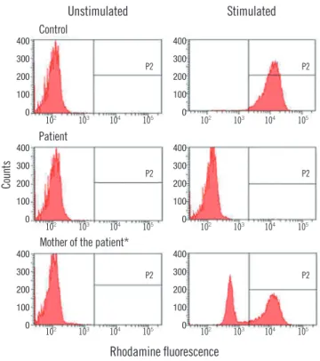
The neutrophil oxidative burst test was performed using the DHR assay before and after stimulation of neutrophils with phor- bol myristate acetate (PMA) (Fig. 1). No DHR fluorescence was detected after stimulation (0.2%), consistent with a phenotype observed in X-linked CGD. Molecular genetic analysis of the CYBB gene encoding the gp91phox mutation, was performed us- ing direct DNA sequencing to confirm the diagnosis of X-linked CGD. A novel frameshift mutation in exon 9 of the CYBB gene was identified (Fig. 2). The DHR assay and genetic tests per- formed on both the mother and brother of the patient revealed that the mother was a carrier of X-linked CGD but his elder brother was not. The family history was reviewed, and we found that the maternal uncle of the patient had died with pneumonia Fig. 1. Neutrophil oxidative burst test using DHR. *The patient’s
mother shows the partial absence of reactivity after PMA stimula- tion, consistent with an X-linked CGD carrier.
Abbreviations: DHR, dihydrorhodamine; PMA, phorbol myristate acetate;
CGD, chronic granulomatous disease.
Rhodamine fluorescence
Stimulated Unstimulated
Control
Patient
Counts
Mother of the patient*
Fig. 2. CYBB gene mutation analysis. A novel mutation (c.1078_1080delGACinsAA in exon 9) results in a frameshift in gp91phox (p.Asp 360Asnfs*26). Patient is homozygous and his mother is a heterozygous carrier of the frameshift mutation.
Wild-type
Patient
Mother of the patient
c.1078_1080delGACinsAA
102
102
102
102
102
102 104
104
104
104
104
104 103
103
103
103
103
103 105
P2
P2
P2
P2
P2
P2 105
105
105
105
105 400
300 200 100 0
400 300 200 100 0
400 300 200 100 0
400 300 200 100 0
400 300 200 100 0
400 300 200 100 0
2 months after birth, and he was, therefore, presumed to have been affected with CGD.
The patient received granulocyte transfusions 9 times at 4-5 months of age. Left hemihepatectomy was performed to remove a liver abscess during the same period; this surgery demon- strated chronic granulomatous inflammation. At the age of 7 months, multiple BCG granulomas, detected to be Mycobacte-
rium tuberculosis by real-time PCR, were developed on the left upper arm as well as the neck and axillary area. Incision and drainage were subsequently performed.
At the age of 13 months, HLA-C 1 allele mismatched (11/12), allogeneic-unrelated, peripheral blood stem cell transplantation (uPBSCT) was performed under a conditioning regimen of bu- sulfan (1.1 mg/kg), fludarabine (40 mg/m2), and rabbit anti-thy- mocyte (2.5 mg/kg). The blood group of the recipient and donor was A/Rh+ and B/Rh+, respectively. Dose of graft mononuclear cells, CD34+ cells, and CD3+ cells were 7.83×10⁸/kg, 2.64×10⁶/ kg, and 2.38×10⁸/kg, respectively. Engraftment of neutrophils and platelets occurred on day 13 and day 43, respectively, after HSCT.
Eighteen days after HSCT, preemptive treatment with ganci- clovir for cytomegalovirus (CMV) viremia was administered. On day 27, amphotericin B treatment for probable invasive aspergil- losis infection was initiated. On day 69 after HSCT, the patient developed grade II acute graft versus host disease of the gut.
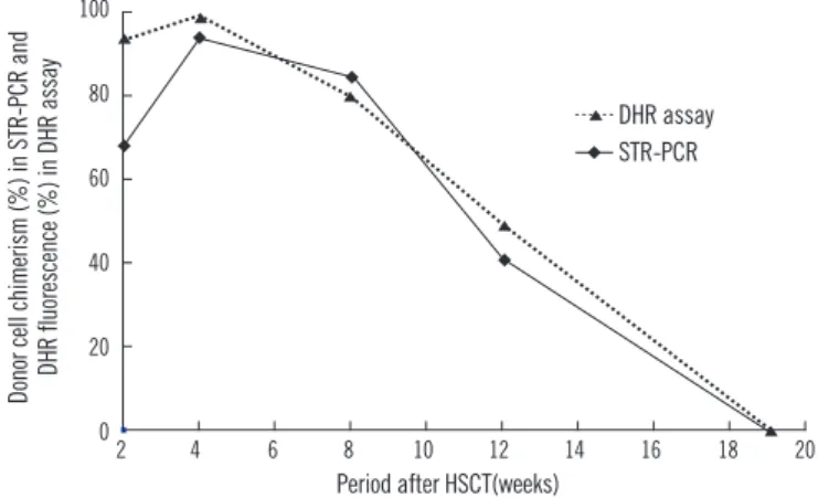
Chimerism status following HSCT was monitored in peripheral blood or bone marrow mononuclear cells using STR-PCR analy- sis. At 2, 4, 8, 12, and 19 weeks after HSCT, donor cell chime- rism was 68.3%, 94.1%, 84.7%, 40.9%, and 0%, respectively.
The DHR assay, which was performed simultaneously, revealed Fig. 3. Chimerism status in STR-PCR analysis and DHR assay dur-
ing the follow-up period.
Abbreviations: HSCT, hematopoietic stem cell transplantation; STR-PCR, short tandem repeat polymerase chain reaction; DHR, dihydrorhodamine.
Donor cell chimerism (%) in STR-PCR and DHR fluorescence (%) in DHR assay
Period after HSCT(weeks) 100
80 60 40 20
02 4 6 8 10 12 14 16 18 20 STR-PCR
DHR assay
Fig. 4. DHR assay after HSCT. The black arrow indicates the small population showing complete lack of DHR fluorescence, which reap- pears 8 weeks after HSCT. The written number (%) is DHR fluorescence (P2) after PMA stimulation.
Abbreviations: HSCT, hematopoietic stem cell transplantation; DHR, dihydrorhodamine; PMA, phorbol myristate acetate.
Rhodamine fluorescence Rhodamine fluorescence
Stimulated Stimulated
Unstimulated Unstimulated
A. 2 weeks after HSCT D. 12 weeks after HSCT
B. 4 weeks after HSCT E. 14 weeks after HSCT
Counts Counts
C. 8 weeks after HSCT F. 19 weeks after HSCT
102 102
102 102
102 102
102 102
102 102
102 102
104 104
104 104
104 104
104 104
104 104
104 104
103 103
103 103
103 103
103 103
103 103
103 103
105 105
P2 P2
P2 P2
P2 P2
P2 P2
93.7% 49.3%
98.8% 71.5%
80.1% 0.0%
P2 P2
P2 P2
105 105
105 105
105 105
105 105
105 105
400 300 200 100 0
400 300 200 100 0
400 300 200 100 0
400 300 200 100 0
400 300 200 100 0
400 300 200 100 0 400
300 200 100 0
400 300 200 100 0
400 300 200 100 0
400 300 200 100 0
400 300 200 100 0
400 300 200 100 0
93.7%, 98.8%, 80.1%, 49.3%, and 0% of reactivity following PMA stimulation (Fig. 3). The fraction of the cell population, which previously showed a complete lack of DHR fluorescence, reappeared at 8 weeks after HSCT, and it gradually increased until defective neutrophils replaced the entire neutrophil popu- lation (Fig. 4). Finally, engraftment failure was determined by the absence of both donor cell chimerism and normal neutro- phil population.
The patient underwent a second HSCT from the same alloge- neic donor after a conditioning regimen of total body irradiation (300 cGy), melphalan (45 mg/m2), and rabbit anti-thymocytes (2.5 mg/kg). Graft mononuclear cell, CD34+ cell, and CD3+ cell doses were 29.57×10⁸/kg, 5.69×10⁶/kg, and 18.53×10⁸/kg, re- spectively. Engraftment of neutrophils and platelets occurred on day 14 and day 46 after HSCT, respectively. One hundred and twenty days after the second HSCT, the donor chimerism level monitored by STR-PCR was 100%, and defective neutrophil ac- tivity was not identified through DHR assay.
DISCUSSION
CGD is diagnosed by an absence or reduction of oxidase activity in stimulated phagocytes, and it is confirmed by genetic testing.
There are various tests for the measurement of neutrophil super- oxide production, which include direct measurement of super- oxide production, cytochrome c reduction assay, chemilumines- cence, nitro blue tetrazolium reduction test, and the DHR assay.
Of these tests, the DHR assay has proved to be a rapid and highly sensitive assay for CGD [6, 7]. This assay provides a clear discrimination between abnormal and normal phagocytes of CGD patients. Quantitative results of cellular responses are dem- onstrated as more or less impaired subgroups. DHR 123 freely enters the cell membrane, localizes in the mitochondria, and is oxidized to rhodamine 123 by ROS in stimulated phagocytes, which then emits a bright fluorescent signal that is measured by flow cytometry [5, 8]. The DHR assay can be helpful in the iden- tification of the X-linked CGD and certain autosomal recessive type of CGD with a p47phox defect [7]. Detection of the X-linked CGD carrier is characterized by a mixture of normal and abnor- mal phagocytes [5]. Because the level of functional defect of neutrophils may differ among various genetic aberrations, espe- cially in autosomal recessive CGD, the DHR assay is not always useful as a diagnostic method [9, 10]. Some exhausted neutro- phils in a systemic inflammatory condition have been shown to have an attenuated response when activated in vitro [11].
There have been consistent advances in antibiotic and anti-
fungal therapy and prophylaxis. However, CGD still causes sig- nificant morbidity and mortality, and the only known cure is suc- cessful allogeneic HSCT [12, 13]. Unrelated HSCT as well as re- lated HSCT are performed more frequently, with improvement in overall survival rates. On the basis of recently reported out- comes, patient survival has increased from approximately 85% prior to 2000 to 90-95% in 2009 [12, 14]. A recent study by Mar- tinez et al, [13] showed 100% survival for 11 patients undergo- ing HSCT, with a median follow-up of 2.5 years.
Analysis of the chimerism status after HSCT is important for evaluating engraftment and graft failure because early detection of imminent relapse provides a guide for intervention [15]. In many patients, the chimerism status is determined by STR-PCR analysis [16, 17]. This is performed through 2 different steps of amplification and capillary electrophoresis of short tandem re- peats, and it has a sensitivity of 0.1–5×10⁻² [17]. To use the STR- PCR for chimerism analysis, initial STR-PCR of the donor and pre-transplant recipient should be performed, and recipient in- formative alleles should be identified. Stutter peaks, an artifact of STR-PCR amplification, may develop, causing difficulty in dis- tinguishing small recipient DNA peaks from stutter peaks [18].
STR-PCR analysis with DNA extracted from whole leukocytes has been shown to be incapable of distinguishing whether mixed chimerism in recipients is caused by the reappearance of nor- mal recipient hematopoiesis or by the reoccurrence of malignant cells [19].
Although complete chimerism of donor cells is always de- sired, especially in malignant disorders, complete replacement of the recipient’s hematopoietic system is not considered nec- essary to improve the underlying state of the disease in non- malignant disorders [20]. The degree of donor cell chimerism required for engraftment and correction of the underlying dis- ease may be host- and disease-specific in patients with non- malignant disorders.
It has been reported that if there are greater than 10% of nor- mal neutrophils, there is typically no evidence of a clinical phe- notype in CGD [21]. Kuhns et al. [3] showed that the ROS pro- duction of phagocytes is a strong predictor of overall survival in CGD, and even small amounts of ROS production can have a significant survival benefit. In this patient, the DHR assay was conducted to monitor the recovery of phagocyte function after HSCT, and ROS production was observed as quantitative re- sults. Additionally, quantitative results of the DHR assay corre- lated well with the donor cell chimerism status of STR-PCR. This finding shows the usefulness of the DHR assay as a monitoring tool supplementary to the PCR-based chimerism assay in post-
HSCT X-linked CGD patients, in addition to highlighting its func- tional aspect as a monitoring tool of ROS production.
Recently, HSCT for CGD is becoming more common due to increased overall transplant success. In these circumstances, the DHR assay would facilitate the rapid determination of a pa- tient’s engraftment status, based on its advantages of simplicity, rapidity, and cost-effectiveness.
Authors’ Disclosures of Potential Conflicts of Interest
No potential conflicts of interest relevant to this article were re- ported.
REFERENCES
1. Winkelstein JA, Marino MC, Johnston RB Jr, Boyle J, Curnutte J, Gallin JI, et al. Chronic granulomatous disease. Report on a national registry of 368 patients. Medicine (Baltimore) 2000;79:155-69.
2. Matute JD, Arias AA, Wright NA, Wrobel I, Waterhouse CC, Li XJ, et al.
A new genetic subgroup of chronic granulomatous disease with autoso- mal recessive mutations in p40 phox and selective defects in neutrophil NADPH oxidase activity. Blood 2009;114:3309-15.
3. Kuhns DB, Alvord WG, Heller T, Feld JJ, Pike KM, Marciano BE, et al.
Residual NADPH oxidase and survival in chronic granulomatous dis- ease. N Engl J Med 2010;363:2600-10.
4. Roos D, Kuhns DB, Maddalena A, Roesler J, Lopez JA, Ariga T, et al.
Hematologically important mutations: X-linked chronic granulomatous disease (third update). Blood Cells Mol Dis 2010;45:246-65.
5. Vowells SJ, Sekhsaria S, Malech HL, Shalit M, Fleisher TA. Flow cyto- metric analysis of the granulocyte respiratory burst: a comparison study of fluorescent probes. J Immunol Methods 1995;178:89-97.
6. Smith JA and Weidemann MJ. Further characterization of the neutro- phil oxidative burst by flow cytometry. J Immunol Methods 1993;162: 261-8.
7. Jirapongsananuruk O, Malech HL, Kuhns DB, Niemela JE, Brown MR, Anderson-Cohen M, et al. Diagnostic paradigm for evaluation of male patients with chronic granulomatous disease, based on the dihydrorho- damine 123 assay. J Allergy Clin Immunol 2003;111:374-9.
8. Elbim C and Lizard G. Flow cytometric investigation of neutrophil oxida- tive burst and apoptosis in physiological and pathological situations. Cy- tometry A 2009;75:475-81.
9. Olsson LM, Nerstedt A, Lindqvist AK, Johansson SC, Medstrand P, Olofs- son P, et al. Copy number variation of the gene NCF1 is associated with rheumatoid arthritis. Antioxid Redox Signal 2012;16:71-8.
10. Roos D, de Boer M, Borregard N, Bjerrum OW, Valerius NH, Seger RA, et al. Chronic granulomatous disease with partial deficiency of cyto- chrome b558 and incomplete respiratory burst: variants of the X-linked, cytochrome b558-negative form of the disease. J Leukoc Biol 1992;51: 164-71.
11. van Eeden SF, Klut ME, Walker BA, Hogg JC. The use of flow cytometry to measure neutrophil function. J Immunol Methods 1999;232:23-43. 12. Kang EM, Marciano BE, DeRavin S, Zarember KA, Holland SM, Malech
HL. Chronic granulomatous disease: overview and hematopoietic stem cell transplantation. J Allergy Clin Immunol 2011;127:1319-26.
13. Martinez CA, Shah S, Shearer WT, Rosenblatt HM, Paul ME, Chinen J, et al. Excellent survival after sibling or unrelated donor stem cell trans- plantation for chronic granulomatous disease. J Allergy Clin Immunol 2012;129:176-83.
14. Ringdén O, Remberger M, Svahn BM, Barkholt L, Mattsson J, Aschan J, et al. Allogeneic hematopoietic stem cell transplantation for inherited disorders: experience in a single center. Transplantation 2006;81:718-25. 15. Thiede C. Diagnostic chimerism analysis after allogeneic stem cell trans- plantation: new methods and markers. Am J Pharmacogenomics 2004; 4:177-87.
16. Lawler M, McCann SR, Marsh JC, Ljungman P, Hows J, Vandenberghe E, et al. Serial chimerism analyses indicate that mixed haemopoietic chimerism influences the probability of graft rejection and disease re- currence following allogeneic stem cell transplantation (SCT) for severe aplastic anaemia (SAA): indication for routine assessment of chimerism post SCT for SAA. Br J Haematol 2009;144:933-45.
17. Koldehoff M, Steckel NK, Hlinka M, Beelen DW, Elmaagacli AH. Quan- titative analysis of chimerism after allogeneic stem cell transplantation by real-time polymerase chain reaction with single nucleotide polymor- phisms, standard tandem repeats, and Y-chromosome-specific se- quences. Am J Hematol 2006;81:735-46.
18. Schichman SA, Suess P, Vertino AM, Gray PS. Comparison of short tan- dem repeat and variable number tandem repeat genetic markers for quantitative determination of allogeneic bone marrow transplant en- graftment. Bone Marrow Transplant 2002;29:243-8.
19. Matsuda K, Yamauchi K, Tozuka M, Suzuki T, Sugano M, Hidaka E, et al. Monitoring of hematopoietic chimerism by short tandem repeats, and the effect of CD selection on its sensitivity. Clin Chem 2004;50:2411-4. 20. Park M, Koh KN, Seo JJ, Im HJ. Clinical implications of chimerism after
allogeneic hematopoietic stem cell transplantation in children with non- malignant diseases. Korean J Hematol 2011;46:258-64.
21. Abraham RS. Relevance of laboratory testing for the diagnosis of prima- ry immunodeficiencies: a review of case-based examples of selected immunodeficiencies. Clin Mol Allergy 2011;9:6.