선천성 외이도 폐쇄증은 신생아 10,000-20,000 명당 1 명 정도로 발생하는 질환으로, 유전성을 나타내는 증후군으로 발 생하기도 하지만 외이도 폐쇄증만 있는 경우에는 유전성이 드 물다 (1). 외이도 폐쇄증은 골성 혹은 막성 폐쇄판에 의한 전 도성 청력 소실을 동반하는데, 외이의 기형이 심할수록 기능상 의 문제가 더 심각하게 나타난다 (2, 3). 청력 검사는 치료 방 침의 결정과 그 효과의 예측에 중요한 전도성 혹은 감각성 난 청의 진단에 도움이 되지만, 골성 및 막성 외이도 폐쇄 모두에 서 폐쇄판에 의한 전도성 청력 소실을 보여 이를 구분할 수 없 고, 또한 내부에 동반되는 기형의 진단에 도움이 되지 않는다.
그러므로 수술 환자의 선택, 수술 방법의 결정, 그리고 치료 후 재활 계획의 수립에 방사선 검사가 중요한 역할을 하며, 그 중 측두골 CT가 가장 유용한 검사법으로 이용된다 (2, 4-9). 수 술을 고려하는 경우 고실 및 유양돌기의 함기 여부, 안면신경
의 주행경로, 이소골의 상태, 그리고 내이의 이상 여부가 수술 효과 및 이환율에 중요한 변수가 된다 (7, 10).
이에 저자들이 경험한 선천성 외이도 폐쇄증 및 협착증 16 예에서 측두골 및 주변 구조물에 동반되는 기형의 측두골 CT 소견을 알아보고, 특히 수술 시에 중요하게 고려되는 고실 및 유돌기의 함기화, 안면신경 주행경로, 이소골 및 내이의 이상 소견을 문헌 고찰과 함께 보고하고자 한다.
대상과 방법
선천성 외이 기형을 주소로 내원하여 측두골 CT를 시행한 15명에서 관찰된 16예의 외이도 폐쇄증(일측성 11예, 양측성 1예) 및 협착증(일측성 3예)을 대상으로 하였다. 남자 8명과 여자 7명으로 연령분포는 1-54세 사이로 평균 연령은 15.8세 였다.
측두골 CT(Siemens Somatom Plus VD 30, Erlagen, Germany)는 안와 하면과 외이도 상연을 지나는 선을 기준으
선천성 외이도 폐쇄증 및 협착증: 측두골 전산화단층촬영 소견1
이동훈・김범수・정소령・김영주・천호종・최규호・박시내2
목적: 선천성 외이도 폐쇄증 및 협착증에서 측두골 및 주변 구조물에 동반되는 이상의 측두골 전산화단층촬영(CT) 소견을 기술하고자 하였다.
대상과 방법: 선천성 외이도 폐쇄증(일측성 11예, 양측성 1예) 및 협착증(일측성3예) 환자 15
명(남자 8명, 여자 7명, 평균연령은 15.8세), 16예에 대하여 시행한 관상면 및 축상면 측두골 CT를 후향적으로 분석하였다. 영상소견은 외이도, 고실 및 이소골, 유돌봉소, 유스타키오관 및 안면신경 주행경로의 이상 여부, 하악과두 및 과상와, S상정맥동 및 경정맥구, 그리고 중두개 와 기저부에 동반된 이상을 관찰하였다.
결과: 13예의 선천성 외이도 폐쇄증 중 골성 폐쇄는 12예, 막성 폐쇄는 1예에서 관찰되었고, 일측성인 경우 좌측보다 우측에(n=8, 73%) 많이 발생하였다. 동반된 소견으로 고실(n=8, 62%), 유돌봉소(n=8, 62%), 하악 과두 및 과상와(n=7, 54%), 유스타키오관(n=7, 54%), 중 두개와 기저부(n=8, 62%) 등에 이상 소견을 보였다. 추골(n=12, 92%), 침골(n=10, 77%) 및 등골(n=6, 46%)의 이상이 동반되었고, 그밖에 안면신경 주행경로의 이상은 4예(31%), 내이 도의 이상이 1예(8%)에서 관찰되었다. 골 외이도 협착증 3예 중 2예에서 이소골의 무형성 혹 은 하악과두 및 과상와의 위치 이상을 보였고 나머지 1예에서는 동반 기형이 없었다.
결론: 선천성 외이도 폐쇄증은 중이강 및 이소골에 이상 소견을 잘 동반하며, 상대적으로 내이 구조물의 이상은 드물었다. 또한 주변의 유돌기, 유스타키오관, 하악과두, 과상와, 정맥동, 안면 신경 주행경로 등에도 이상이 동반되며, 측두골 CT는 이러한 소견을 평가하는데 유용하다.
1가톨릭대학교 의과대학 방사선과학교실
2가톨릭대학교 의과대학 이비인후과학교실
이 논문은 2001년 10월 29일 접수하여 2001년 11월 21일에 채택되었음.
상, 안면신경 주행경로, 그리고 내이를 포함한 주변 구조물의 위치 및 형태이상에 대하여 두 명의 방사선과의사가 합의 하 에 후향적으로 분석하였으며 정상측과 비교하여 이상유무를 판 단하였다. 외이도의 형태는 골 폐쇄 및 연부 조직에 의한 폐쇄, 그리고 협착 유무를 관찰하였고, 유돌부위의 소견은 유돌 봉소 의 함기화 정도를 평가하였으며, 고실부에 대한 분석은 고실의 위축 유무, 이소골 이형성 혹은 무형성, 안면신경 주행 경로의 이상 여부를 관찰하였다. 그리고 주변 구조물로는 하악과두 및 과상와, S상 정맥동과 경정맥구, 유스타키오관, 그리고 내이의 이상 여부에 대하여 각각 알아보았다.
결 과
외이도 폐쇄증은 12명 중 일측성인 경우가 오른쪽 8명, 왼 쪽 3명이었고 (Fig. 1), 양측성인 경우는 한명으로 (Fig. 2) 총 13예였다. 일측성인 경우 좌측보다 우측에(n=8, 73%) 많이 발생하였다. 외이도 폐쇄증은 CT상 골 외이도가 관찰되지 않 고 고실 외측에 비교적 두터운 폐쇄판이 관찰된 골성 폐쇄증 이 12예, 외이도를 채우는 연부 조직 음영에 의한 막성 폐쇄 증이 1예에서 관찰되었다. 외이도 폐쇄와 함께 측두골 및 주변 구조물에 다양한 기형이 동반되어 관찰되었다 (Table 1). 측 두골에 동반된 이상 소견으로 상고실 및 중고실의 위축(n=8, 62%) 및 유돌 봉소의 함기화 소실(n=8, 62%)이 흔히 관찰되 었으며, 이소골의 기형으로는 막성 폐쇄를 제외한 골성 폐쇄 12예 모두에서 추골의 이형성(n=8, 62%) 혹은 무형성(n=4, 31%)이 관찰되었고, 10예(77%)에서 침골의 이형성(n=5, 38%), 위치 이상(n=2, 15%), 혹은 무형성(n=3, 23%)이 관 찰되었다. 등골은 5예(38%)에서 관찰되지 않았고, 위치 이상
반된 경우가 1예(8%)였다. 측두골 주변 구조물에 동반된 이상 소견으로 하악 과두의 상방 전위(n=2, 15%) 및 과상와의 후 방 전위(n=6, 46%), S상 정맥동 (n=3, 25%) 및 경정맥구 (n=6, 46%)의 상방 전위, 유스타키오관의 확장(n=7, 54%), 중두개와 기저부의 하방 전위(n=8, 62%)등이 관찰되었다.
골 외이도의 협착을 보였던 외이도 협착증은 외이도 폐쇄증 에 비해 동반된 기형이 적었다. 외이도 협착증3 예 중 고실의 위축을 보였던 1예에서 이소골의 무형성이 동반되었고 (Fig.
3), 1예에서는 하악과두 및 과상와의 위치 이상이 관찰되었으 나, 나머지 1예에서는 동반된 이상 소견이 없었다.
A B
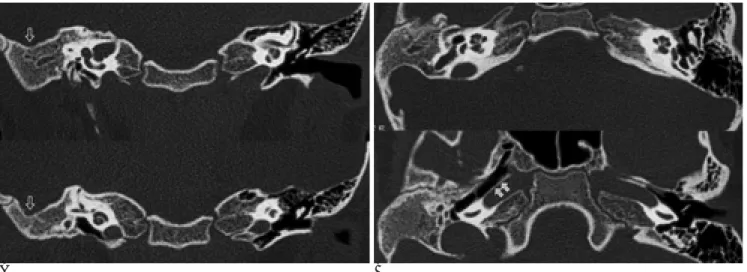
Fig. 1. An eight-year-old boy with right EAC atresia.
A. Coronal images show bony atretic plate with small middle ear cavity. Middle ear ossicles are absent and mastoid process is acel- lular. There is inferior displacement of middle cranial fossa base (open arrows). No inner ear abnormality is associated.
B. Axial image shows patulous ipsilateral Eustachian tube (arrows).
Fig. 2. A five-year-old girl with bilateral EAC atresia.
Coronal images show bilateral EAC atresia with bony atretic plates. Ossicles are bilaterally dysplastic (open arrows) and pneumatization is decreased in left mastoid process (arrows).
고 찰
측두골의 발생은 세 배아층의 복합적인 상호 작용에 의하여 이루어진다. 그 중 외이도의 발생은 첫번째와 두 번째 아가미 궁(arch) 사이의 첫번째 아가미구(groove)가 태생기 9주경에 깊어지면서 시작되며, 골성 외이도의 개구는 내이, 중이, 외이 의 분화가 이루어진 후인 태생기 30주경에 이루어진다. 이 첫 번째 아가미구의 표피세포가 서로 분리되지 못하는 경우 그 시 기 및 정도에 따라 골성 및 막성 외이도 폐쇄증 혹은 협착증
이 발생하게 된다 (3, 11-13). 외이도 폐쇄증은 남자에 호발 하며, 일측성인 경우 우측에 잘 발생하는데 (2, 3), 저자들의 증례에서도 11예의 일측성 폐쇄증 중 8예가 우측에 발생하였 다. 골성 폐쇄증에 비하여 현저히 발생 빈도가 낮은 막성 폐쇄 증은 저자들의 경우 1예 (8%)에서 관찰되었다.
한편 중이의 이소골은 첫번째와 두 번째 아가미궁에서 발생 하는데, 추골(malleus)의 머리, 고실 장근(tensor tympani) 근 육과 건, 그리고 침골(incus)의 몸체와 단돌기는 첫번째 궁 (Meckel cartilage)에서 생기고, 등골 근육과 건, 그리고 등골 저(footplate)를 제외한 나머지 이소골은 두 번째 궁 (Reichert cartilage)에서 생기므로, 인접한 첫번째 아가미구 기원의 외이 도 폐쇄증에 흔히 중이강 구조물의 기형이 동반되며 (3, 5, 8, 13), 저자들의 증례에서도 추골(12/13, 92%) 및 침골 (11/13, 85%) 기형이 외이도 폐쇄증에 매우 높게 동반됨을 확인하였 다. 등골의 전위는 1예 (8%)에서 관찰되었고, 5예 (38%)에서 는 등골을 확인할 수 없었다. 수술소견에 의한 확진이 불가능 하여 등골의 무형성에 의한 소견 이외에 영상조건(절편두께 및 각도)에 연관된 위양성 소견이 포함되었을 가능성은 있으나, Mayer등의 보고 (5)에서 경증 및 중증 소이증(microtia)에 추 골-침골의 이상이 56-98%, 등골의 이형성이 15.5-17.2%, 무형성이 34.5-56%에서 동반된 것을 고려할 때, 저자들의 결 과와 큰 차이를 보이지 않아 위양성 소견의 영향은 적을 것으 로 생각된다.
외이도 폐쇄증에 대한 수술의 주된 목적이 청력의 개선이며, 세 이소골 존재여부가 수술 후 청력 개선에 큰 영향을 미친다.
그러므로 CT 진단에 있어서 특히 등골을 포함한 이소골의 이 형성 및 무형성의 주의 깊은 관찰이 요구되며, 그 빈도 및 소 견을 유념하여야 한다 (6, 7).
하악과두, 경상돌기(styloid process), 그리고 안면신경관 또 한 두 번째 궁에서 생기므로 이 구조물들의 기형이 함께 동반 되는 경우가 흔하다. 또한, 외이도의 선천성 폐쇄와 함께 주변 구조물들은 외이도가 위치할 공간 방향으로 전위되는 변화를 Table 1. External Auditory Canal Atresia and Stenosis:
Associated Abnormalities
Associated Abnormalities. Atresia (n=13) Stenosis (n=3)
Small Tympanic cavity 8 1
Malleus
dysplastic 8 1
displaced 0 0
absent 4 0
Incus
dysplastic 5 1
displaced 2 0
absent 3 0
Stapes
dysplastic 1 1
displaced 0 0
absent 5 0
Decreased mastoid pneumatization 8 0
Abnormal facial nerve course 4 0
Displacement of
Mandibular condyle, posterior 2 1
Condylar fossa, posterior 6 1
Sigmoid sinus, anterior 3 0
Jugular bulb, superior 6 0
Middle cranial fossa base, inferior 8 1
Widening of Eustachian tube 7 0
Internal auditory canal abnormality 1 0
A B
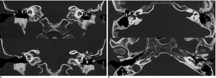
Fig. 3. A 27 year-old-woman with left EAC stenosis.
A. Coronal images show hypoplasia of left middle ear cavity and dysplastic ossicles (arrows).
B. Stenotic change involves the left EAC on axial images (open arrow). No other abnormality is found.
Swartz 등 (8)은 15예의 외이도 폐쇄증에 대한 분석에서 고 실의 위축(n=7), 하악 과두의 전위(n=7), 안면신경 유돌기부 위의 이상 주행경로(n=8), 유돌 봉소의 함기화 소실 (n=5)등 이 흔히 동반되는 소견으로 보고하였다. 본 연구에서도 고실의 위축(62%), 하악과두 및 과상와의 전위(54%), 유돌봉소의 함 기화 이상(62%)이 높은 빈도로 관찰되어 Swartz등의 결과와 일치하였다.
저자들의 증례에서 안면신경 주행경로의 이상은 미로부위에 서 관찰되지 않았고, 고실부의 외측 전이를 보인 경우가 1예, 유돌부위의 저형성 및 전방전위를 보인 경우가 3예였다. 이는 Mayer등의 보고(5)에서 심한 소이증(major microtia)에 동반 된 안면신경 주행경로의 이상이 미로부위(14.3%) 혹은 고실 부위 (28.6%)에 비하여 유돌부위(73.8%)에 높게 나타나는 경 향을 보였던 것과 유사하지만, 이들에 비하여 안면신경 유돌부 위의 주행경로 이상의 빈도는 낮게 관찰되었다. 이는 저자들의 증례에서 고실 및 유양동의 위축과 함께 유돌기의 전방전위가 동반되는 등 해부학적 지표가 변형된 상태에서 안면신경 유돌 부위의 평가가 어려운 증례가 있었던 것과, 인용된 Mayer등의 결과가 중증 소이증에 동반된 기형이므로 상대적으로 높은 빈 도를 보였을 가능성이 있다. 한편, 외이도 폐쇄증의 수술 후에 발생하는 중한 합병증인 안면신경 마비가 대개 난원창에 대한 접근 과정에서 안면신경의 전위(transposition)로부터 일어나 는 점을 고려할 때 (15, 16), 외이도 폐쇄증의 CT 진단에 있 어서 이러한 안면신경 주행경로 중 특히 고실부와 유돌부에 유 의하여 관찰하는 것이 중요하겠다.
외이도 폐쇄증에 대한 수술에 있어서 중이강 및 유양동의 크 기와 함기화 여부는 수술 방법의 결정 뿐만 아니라 수술 후 예 후에도 큰 변수로 작용한다 (7). 저자들의 증례에서 62%가 고 실의 위축 및 유돌기의 함기화 소실을 보였는데, 수술의 효과 를 얻으려면 일측성 외이도 폐쇄증의 경우 중이강 및 유양동 의 크기가 정상측의 2/3 이상은 되어야 하며, 수술이 보다 적 극적으로 권고되는 양측성의 증례에 대하여도 고실의 크기가 정상의 절반 이상이 되는 경우 수술 효과를 기대할 수 있다.
외이 및 중이의 발생이 서로 깊은 연관을 보이는데 반해 내 이의 발생은 독립적으로 일어난다. 그러므로 외이도 폐쇄증 및 협착증에 동반되는 내이의 이상이 빈도가 낮을 것으로 기대되 지만 전혀 없지는 않아 문헌에 따라 4% 내지 30%에서 동반 되는 것으로 보고된다 (1, 17). 저자들의 증례에서 13예의 폐 쇄증 및 3예의 협착증 중 1예에서 외이도의 입사각 변화를 보 였다. 그러므로 외이도 폐쇄증에 대한 CT 평가에 있어 낮은 빈도이지만 내이 구조물의 기형이 동반될 수 있음을 유념하여 야 하며, 이는 특히 수술 전 검사의 평가에 중요하다.
결론적으로 선천성 외이도 폐쇄증은 그 발생에 깊이 연관된
있어 측두골 CT는 유용하며, 특히 수술적 치료를 위한 환자 선택에 중요한 변수인 이소골 존재여부, 고실의 크기 및 유돌 기의 함기화, 안면신경 주행경로의 평가, 그리고 드물게 발견 되는 내이 구조물의 동반기형을 포함한 전반적인 측두골 주변 구조물에 대한 관찰이 필수적으로 요구된다.
참 고 문 헌
1. Jafek BW, Nager GT, Strife J, Gayler RW. Congenital aural atresia:
An analysis of 311 cases. Trans Am Acad Ophthalmol Otolaryngol 1975;80:588-595
2. Takegoshi H, Kaga K, Kikuchi S, Ito K. Mandibulofacial dysostosis:
CT evaluation of the temporal bones for surgical risk assessment in patients of bilateral aural atresia. Int J Pediatr Otorhinolaryngol 2000;54:33-40
3. Bellucci RJ. Congenital aural malformations: diagnosis and treat- ment. Otolaryngol Clin North Am 1981;14:95-124
4. Swartz JD, Wolfson RJ, Marlowe FI, et al. External auditory canal dysplasia: CT evaluation. Laryngoscope 1985;95:841-845
5. Mayer TE, Brueckmann H, Siegert R, Witt A, Weerda H. High-res- olution CT of the temporal bone in dysplasia of the auricle and ex- ternal auditory canal. AJNR Am J Neuroradiol 1997;18:53-65 6. Yeakley JW, Jahrsdoerfer RA. CT evaluation of congenital aural
atresia: What the radiologist and surgeon need to know. J Comput Assist Tomogr 1996;20:724-731
7. Mehra YN, Dubey SP, Mann SB, Suri S. Correlation between high- resolution computed tomography and surgical findings in congeni- tal aural atresia. Arch Otolaryngol Head Neck Surg 1988;114:137- 141
8. Swartz JD, Faerber EN. Congenital malformations of the external and middle ear: High-resolution CT findings of surgical import.
AJR Am J Roentgenol 1985;144:501-506
9. Virapongse C, Sarwar M, Sasaki C, Kier EL. High resolution com- puted tomography of the osseous external auditory canal: 2.
Pathology. J Comput Assist Tomogr 1983;7:493-497
10. De la Cruz A, Linthicum FH, Jr., Luxford WM. Congenital atresia of the external auditory canal. Laryngoscope 1985;95:421-427 11. Siegert R, Weerda H, Remmert S. Embryology and surgical anato-
my of the auricle. Facial Plast Surg 1994;10:232-243
12. Jahrsdoerfer RA. Congenital atresia of the ear. Laryngoscope 1978;
88:1-48
13. Sando I, Wood RP. Congenital middle ear anomalies. Otolaryngol Clin North Am 1971;4:291-318
14. Jahrsdoerfer RA, Lambert PR. Facial nerve injury in congenital au- ral atresia surgery. Am J Otol 1998;19:283-287
15. Jahrsdoerfer RA, Hall JW, III. Congenital malformations of the ear.
Am J Otol 1986;7:267-269
16. Schuknecht HF. Congenital aural atresia. Laryngoscope 1989;99:
908-917
17. Patterson ME, Linthicum FH, Jr. Congenital hearing impairment.
Otolaryngol Clin North Am 1970;3:201-219
J Korean Radiol Soc 2002;46:315-319
Address reprint requests to : Ho Jong Chun, M.D., Department of Radiology, Kangnam St. Mary’s Hospital College of Medicine, The Catholic University of Korea 505 Banpo-dong, Seocho-gu, Seoul 137-040, Korea.
Tel. 82-2-590-1588 Fax. 82-2-599-6771 E-mail: [email protected]
Congenital External Auditory Canal Atresia and Stenosis:
Temporal Bone CT Findings
1Dong Hoon Lee, M.D., Bum Soo Kim, M.D., So Lyung Jung, M.D.,
Young Joo Kim, M.D., Ho Jong Chun, M.D., Kyu Ho Choi, M.D., Shi-Nae Park, M.D.2
1Department of Radiology, College of Medicine, Catholic University of Korea
2Department of Otolaryngology, College of Medicine, The Catholic University of Korea
Purpose: To determine the computed tomographic (CT) findings of atresia and stenosis of the external audito- ry canal (EAC), and to describe associated abnormalities in surrounding structures.
Materials and Methods: We retrospectively reviewed the axial and coronal CT images of the temporal bone in 15 patients (M:F=8:7; mean age, 15.8 years) with 16 cases of EAC atresia (unilateral n=11, bilateral n=1) and EAC stenosis (unilateral n=3). Associated abnormalities of the EAC, tympanic cavity, ossicles, mastoid air cells, eustachian tube, facial nerve course, mandibular condyle and condylar fossa, sigmoid sinus and jugular bulb, and the base of the middle cranial fossa were evaluated.
Results: Thirteen cases of bony EAC atresia (one bilateral), with an atretic bony plate, were noted, and one case of unilateral membranous atresia, in which a soft tissue the EAC. A unilateral lesion occurred more fre- quently on the right temporal bone (n=8, 73%). Associated abnormalities included a small tympanic cavity (n=8, 62%), decreased mastoid pneumatization (n=8, 62%), displacement of the mandibular condyle and the posterior wall of the condylar fossa (n=7, 54%), dilatation of the Eustachian tube (n=7, 54%), and inferior dis- placement of the temporal fossa base (n=8, 62%). Abnormalities of ossicles were noted in the malleolus (n=12, 92%), incus (n=10, 77%) and stapes (n=6, 46%). The course of the facial nerve was abnormal in four cases, and abnormality of the auditory canal was noted in one. Among three cases of EAC stenosis, ossicular aplasia was observed in one, and in another the location of the mandibular condyle and condylar fossa was ab- normal. In the remaining case there was no associated abnormality.
Conclusion: Atresia of the EAC is frequently accompanied by abnormalities of the middle ear cavity, ossicles, and adjacent structures other than the inner ear. For patients with atresia and stenosis of this canal, CT of the temporal bone is essentially helpful in evaluating these associated abnormalities.
Index words :Computed tomography (CT) Temporal bone, abnormalities Temporal bone, CT
Ear, abnormalities