Melorheostosis is a rare nonhereditary sclerosing mes- enchymal dysplasia of bone that is diagnosed by a char- acteristic linear cortical hyperostosis with a flowing can- dle wax appearance (1-3). It commonly affects long bones, usually in the lower limb and on one side of the body. The involvement of flat bones is rare (1, 2). In ad- dition, bilateral involvement is extremely rare, with on- ly one case of long tubular bones with a symmetric dis- tribution reported to date. We present the first case of melorheostosis with multiple flat bone involvement and symmetric distribution of the lesions.
Case Report
A 55-year-old man was diagnosed with sclerotic ribs
and scapulas on a chest radiograph (Fig. 1A) as part of a routine check-up. Neither the patient nor his family members had a history of significant disease. The pa- tient’s blood chemistry and physical examination re- vealed no significant abnormalities. A chest CT showed uneven cortical thickening in multiple ribs, both scapu- las, and thoracic vertebral bodies. The cortical thicken- ing of the ribs and scapulas was bilateral and symmetri- cal in distribution (Fig. 1B). Our differential diagnosis in- cluded the polyostotic form of melorheostosis, bilateral osteoma, and osteoblastic metastasis. For further evalu- ation, using the acquired volume data, we obtained mul- tiplanar reconstruction (MPR) images, which revealed that cortical thickening was seen along the lower aspects of the ribs, infraspinatus fossas, the spines of both scapulas, and the anterior column of the thoracic verte- bral bodies (Figs. 1C, D).
Discussion
Melorheostosis is a rare, osteosclerotic dysplasia with an unknown etiology and first described in 1922 by Leri and Joanny (1, 2). The diagnosis is based on the clinical
J Korean Soc Radiol 2010;63:185-187
─ 185 ─
Symmetric Presentation of Melorheostosis Involving Multiple Flat Bones in the Thorax: A Case Report1
Jung Suk Oh, M.D., Hyun Jin Park, M.D., Jee Young Kim, M.D., Ki Jun Kim, M.D.2, Won Sang Jung, M.D.
1Department of Diagnostic Radiology, St. Vincent’s Hospital, The Catholic University of Korea
2Department of Diagnostic Radiology, Incheon St. Mary’s Hospital, The Catholic University of Korea
Received December 15, 2009 ; Accepted April 20, 2010
Address reprint requests to : Won Sang Jung, M.D., Department of Diagnostic Radiology, St. Vincent’s Hospital, The Catholic University of Korea, 93-6 Ji-dong Paldal-gu, Suwon-si, Gyeonggi-do 442-723, Korea.
Tel. 82-31-249-7492 Fax. 82-31-247-5713 E-mail: [email protected]
Melorheostosis is a rare bone dysplasia of unknown etiology that usually affects a single limb and is characterized by cortical thickening, with a flowing candle wax ap- pearance, extending vertically along the surface of the long bone. We report a case of polyostotic melorheostosis symmetrically involving multiple flat bones, ribs, and the scapulas. This is the first case of melorheostosis symmetrically involving multiple flat bones.
Index words :Melorheostosis Thorax
Tomography, X-Ray Computed
and radiological findings. The radiological appearance of melorheostosis is a characteristic wavy cortical hyperos- tosis along the surfaces of the long bone with vertical ex- tension, and resembles melted wax dripping down one side of a candle; the so called flowing candle wax ap- pearance (2, 4). Melorheostosis frequently involves the long tubular bones. By far, the most common sites af- fected include the long bones of the lower limbs (4). This disorder tends to be segmental and unilateral, and based on the extension of the bone involvement of the lesion, it may be monostotic, monomelic, or polyostotic (4). A bone scintigraphy is invariably positive in patients with melorheostosis, revealing a moderate increase in uptake of tracer, predominantly localized to the cortex (3, 4).
Computed tomography (CT) scanning more effectively reveals a clear demarcation from normal bone than standard radiographs (4). Abnormalities of the soft tis- sues overlying the bone and joint lesions are common, and in many cases, associated scleroderma have been reported (3, 4).
Most of the reported cases showed distribution of one long bone or one limb with a scleroderma distribution
(5). This characteristic distribution pattern and radiologi- cal features differentiates melorheostosis from other os- teosclerotic dysplasias. Isolated flat bone involvement is uncommon (3), and multiple flat bone involvement without long bone involvement, as seen in our patient, is exceedingly rare. Moreover, symmetric involvement of multiple flat bones has not been previously reported.
Melorheostosis is a rare dysplasia with characteristic cortical thickening, commonly involving the long bones of one limb. Here we report the first case of melorheostosis with the characteristic CT findings and an unusual distribution pattern with multiple flat bones that were symmetrically involved without long bone in- volvement.
References
1. Motimaya AM, Meyers SP. Melorheostosis involving the cervical and upper thoracic spine: radiographic, CT, and MR imaging find- ing. AJNR Am J Neurotadiology 2006;27:1198-1200
2. Nasu K, Kuroki Y, Nawano S, Murakami K, Kuroki S, Sekiguchi R, et al. Thoracic cage extent of melorheostosis depicted by multislice CT. Skeletal Radiol 2002;31:464-466
Jung Suk Oh, et al: Symmetric Presentation of Melorheostosis Involving Multiple Flat Bones in the Thorax
─ 186 ─
A B
C D
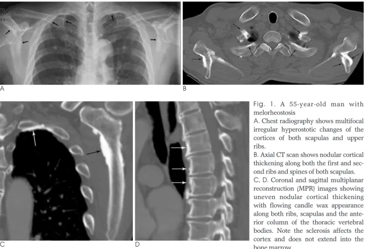
Fig. 1. A 55-year-old man with melorheostosis
A. Chest radiography shows multifocal irregular hyperostotic changes of the cortices of both scapulas and upper ribs.
B. Axial CT scan shows nodular cortical thickening along both the first and sec- ond ribs and spines of both scapulas.
C, D. Coronal and sagittal multiplanar reconstruction (MPR) images showing uneven nodular cortical thickening with flowing candle wax appearance along both ribs, scapulas and the ante- rior column of the thoracic vertebral bodies. Note the sclerosis affects the cortex and does not extend into the bone marrow.
3. Biaou O, Avimadje M, Guira O, Adjagba A, Zannou M, Hauzeur JP. Melorheostosis with bilateral involvement in a black African patient. Joint Bone Spine 2004;71:70-72
4. Greenspan A, Azouz EM. Bone dysplasia series. Melorheostosis:
review and update. Can Asso Radiol J 1999;50:324-330
5. Rhys R, Davies AM, Mangham DC, Grimer RJ. Sclerotome distrib- ution of melorheostosis and multicentric fibromatosis. Skeletal Radiol 1998;27:633-636
J Korean Soc Radiol 2010;63:185-187
─ 187 ─
대한영상의학회지 2010;63:185-187
흉곽에서 대칭적으로 편평골을 침범한 다발성 유선상과골증: 1예 보고1
1가톨릭의대 성빈센트병원 영상의학과
2가톨릭의대 인천성모병원 영상의학과
오정석∙박현진∙김지영∙김기준2∙정원상
유선상과골증은 원인 미상의 드문 골 이형성증이다. 대개 단일 사지를 침범하며, 장골의 표면을 따라 수직적으로 흐르는 촛농 모양의 피질비대가 특징적이다. 저자들은 다골성 유선상과골증이 늑골과 견갑골과 같은 편평골을 대칭 적이고 다발성으로 침범한 예를 보고하고자 한다.