Introduction
Midline splitting cervical laminoplasty (MSCL) has been considered an effective and safe method for decompressing multisegmental cervical lesions caused by cervical spondy-
lotic myelopathy (CSM) or ossification of the posterior lon- gitudinal ligament (OPLL) as well as for achieving immedi- ate stability of these lesions.4,5)
In the beginning of this procedure, autologous grafts from the iliac crest were used to keep the opened lamina split- ted.2) However, pain and discomfort at the donor site of the iliac crest were not uncommon, probably due to injury to the superior gluteal nerve,11) and sunken-down of grafted bone spacer occurred. And then hydroxyapatite (HA) was introduced as a spacer material, but the fusion rate between HA and cervical lamina is not good,9) which may cause sunk- en-down or pull out. In addition, HA has the higher potency
Midline Splitting Cervical Laminoplasty Using Allogeneic Bone Spacers: Comparison of Fusion Rates between Cervical Spondylotic Myelopathy and Ossification of Posterior Longitudinal Ligament
Jae Jon Sheen, MD and Sang Ryong Jeon, MD, PhD
Department of Neurological Surgery, Asan Medical Center, University of Ulsan College of Medicine, Seoul, Korea
Objective: To analyze factors associated with fusion using allogeneic bone spacers for midline splitting cervical laminoplas- ty (MSCL).
Methods: During April 2012 and September 2013, seventeen patients with cervical spondylotic myelopathy (CSM) or ossi- fication of posterior longitudinal ligament (OPLL) underwent MSCL with allogeneic bone spacers by a single surgeon.
Mean follow up periods was 11.3 months (range, 6-19 months). Clinical outcomes were evaluated by the Japanese Orthope- dic Association (JOA) scores at preoperative and postoperative 6 months. Simple cervical X-rays were taken preoperatively, immediate postoperatively, 3, and 6 months after operation. Computed tomography (CT) scans were performed preopera- tively, immediate postoperatively and 6 months postoperatively. The differences between two diseases were analyzed on cer- vical lordosis, canal dimension, anteroposterior (AP) distance, fusion between lamina and allogeneic bone spacer and affect- ing factors of fusion.
Results: All surgeries were performed on 59 levels. There were no significant differences on the changes of lordosis (p=
0.602), canal dimension (p=0.554), and AP distance (p=0.924) as well as JOA scores (p=0.257) between CSM and OPLL groups. Overall fusion rate was 51%. Multivariate analysis on the factor for the fusion rates between lamina and spacers showed that the immediate postoperative contact status between lamina and spacers in CT as significant factor of fusion (p=0.024).
Conclusion: The present study suggests that CSM and OPLL did not show difference of surgical outcome in MSCL using allogeneic bone spacer. In addition, we should consider the contact status between lamina and bone spacer for the better
fusion rates for this surgery. (Korean J Neurotrauma 2014;10(2):60-65)
KEY WORDS: Allografts ㆍSpondylosis ㆍSpinal fusion ∙ Spinal cord diseases ㆍOssification of posterior longitudinal ligament.
Received: June 11, 2014 / Revised: September 3, 2014 Accepted: October 6, 2014
Address for correspondence: Sang Ryong Jeon, MD, PhD Department of Neurological Surgery, Asan Medical Center, Univer- sity of Ulsan College of Medicine, 88 Olympic-ro 43-gil, Songpa- gu, Seoul 138-736, Korea
Tel: +82-2-3010-3550, Fax: +82-2-476-6738 E-mail: [email protected]
CLINICAL ARTICLE
Korean J Neurotrauma 2014;10(2):60-65
pISSN 2234-8999 / eISSN 2288-2243 http://dx.doi.org/10.13004/kjnt.2014.10.2.60
of infection than autologous grafts.7)
In contrast, allogeneic bone grafts can avoid the compli- cations associated with harvesting autologous bone. Allograft materials are obtained from deceased human donors, under- goes rigorous safety screening, and can be transplanted into the patient at the time of surgery. The shape of allografts is well developed and capability to keep splitted lamina also increased.6) However, the fusion rate of MSCL using alloge- neic bone spacers has not been studied well.
The purpose of this study was to examine the clinical and radiological outcomes in patients undergoing MSCL using allogeneic bone spacers, especially compared the results be- tween CSM and OPLL.
Materials and Methods
A total of 27 consecutive patients with compressive cer- vical myelopathy including CSM and OPLL underwent MSCL using allogeneic bone spacers, Allo-Spine® LAMI- NA SPACER (CG Bio, Seoul, Korea) between April 2012 and September 2013. Of these 27 patients, 10 patients were lost to follow-up. Patients with cervical kyphosis, evidence of cervical instability and serious medical problems were excluded for this surgery.
Therefore, we retrospectively analyzed the outcomes of 17 patients (4 men, 13 women). Mean follow-up duration was 11.3 months (range, 6-19 months). Of these patients, 6 were diagnosed as CSM and 11 as OPLL. Three patients of CSM had myelopathy due to dynamic factor such as fall- down and traffic accident, and the others due to static factor such as soft disc herniation and congenital canal stenosis.
Fifty nine spacers were used in total. The numbers and loca- tions of spacers were: 11 at C3 level, 17 at C4, 16 at C5, 12 at C6, and 3 at C7.
A single surgeon performed all laminoplasties. He made gutters at the bilateral laminofacet junctions with a 3 mm diameter diamond type burr, and split the midline spinous process with a 2 mm diameter diamond type burr. The allo- geneic bone spacers were inserted between the splitted spi- nous processes and fixed with 1-0 black silk.
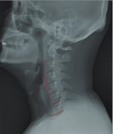
Cervical X-rays were taken preoperatively, immediate post-operatively, and after 3, 6 months. Computed tomog- raphy (CT) was performed preoperatively, immediate post- operatively, and after 6 months. Cervical lordosis was de- fined as the angle between C2 and C7 inferior endplates.
Change in overall cervical lordosis was assessed on X-ray images (Figure 1). Spinal canal dimension and anteroposte- rior (AP) distance between the posterior surface of the ver- tebral body and the anterior surface of allogeneic bone spac-
er were assessed on axial CT images, which were measured at the level of the vertebral pedicles.
The primary clinical outcome was the rate of change in the Japanese Orthopedic Association (JOA) scores (0-17) be- tween preoperative and postoperative 6 month period. Re- covery from myelopathy at 6 months was calculated using the formula: (6 month JOA score-pre operative JOA score)/
(17-pre operative JOA score).
Contact status between lamina and allogeneic bone spac- FIGURE 1. Cervical lordosis, defined as the angle between C2 and C7 inferior end plates.
A
C
B
D
FIGURE 2. Four categories of immediate post-operative con- tact status between the spacer and the lamina. A: Excellent:
complete touch on both sides of the spacer to the splitted spi- nous process. B: Good: complete touch on one side and more than half touch on the other side. C: Fair: more than half touch on both sides. D: Poor: half or less touch on at least one side.
er immediately after surgery, and fusion between lamina and allogeneic bone spacer at 6 months were assayed as Hi- rabayashi method and Ichikawa classification (Figures 2 and 3).2,3) D or E statuses were classified as fused status.
The factors that affected the rate of fusion between lam- ina and allogeneic bone spacer were analyzed by uni- and multivariate logistic regression. Age, sex, type of disease (CSM or OPLL), level of operation, contact status, smoking, diabetes mellitus (DM) and hypertension were included in these analyses. Groups were compared using the paired t- test, chi square test, Fisher’s exact test, Mann-Whitney test, Linear mixed model, and uni- and multivariate logistic re- gression. We defined statistical significance as p value <0.05.
Results
Patients were classified into two groups: CSM and OPLL.
The baseline characteristics of the groups are shown in Ta- ble 1. Of the 17 included patients, 6 were CSM and 11 were OPLL. Surgeries were performed on 59 levels, 21 for CSM and 38 for OPLL. There was no significant difference be- tween those two groups in baseline characteristics.
Mean cervical lordosis, which was defined as the angle be- tween C2 and C7 inferior endplates, changed from 10.3±8.7 to 15.0±12.6 degrees in the CSM group and from 11.7±11.1 to 13.6±11.0 degrees in the OPLL group (p=0.602)(Figure 4).
FIGURE 3. Fusion status between lamina and allogeneic bone spacer (Ichikawa classification). A: Bone resorption occurs around the implant. B: A clear space is present between the spacer and the bone without new bone formation. C: There is a space between the implant and the bone; however, new laminar bone formation is observed at the inner surface of the spinal canal. D: No space at the interface with new bone formation. E: Bridging of the new bone at the inner surface of the canal. When the different types were present at both interfaces, a lower degree of classification was applied.
E D
C B
A
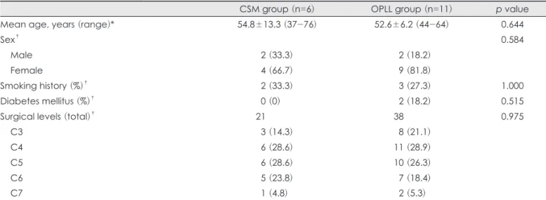
TABLE 1. Characteristics of the two patient groups
CSM group (n=6) OPLL group (n=11) p value
Mean age, years (range)* 54.8±13.3 (37-76) 52.6±6.2 (44-64) 0.644
Sex† 0.584
Male 02 (33.3) 02 (18.2)
Female 04 (66.7) 09 (81.8)
Smoking history (%)† 02 (33.3) 03 (27.3) 1.000
Diabetes mellitus (%)† 00 (0) 02 (18.2) 0.515
Surgical levels (total)† 21 38 0.975
C3 03 (14.3) 08 (21.1)
C4 06 (28.6) 11 (28.9)
C5 06 (28.6) 10 (26.3)
C6 05 (23.8) 07 (18.4)
C7 01 (4.8) 02 (5.3)
*continuous variables: t-test, †categorical variables: Fisher’s exact test. CSM: cervical spondylotic myelopathy, OPLL: ossifica- tion of posterior longitudinal ligament
FIGURE 4. Changes in cervical lordosis in the cervical spondy- lotic myelopathy and ossification of posterior longitudinal liga- ment groups. AP: anteroposterior, CSM: cervical spondylotic my- elopathy, OPLL: ossification of posterior longitudinal ligament, Preop: preoperative, Postop: postoperative.
30 25 20 15 10 5 0
-5
Preop Postop 3 m 6 m
AP distance
CSM OPLL
These data reveal the mean angle of cervical lordosis was not changed significantly and the occurrence of post-operative kyphosis was negligible. The spinal canal dimension was significantly increased after surgery from 183.1±41.1 mm2 to 295.6±46.2 mm2 in the CSM group (p<0.001) and from 145.5±38.7 mm2 to 255.8±56.6 mm2 in the OPLL group (p<0.001). In addition, the dimensions were evaluated as 299.0±34.2 mm2 (p=0.785) and 248.2±52.2 mm2 (p=0.216), respectively in post-operative 6 months. The spinal canal dimensions were increased with statistical significance after MSCL surgery in both CSM and OPLL, but there was no statistical difference between two groups (p=0.554) (Figure 5). The AP distance of the spinal canal on axial CT images was increased immediately after surgery from 9.6±
1.6 mm to 17.9±2.0 mm in the CSM group (p<0.001) and from 6.5±1.6 mm to 15.0±2.7 mm in the OPLL group (p<
0.001), but slightly decreased to 17.7±2.1 mm (p=0.400) and 14.8±2.8 mm (p=0.312), respectively at 6 months (Figure 6). There was also no statistical difference in increase of AP distance between two groups (p=0.924).
Mean JOA scores changed from 14.3±2.0 to 16.3±0.5 in the CSM group and from 12.0±4.9 to 14.6±2.8 in the OPLL group, with mean calculated recovery rates of 76.4±22.6%
and 62.5±27.2%, respectively (p=0.257)(Table 2). There was no post-operative infection.
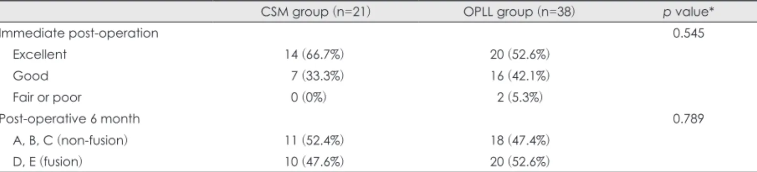
Excellent contact was achieved with 9.1% of spacers at C3, 35.3% at C4, 81.3% at C5, 100% at C6, and 66.7% at C7 (p<0.001). Contacts between the spacer and the lamina become better in lower cervical level except C7 (Table 3). In the respect of immediate post-operative contact status for CSM and OPLL, excellent in 14 levels (66.7%) and 20 lev- els (52.6%), good in 7 (33.3%) and 16 (42.1%), and fair at 0 (0%) and 2 (5.3%), respectively (p=0.545). There was no poor status. The post-operative 6 month fusion status be- tween lamina and allogeneic bone spacer, classified as D FIGURE 5. Changes of spinal canal dimension in the cervical
spondylotic myelopathy and ossification of posterior longitudinal ligament groups. CSM: cervical spondylotic myelopathy, OPLL:
ossification of posterior longitudinal ligament, Preop: preopera- tive, Postop: postoperative.
350
300
250
200
150
100
Preop Postop 6 m
Spinal canal dimension
CSM OPLL
FIGURE 6. Changes of AP distance of the spinal canal in the cervical spondylotic myelopathy and ossification of posterior longitudinal ligament groups. AP: anteroposterior, CSM: cervi- cal spondylotic myelopathy, OPLL: ossification of posterior lon- gitudinal ligament, Preop: preoperative, Postop: postoperative.
20
15
10
5
Preop Postop 6 m
AP distance
CSM OPLL
TABLE 2. JOA score changes in the two groups
Pre-operative JOA score Post-operative 6 month JOA score Recovery rate (%) p value*
CSM group (n=6) 14.3±2.0 16.3±0.5 76.4±22.6 0.257
OPLL group (n=11) 12.0±4.9 14.6±2.8 62.5±27.2
*Mann-Whitney test. CSM: cervical spondylotic myelopathy, OPLL: ossification of posterior longitudinal ligament, JOA: Japa- nese Orthopedic Association
TABLE 3. Number of spacers in each category of contact status between lamina and allogeneic bone spacer according to spinal levels
Category of contact status
Spinal level
C3 (n=11) C4 (n=17) C5 (n=16) C6 (n=12) C7 (n=3) p value*
Excellent 1 (9.1%) 06 (35.3%) 13 (81.3%) 12 (100%) 2 (66.7%) <0.001
Good 9 (81.8%) 10 (58.8%) 03 (18.8%) 00 (0%) 1 (33.3%)
Fair 1 (9.1%) 01 (5.9%) 00 (0%) 00 (0%) 0 (0%)
*Fisher’s exact test
or E status were 30 (51%) in total, 10 (47.6%) in CSM, and 20 (52.6%) in OPLL (p=0.789). There was no statistical dif- ference in contact and fusion status between two groups.
Excellent immediate post-operative contact status between lamina and allogeneic bone spacer had a significantly higher probability of fusion (Table 4). In the respect of immediate post-operative bonding state affecting fusion rate, excellent group showed fusion in 24 (71.0%), good in 5 (22.0%), and fair in 1 (50.0%). Overall fusion rate was 51% (Table 5).
In univariate logistic regression analysis, surgical level of allogeneic bone spacer (p=0.043), immediate post-op- erative contact status of spacer (p=0.001), and absence of DM (p=0.048) were significant factors. However, multivar- iate logistic regression analysis showed only contact status
of lamina and bone spacer as significant factor of fusion (p=
0.024)(Table 6).
Discussion
There was no significant difference in clinical recovery rate between CSM and OPLL in previous studies.1,12) Our results in this present study also support the data, in which post-operative clinical outcomes were significantly improved in CSM as well as OPLL, but no significant difference be- tween two groups. The reason why there was no difference in clinical outcome between two groups could be primarily because this clinical outcome was affected by decompres- sion of the spinal cord and post-operative expansion of spi- TABLE 4. Contact status and fusion status between lamina and allogeneic bone spacer in the two groups
CSM group (n=21) OPLL group (n=38) p value*
Immediate post-operation 0.545
Excellent 14 (66.7%) 20 (52.6%)
Good 07 (33.3%) 16 (42.1%)
Fair or poor 00 (0%) 02 (5.3%)
Post-operative 6 month 0.789
A, B, C (non-fusion) 11 (52.4%) 18 (47.4%)
D, E (fusion) 10 (47.6%) 20 (52.6%)
*Fisher’s exact test. CSM: cervical spondylotic myelopathy, OPLL: ossification of posterior longitudinal ligament TABLE 5. Relationship between immediate post-operative contact status and fusion status after 6 months
Non-fusion (A, B, C)(n=29) Fusion (D, E)(n=30) p value*
Excellent 10 (29.0%) 24 (71.0%) <0.001
Good 18 (78.0%) 05 (22.0%)
Fair (no poor) 01 (50.0%) 01 (50.0%)
A-E: fusion status by Ichikawa classification. *Fisher’s exact test
TABLE 6. Logistic regression of factors affecting fusion between lamina and allogeneic bone spacer
Univariate analysis Multivariate analysis
Odds ratio 95% CI p value Odds ratio 95% CI p value
Age 1.054 0.972-1.143 0.206
Sex Male 1.045 0.262-4.178 0.950
Female 1
Diagnosis OPLL 1.222 0.350-4.264 0.753
CSM 1
Surgical level C3 1 0.043 1 0.318
C4 5.062 1.211-21.157 0.026 3.474 0.709-17.023 0.125
C5 5.786 1.245-26.896 0.025 1.324 0.156-11.268 0.797
C6/C7 9.000 1.778-45.546 0.008 1.311 0.186-9.2390 0.786
Contact status Excellent 7.600 2.264-25.518 0.001 9.333 1.349-64.573 0.024
Good/fair 1 1
Smoking Y 0.498 0.182-1.361 0.174
DM Y 0.166 0.028-0.985 0.048 0.215 0.026-1.763 0.152
Hypertension Y 0.950 0.275-3.287 0.935
Logistic regression using generalized estimating equations that accounted for the clustering of same patient. OPLL: ossifica- tion of posterior longitudinal ligament, CSM: cervical spondylotic myelopathy, DM: diabetes mellitus, CI: confidence interval
nal canal dimensions were similarly increased in two groups.
In the previous report on MSCL using HA spacer,9) the overall fusion rate between lamina and HA was 27.46% and wingless type HA, which is similar shape as the allogeneic bone spacer used in this study showed 48.8%.9) In our pres- ent study, the fusion rate was 51%, which is slightly higher than wingless type HA. Allogeneic bone material has phys- iologic matrix structure and porosity compared to HA ma- terial. Therefore, allogeneic bone spacer seems to have high- er potential for bone fusion even though it was sterilized with antibiotics, chemicals, and gamma ray. But it had the potential of disease transmission such as viral hepatitis, tuberculosis, syphilis, septicemia and malignancy.10)
In multivariate analysis, initial contact status between lamina and allogeneic bone spacer significantly affected fusion status. Nagashima et al.8) demonstrated in animal experiments that a high degree of new bone formation oc- curred at the interface between the spacer and the spinous process. This finding is consistent with our study. We think that the splitted spinous process becomes higher in lower cervical levels and contacts between the spacer and the lam- ina become better in lower cervical level which explains the significance in univariate analysis.
The limitations of our present study are that this study is not prospective analysis and there were small cases in some classified groups such as C7 level and good/fair contact sta- tus. To study further results and outcome about MSCL us- ing allogeneic bone spacer, additive analysis may be need- ed with prospective, larger cases and longer follow-up duration.
Conclusion
This present study suggests that CSM and OPLL did not show difference of surgical outcome in MSCL using allo- geneic bone spacer. In addition, we should consider the con- tact status between lamina and bone spacer for the better fu-
sion rates for this surgery.
■ The authors have no financial conflicts of interest.
REFERENCES
1) Fujiwara K, Yonenobu K, Ebara S, Yamashita K, Ono K. The prog- nosis of surgery for cervical compression myelopathy. An analysis of the factors involved. J Bone Joint Surg Br 71:393-398, 1989 2) Hirabayashi S, Kumano K. Contact of hydroxyapatite spacers with
split spinous processes in double-door laminoplasty for cervical myelopathy. J Orthop Sci 4:264-268, 1999
3) Iguchi T, Kanemura A, Kurihara A, Kasahara K, Yoshiya S, Doita M, et al. Cervical laminoplasty: evaluation of bone bonding of a high porosity hydroxyapatite spacer. J Neurosurg 98(2 Suppl):
137-142, 2003
4) Iwasaki M, Kawaguchi Y, Kimura T, Yonenobu K. Long-term re- sults of expansive laminoplasty for ossification of the posterior lon- gitudinal ligament of the cervical spine: more than 10 years follow up. J Neurosurg 96(2 Suppl):180-189, 2002
5) Kimura I, Shingu H, Nasu Y. Long-term follow-up of cervical spon- dylotic myelopathy treated by canal-expansive laminoplasty. J Bone Joint Surg Br 77:956-961, 1995
6) Miller LE, Block JE. Safety and effectiveness of bone allografts in anterior cervical discectomy and fusion surgery. Spine (Phila Pa 1976) 36:2045-2050, 2011
7) Moreira-Gonzalez A, Jackson IT, Miyawaki T, Barakat K, DiNick V. Clinical outcome in cranioplasty: critical review in long-term follow-up. J Craniofac Surg 14:144-153, 2003
8) Nagashima T, Ohshima Y, Takeuchi H. [Osteoconduction in po- rous hydroxyapatite ceramics grafted into the defect of the lamina in experimental expansive open-door laminoplasty in the spinal canal]. Nihon Seikeigeka Gakkai Zasshi 69:222-230, 1995 9) Park JH, Jeon SR. Midline-splitting open door laminoplasty using
hydroxyapatite spacers: comparison between two different shaped spacers. J Korean Neurosurg Soc 52:27-31, 2012
10) Putzier M, Strube P, Funk JF, Gross C, Mönig HJ, Perka C, et al.
Allogenic versus autologous cancellous bone in lumbar segmental spondylodesis: a randomized prospective study. Eur Spine J 18:687- 695, 2009
11) Russell JL, Block JE. Surgical harvesting of bone graft from the ilium: point of view. Med Hypotheses 55:474-479, 2000 12) Seo DK, Park JH, Jeon SR. The comparison of clinical and radio-
logical long-term outcomes between ossification of posterior lon- gitudinal ligament and cervical spondylotic myelopathy after modi- fied midline splitting cervical laminoplasty. Korean J Neurotrauma 8:26-31, 2012