www.jpis.org
pISSN 2093-2278 eISSN 2093-2286 Copyright © 2011 Korean Academy of PeriodontologyThis is an Open Access article distributed under the terms of the Creative Commons Attribution Non-Commercial License (http://creativecommons.org/licenses/by-nc/3.0/).
Synergic induction of human periodontal ligament fibroblast cell death by nitric oxide and N-methyl-D-aspartic acid receptor antagonist
Taegun Seo1, Seho Cha1, Kyung Mi Woo2, Yun-Soo Park3, Yun-Mi Cho3, Jeong-Soon Lee3, Tae-Il Kim3*
1Department of Life Science, Dongguk University-Seoul, Seoul, Korea
2Department of Cell and Developmental Biology, Seoul National University School of Dentistry, Seoul, Korea
3Department of Periodontology, Dental Research Institute, Seoul National University School of Dentistry, Seoul, Korea
Purpose: Nitric oxide (NO) has been known as an important regulator of osteoblasts and periodontal ligament cell activity.
This study was performed to investigate the relationship between NO-mediated cell death of human periodontal ligament fi- broblasts (PDLFs) and N-methyl-D-aspartic acid (NMDA) receptor antagonist (+)-5-methyl-10, 11-dihydro-5H-dibenzo[a,d]cy- clohepten-5, 10-imine hydrogen maleate (MK801).
Methods: Human PDLFs were treated with various concentrations (0 to 4 mM) of sodium nitroprusside (SNP) with or with- out 200 μM MK801 in culture media for 16 hours and the cell medium was then removed and replaced by fresh medium con- taining MTS reagent for cell proliferation assay. Western blot analysis was performed to investigate the effects of SNP on the expression of Bax, cytochrome c, and caspase-3 proteins. The differences for each value among the sample groups were com- pared using analysis of variance with 95% confidence intervals.
Results: In the case of SNP treatment, as a NO donor, cell viability was significantly decreased in a concentration-dependent manner. In addition, a synergistic effect was shown when both SNP and NMDA receptor antagonist was added to the medi- um. SNP treated PDLFs exhibited a round shape in culture conditions and were dramatically reduced in cell number. SNP treatment also increased levels of apoptotic marker protein, such as Bax and cytochrome c, and reduced caspase-3 in PDLFs.
Mitogen-activated protein kinase signaling was activated by treatment of SNP and NMDA receptor antagonist.
Conclusions: These results suggest that excessive production of NO may induce apoptosis and that NMDA receptor may mod- ulate NO-induced apoptosis in PDLFs.
Keywords: Cell proliferation, Mitogen-activated protein kinase, N-methyl-D-aspartate receptor, Periodontal ligament.
INTRODUCTION
Nitric oxide (NO), a free radical synthesized from L-argi- nine by NO synthases (NOS), can modulate various tissue and cell activities, including vasodilation, neurotransmission,
immune responses, and death control [1,2]. NO is also involved in the regulation of bone metabolism through the several bi- ological process [3]. Bone forming osteoblasts constitutively produce a small amount of NO which regulates proliferation and differentiation of osteoblasts [4,5]. Overproduction of
Received: Oct. 7, 2010; Accepted: Jan. 12, 2011
*Correspondence: Tae-Il Kim
Department of Periodontology, Dental Research Institute, Seoul National University Dental Hospital, Seoul National University School of Dentistry, 28 Yeongeon-dong, Jongno-gu, Seoul 110-749, Korea
E-mail: [email protected], Tel: +82-2-2072-2642, Fax: +82-2-744-1349
chanical stress of osteoblasts, may lead to cell damage and initiate and regulate apoptosis [4,6,7]. In inflammation-in- duced osteoporosis, elevated levels of NO have been shown to cause apoptosis and to increase bone-resorption by osteo- clasts, resulting in decreased bone mineral density [8].
In the central nervous system, NO production is closely as- sociated with N-methyl-D-aspartic acid (NMDA) receptors [9]. NMDA receptors, as glutamate receptors residing in pre- synaptic and postsynaptic neurons, have been known to con- trol synaptic plasticity and memory formation in the central nervous system [10]. Upon stimulation of NMDA receptors by an external signal, Ca2+ enters the cell through the activat- ed ionotropic NMDA receptor and then NO is produced by NOS which translocates to the membrane and is activated by Ca2+ [11]. An increase in NO induces excitotoxic cell death or apoptotic events in afflicted neurons.
Previous studies have revealed that glutamate NMDA re- ceptors in osteoblasts may be involved in regulation of bone formation [12,13]. Recently, we have also reported that NMDA receptors are involved in periodontal ligament fibroblast (PDLF) differentiation but not their proliferation [14]. PDL cells form the connective tissue between the tooth cemen- tum and alveolar bone to anchor teeth and maintain struc- tural integrity. In addition, PDLFs have the potential to dif- ferentiate into cementoblasts and osteoblasts needed for ce- mentum and alveolar bone formation, respectively [15-17].
Mechanical stress has been shown to promote an increase in NO production in cultured human PDL cells [18]. Consti- tutive NO production arising from chewing or other mechan- ical-stress of teeth may play several roles in periodontal tis- sues, such as serving to maintain structural integrity of PDL [19] and regulating PDL response to various stimuli like pro- inflammatory cytokines, mechanical strain, and sex hormones [6,20,21]. However, excessively produced NO may lead to cell damage in periodontal tissues similar to that in endothelial cells and osteoblasts [22,23]. For example, lipoplysaccharide (LPS)-induced nitrite synthesis in PDLFs and osteoblasts led to cell apoptosis and increased pro-apoptotic responses to cytokines [24,25]. This has been shown in the cell types such as human monocytes, endothelial cells, macrophages, and gingival fibroblasts, which produce inflammatory cytokines via stimuli of bacteria or bacterial components like LPS [26].
However, the function of NMDA receptor in NO-mediat- ed PDLF cell damage has not been well characterized. We in- vestigated the relationship between the effect of excessively produced NO using sodium nitroprusside (SNP) and NMDA receptor antagonist.
Chemicals and antibodies
(+)-5-methyl-10, 11-dihydro-5H-dibenzo[a,d]cyclohepten-5, 10-imine hydrogen maleate (MK801) and SNP were pur- chased from Sigma Aldrich (St. Louis, MO, USA). 3-(4,5-di- methylthiazol-2-yl)-5-(3-carboxymethoxyphenyl)-2-(4-sul- fophenyl)-2H-tetrazolium, inner salt (MTS) was purchased from Promega (Madison, WI, USA ). Cell culture reagents were obtained from Hyclone (Logan, UT, USA) and Gibco (Paisley, Scotland). Protease inhibitor cocktail was purchased from Roche (Rotkreuz, Switzerland). Primary antibodies of caspase-3, Bax, cytochrome c, c-Jun N-terminal kinase/stress- activated protein kinase (JNK/SARK), mitogen-activated pro- tein kinases (p38) and anti-rabbit, anti-mouse second anti- bodies were obtained from Santa Cruz Biotechnology (Santa Cruz, CA, USA). Anti-phosphorylated extracellular signal-reg- ulated kinases (pERK), ERK rabbit polyclonal antibodies were prepared by Cell Signaling Technology (Beverly, MA, USA).
β-actin mouse monoclonal antibody was purchased from Sigma Aldrich.
Cell culture and chemical treatment
Human PDLFs were purchased from ScienCell (San Diego, CA, USA). They were cultured in α-MEM supplemented with 10% fetal bovine serum, 100 U/mL penicillin and 100 μg/mL streptomycin at 37°C in a 5% CO2 humidified atmosphere.
SNP was dissolved in phosphate buffered saline (PBS) buffer to 1 M as stock solution, stored frozen at -20°C and protected from light for use in related experiments. PDLFs were treated with various concentrations (0, 0.5, 1, 1.5, 2, 2.5, 3, 3.5, and 4 mM) of SNP with or without 200 μM MK801 in culture media for 16 hours and PDLF morphologies were observed and photo- graphed. All of the media were changed every 3 days.
Cell proliferation assay
After the cells were treated with SNP at the various concen- trations for 16 hours, the cell medium was removed and then replaced by fresh medium containing MTS reagent. The cells were incubated at 37°C in a 5% CO2 for 3 hours, and then the absorbance at 490 nm was measured on a microplate reader (Molecular Devices, Sunnyvale, CA, USA).
Western blot analysis
Human PDLFs were washed with PBS and collected in ra- dioimmunoprecipitation assay buffer (150 mM sodium chlo- ride, 1% Triton X-100, 1% sodium deoxycholate, 0.1% sodium dodecyl sulfate [SDS], 50 mM Tris-HCl, pH 7.5, and 2 mM eth- ylenediaminetetraacetic acid) with protease inhibitor cocktail (Roche, Rotkreuz, Switzerland). Equal amounts of proteins
were separated by SDS-polyacrylamide gel electrophoresis and transferred to polyvinylidine fluoride membrane. The membrane was incubated with blocking buffer containing 5% skim milk in tris buffered saline for 2 hours. After block- ing, the membranes were incubated for 2 hours with primary antibody, 1:1,000 anti-pERK, anti-ERK, anti-caspase-3 rabbit polyclonal antibodies, and anti-Bax, anti-cytochrome c, anti- β-actin mouse monoclonal antibodies, followed by incuba- tion for 1 hour with 1:5,000 anti-rabbit and anti-mouse sec- ondary antibodies, respectively. The membrane was detected with the aid of electrochemiluminescence detection reagents (Takara, Kyoto, Japan) using a fluorescence imaging system (Fuji Film, Tokyo, Japan).
Data analysis
A statistical software package (SPSS Inc., Chicago, IL, USA) was used to evaluate the data on cell proliferation. The differ- ences for each value among the sample groups were com- pared using analysis of variance. The statistical significance of the results was analyzed using 95% confidence intervals.
RESULTS
Effect of NO and NMDA receptor antagonist on the viabil- ity in PDLFs
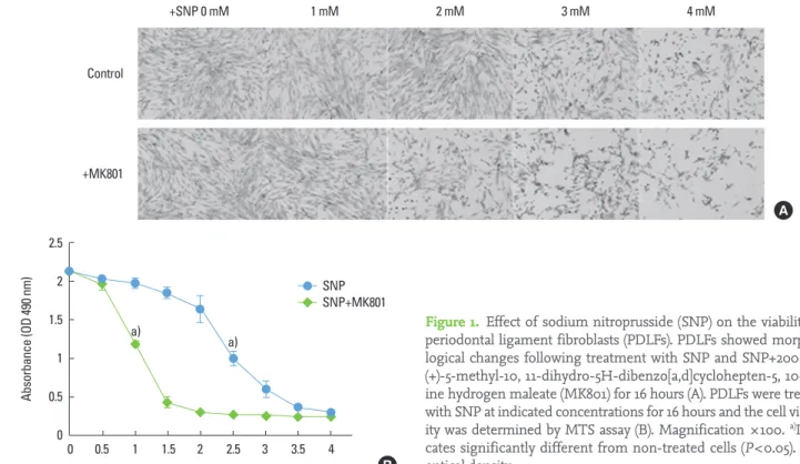
To determine the effect of SNP on the PDLFs, we first ob- served morphological changes. PDLF morphologies were
observed and photographed following treatments of 0 to 4 mM SNP with or without 200 μM MK801, the NMDA recep- tor antagonist, for 16 hours. There was alteration of PDLF cell morphology in a SNP concentration-dependent manner.
The 3 mM SNP treated PDLFs led to a dramatic reduction in the numbers of flat, spread, and spindle-shaped fibroblasts and loss of cell-cell contact, and increased cellular debris from apparently dead/dying cells (Fig. 1A). The PDLF morpholo- gies were largely unaltered when they were cultured with 0 to 2 mM SNP. Interestingly, the PDLF morphology and via- bility were more sensitive to SNP in the presence of MK801, the blocker of NMDA receptors; the rounded and floating cells, and large loss of flat and spread PDLFs were observed at 2 mM SNP (Fig. 1A). We determined cell viability by MTS assay after SNP and MK801 exposure. Exposure of PDLFs to 0 and 0.5 mM SNP for 16 hours did not affect cell viability. In the 2.5 mM SNP treated PDLFs, significantly decreased opti- cal density (A490) was determined by MTS assay, and in the presence of MK801, 1 mM SNP treated PDLFs showed simi- larly significant reduction compared to the untreated control (Fig. 1B), suggesting that PDLF viability is sensitive to NO and MK801.
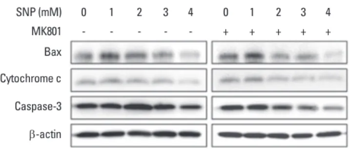
Effects of NO and NMDA antagonist on the expression of apoptotic proteins
Fig. 2 shows the effects of SNP on Bax, cytochrome c, and caspase-3 proteins. Administration of SNP in PDLFs increased
Control
+MK801
A
Figure 1. Effect of sodium nitroprusside (SNP) on the viability of periodontal ligament fibroblasts (PDLFs). PDLFs showed morpho- logical changes following treatment with SNP and SNP+200 μM (+)-5-methyl-10, 11-dihydro-5H-dibenzo[a,d]cyclohepten-5, 10-im- ine hydrogen maleate (MK801) for 16 hours (A). PDLFs were treated with SNP at indicated concentrations for 16 hours and the cell viabil- ity was determined by MTS assay (B). Magnification ×100. a)Indi- cates significantly different from non-treated cells (P<0.05). OD:
optical density.
2.5 2
1.5
1 0.5
0 0 0.5 1 1.5 2 2.5 3 3.5 4 SNP SNP+MK801
Absorbance (OD 490 nm)
SNP concentration (mM)
a) a)
B
Bax and cytochrome c production. However caspase-3 pro- tein was decreased. Bax protein production was increased in 2 mM SNP treated PDLFs, and was highly expressed at 1 mM SNP treated cells in the presence of MK801. Cytochrome c protein represented the high expression in 2 mM SNP treat- ed PDLFs. In MK801 treated PDLFs showed that cytochrome c was highly expressed at 1 mM SNP treatment. Caspase-3 protein was decreased in 3 mM SNP treated PDLFs and 2 mM SNP treated PDLFs in the presence of MK801. These re- sults suggest that PDLFs reacted more sensitively in the presence of MK801 compared to the group treated with SNP alone.
Effects of NO and NMDA antagonist on the expression of apoptotic proteins and MAPK pathway proteins
SNP treated PDLFs increased the phosphorylation of mito- gen-activated protein kinase (MAPK) pathway proteins such as JNK/SARK, ERK, and p38 (Fig. 3). Western blot results re- vealed that phosphorylation of JNK/SARK and ERK proteins were induced from 3 mM SNP treated PDLFs. In the presence of MK801, increased phosphorylation of these proteins was shown in 2 mM SNP treated PDLFs. Phosphorylation of p38 was detected in 4 mM SNP treated PDLFs and revealed with 3 mM SNP in the presence of MK801. Notably, pERK was significantly increased in the presence of SNP and MK801.
DISCUSSION
Many of the pathogenic effects of bacteria in periodontitis involving alveolar bone destruction are mediated by bacterial products including endotoxin or LPS [27,28]. LPS can directly induce cell death or apoptosis in many cell types, and by stim- ulating NO production in macrophages, epithelial cells, and fibroblasts [29-31]. The production of high levels of NO in various cells is associated with autocytotoxicity, suppression
of carcinogenesis, and inhibition of metastasis that result partially from induction of apoptosis [32].
In central neurons, NO is produced by elevated cytosolic Ca2+ which constitutively activates neuronal NOS (nNOS) via the activation of glutamate NMDA receptors [9]. There are two NOS subtypes: inducible NOS (iNOS) found in fibro- blasts and macrophages and endothelial NOS (eNOS) in blood vessel walls [33]. PDL cells express both eNOS and iNOS, and NO products by eNOS, and iNOS modulates the function of PDL cells [19] and by occlusal stimuli in the rat PDL promotes PDL healing in transplanted teeth [34]. In this study we fo- cused on the relation of NO and NMDA receptors in PDLFs.
NO released by SNP, an extracellular NO donor, has similar cytotoxic effects on PDLFs to induction of intracellular NO which is produced by NOS. A high level of NO induces oxi- dative stress in cells that may lead to apoptosis accompanied by changes in gene expression, and cell dysfunction and damage. As shown in Fig. 1, SNP treated PDLFs are associat- ed with cell viability as evidenced by changes in their mor- phology into shrunken and round from spindle-shaped, and by the decrease in cell number and reduction in response to MTS assay. When PDLFs were treated with NMDA receptor antagonist, cell death of PDLFs was promoted at a lower con- centration than only administration of SNP. In addition, our results showed that the oxidative stress by SNP-induced NO increased expression of apoptotic marker proteins such as Bax and cytochrome c. Interestingly, MK801 treatment of SNP stimulated PDLFs further enhanced the apoptotic re- sponses of these cells in morphology and apoptosis marker protein expression than SNP treatment alone. Our results
MK801 - - - - - + + + + +
Bax Cytochrome c Caspase-3 β-actin
Figure 2. Sodium nitroprusside (SNP) altered the expression of apoptosis marker proteins. Periodontal ligament fibroblasts were treated with SNP and (+)-5-methyl-10, 11-dihydro-5H-dibenzo[a,d]
cyclohepten-5, 10-imine hydrogen maleate (MK801) for 16 hours.
The cell lysates were performed to Western blot analysis using anti- Bax, anti-cytochrome c, anti-caspase-3, and anti-β-actin antibodies.
β-actin was used as internal control.
MK801 - - - - - + + + + +
JNK/SARK P-JNK/SARK ERK P-ERK p38 P-p38
Figure 3. Sodium nitroprusside (SNP) induced phosphorylation of mitogen-activated protein kinases in periodontal ligament fibro- blasts (PDLFs). PDLFs were treated with SNP and (+)-5-methyl-10, 11-dihydro-5H-dibenzo[a,d]cyclohepten-5, 10-imine hydrogen ma- leate (MK801) for 16 hours. The cell lysates were performed to West- ern blot analysis using anti-c-Jun N-terminal kinase/stress-activat- ed protein kinase (JNK/SARK), anti-extracellular-signal-regulated kinase (ERK), anti-mitogen-activated protein kinases (p38), anti- phosphorylated JNK/SARK, anti-phosphorylated ERK or anti-phos- phorylated p38 antibodies.
tein-induced PDLF cell death.
De novo synthesis of Bax protein by SNP has been shown to be proapoptotic in NO-induced osteoblast death [35], sug- gesting that increased Bax protein levels may be a critical trigger of apoptosis induced by oxidative stress in osteoblasts [36]. Cytochrome c is another well-known marker protein of apoptosis that may be released from mitochondria to the cy- toplasm in osteoblasts under oxidative stress and associated depolarization of the mitochondrial membrane potential [37].
Decreased levels of caspase-3 expression in SNP treated PDLFs are also consistent with conversion and activation from the inactive form of caspase-3, which is specifically la- beled as anti-caspase-3 antibody in the immunoblot analysis shown in Fig. 2. Activation of caspase-3 can cause the diges- tion of key cellular proteins and induce DNA fragmentation and cell apoptosis [38,39]. Possibly, similar proapoptotic alter- ations in Bax levels, cytochrome c-mediated activation of caspase-3 activity, and DNA fragmentation may have oc- curred in PDLFs treated with SNP in our study.
In most cells, NO-mediated apoptosis typically involves ac- tivation by phosphorylation of the JNK/SARK, ERK, and p38 groups of MAPKs [40]. We monitored the phosphorylation of ERK, p38, and JNK following treatment with SNP and SNP/
MK801 to determine whether these kinases were involved in the signaling pathway of NO-mediated apoptosis in PDLFs.
In NO induced PDLFs, phosphorylation of JNK/SARK, ERK and p38 was increased compared to the control PDLFs. In PDLFs treated with SNP and MK801, the phosphorylation of the MAPK panel occurred at lower concentrations of SNP that also produced morphologic and biochemical differences as shown in Fig. 1. Overall, these results indicate that in PDLFs, NO overproduction may induce an apoptosis signal via the MAPK pathway, and suggest that activated glutamate NMDA receptors in PDLFs may inhibit cell death and apoptosis, which are induced by conditions that stimulate excessive NO pro- duction, such as periodontal bacterial infections.
CONFLICT OF INTEREST
The authors have no conflicts of interests related to this study.
ACKNOWLEDGMENTS
This work was supported by a Korea Research Foundation (KRF) grant funded by the Korean government (MEST) (No.
2009-0077792) and a grant of the Korean Health Technology R&D Project (A101768), Ministry for Health, Welfare & Family Affairs, Republic of Korea.
1. Chang H, Tsai SY, Chang Y, Chen TL, Chen RM. Therapeu- tic concentrations of propofol protects mouse macro- phages from nitric oxide-induced cell death and apopto- sis. Can J Anaesth 2002;49:477-80.
2. Moncada S, Palmer RM, Higgs EA. Nitric oxide: physiolo- gy, pathophysiology, and pharmacology. Pharmacol Rev 1991;43:109-42.
3. van’t Hof RJ, Ralston SH. Nitric oxide and bone. Immu- nology 2001;103:255-61.
4. Mancini L, Moradi-Bidhendi N, Becherini L, Martineti V, MacIntyre I. The biphasic effects of nitric oxide in primary rat osteoblasts are cGMP dependent. Biochem Biophys Res Commun 2000;274:477-81.
5. Koyama A, Otsuka E, Inoue A, Hirose S, Hagiwara H. Ni- tric oxide accelerates the ascorbic acid-induced osteoblas- tic differentiation of mouse stromal ST2 cells by stimulat- ing the production of prostaglandin E(2). Eur J Pharmacol 2000;391:225-31.
6. Ralston SH, Todd D, Helfrich M, Benjamin N, Grabowski PS. Human osteoblast-like cells produce nitric oxide and express inducible nitric oxide synthase. Endocrinology 1994;135:330-6.
7. Damoulis PD, Hauschka PV. Cytokines induce nitric oxide production in mouse osteoblasts. Biochem Biophys Res Commun 1994;201:924-31.
8. Armour KE, Van’T Hof RJ, Grabowski PS, Reid DM, Ralston SH. Evidence for a pathogenic role of nitric oxide in in- flammation-induced osteoporosis. J Bone Miner Res 1999;
14:2137-42.
9. Brunetti L. Nitric oxide: a gas as a modulator of neuroen- docrine secretions. Clin Ter 1994;144:147-53.
10. Contestabile A. Roles of NMDA receptor activity and ni- tric oxide production in brain development. Brain Res Brain Res Rev 2000;32:476-509.
11. Arundine M, Sanelli T, Ping He B, Strong MJ. NMDA in- duces NOS 1 translocation to the cell membrane in NGF- differentiated PC 12 cells. Brain Res 2003;976:149-58.
12. Hinoi E, Fujimori S, Yoneda Y. Modulation of cellular dif- ferentiation by N-methyl-D-aspartate receptors in osteo- blasts. FASEB J 2003;17:1532-4.
13. Taylor AF. Osteoblastic glutamate receptor function regu- lates bone formation and resorption. J Musculoskelet Neuronal Interact 2002;2:285-90.
14. Yu JH, Lee SP, Kim TI, Jang JH. Identification of N-meth- yl-D-aspartate receptor subunit in human periodontal ligament fibroblasts: potential role in regulating differen- tiation. J Periodontol 2009;80:338-46.
15. Tomokiyo A, Maeda H, Fujii S, Wada N, Shima K, Akamine
tal ligament cell line. Differentiation 2008;76:337-47.
16. Basdra EK, Komposch G. Osteoblast-like properties of hu- man periodontal ligament cells: an in vitro analysis. Eur J Orthod 1997;19:615-21.
17. Seo BM, Miura M, Gronthos S, Bartold PM, Batouli S, Bra- him J, et al. Investigation of multipotent postnatal stem cells from human periodontal ligament. Lancet 2004;364:
149-55.
18. Kikuiri T, Hasegawa T, Yoshimura Y, Shirakawa T, Oguchi H. Cyclic tension force activates nitric oxide production in cultured human periodontal ligament cells. J Periodontol 2000;71:533-9.
19. Watarai H, Warita H, Soma K. Effect of nitric oxide on the recovery of the hypofunctional periodontal ligament. J Dent Res 2004;83:338-42.
20. Armour KE, Ralston SH. Estrogen upregulates endothelial constitutive nitric oxide synthase expression in human osteoblast-like cells. Endocrinology 1998;139:799-802.
21. Klein-Nulend J, Helfrich MH, Sterck JG, MacPherson H, Joldersma M, Ralston SH, et al. Nitric oxide response to shear stress by human bone cell cultures is endothelial ni- tric oxide synthase dependent. Biochem Biophys Res Com- mun 1998;250:108-14.
22. Chen RM, Chen TL, Chiu WT, Chang CC. Molecular mech- anism of nitric oxide-induced osteoblast apoptosis. J Or- thop Res 2005;23:462-8.
23. Palmer RM, Bridge L, Foxwell NA, Moncada S. The role of nitric oxide in endothelial cell damage and its inhibition by glucocorticoids. Br J Pharmacol 1992;105:11-2.
24. Lin SK, Kok SH, Lin LD, Wang CC, Kuo MY, Lin CT, et al.
Nitric oxide promotes the progression of periapical lesion via inducing macrophage and osteoblast apoptosis. Oral Microbiol Immunol 2007;22:24-9.
25. Thammasitboon K, Goldring SR, Boch JA. Role of macro- phages in LPS-induced osteoblast and PDL cell apoptosis.
Bone 2006;38:845-52.
26. Alexander MB, Damoulis PD. The role of cytokines in the pathogenesis of periodontal disease. Curr Opin Periodon- tol 1994:39-53.
27. Rogers JE, Li F, Coatney DD, Rossa C, Bronson P, Krieder JM, et al. Actinobacillus actinomycetemcomitans lipo- polysaccharide-mediated experimental bone loss model
28. Reddi K, Meghji S, Wilson M, Henderson B. Comparison of the osteolytic activity of surface-associated proteins of bacteria implicated in periodontal disease. Oral Dis 1995;
1:26-31.
29. Sosroseno W, Bird PS, Seymour GJ. Nitric oxide produc- tion by a human osteoblast cell line stimulated with Ag- gregatibacter actinomycetemcomitans lipopolysaccha- ride. Oral Microbiol Immunol 2009;24:50-5.
30. Albina JE, Cui S, Mateo RB, Reichner JS. Nitric oxide-me- diated apoptosis in murine peritoneal macrophages. J Im- munol 1993;150:5080-5.
31. Tiwari MM, Messer KJ, Mayeux PR. Inducible nitric oxide synthase and apoptosis in murine proximal tubule epithe- lial cells. Toxicol Sci 2006;91:493-500.
32. Xie K, Huang S, Dong Z, Juang SH, Wang Y, Fidler IJ. De- struction of bystander cells by tumor cells transfected with inducible nitric oxide (NO) synthase gene. J Natl Cancer Inst 1997;89:421-7.
33. Stuehr DJ. Mammalian nitric oxide synthases. Biochim Biophys Acta 1999;1411:217-30.
34. Chen CC, Kanno Z, Soma K. Occlusal forces promote peri- odontal healing of transplanted teeth with enhanced ni- tric oxide synthesis. J Med Dent Sci 2005;52:59-64.
35. Chen RM, Liu HC, Lin YL, Jean WC, Chen JS, Wang JH. Ni- tric oxide induces osteoblast apoptosis through the de novo synthesis of Bax protein. J Orthop Res 2002;20:295-302.
36. Srivastava RK, Sollott SJ, Khan L, Hansford R, Lakatta EG, Longo DL. Bcl-2 and Bcl-X(L) block thapsigargin-induced nitric oxide generation, c-Jun NH(2)-terminal kinase ac- tivity, and apoptosis. Mol Cell Biol 1999;19:5659-74.
37. Ho WP, Chen TL, Chiu WT, Tai YT, Chen RM. Nitric oxide induces osteoblast apoptosis through a mitochondria-de- pendent pathway. Ann N Y Acad Sci 2005;1042:460-70.
38. Earnshaw WC, Martins LM, Kaufmann SH. Mammalian caspases: structure, activation, substrates, and functions during apoptosis. Annu Rev Biochem 1999;68:383-424.
39. Adrain C, Martin SJ. The mitochondrial apoptosome: a killer unleashed by the cytochrome seas. Trends Biochem Sci 2001;26:390-7.
40. Brüne B, von Knethen A, Sandau KB. Nitric oxide (NO): an effector of apoptosis. Cell Death Differ 1999;6:969-75.