I. Introduction
As we all know, periodontal disease is character- ized by inflammation that can lead to periodontal attachment loss and bone destruction and is largely initiated by bacterial plaque. It is now recognized that during active periodontitis, degradation of the collagenous matrices which support the skeletal framework of gingival complex is due in part to matrix metalloproteinase (MMPs) expressed in situ by inflammatory cells(monocytes, macro- phages, lymphocytes, polymorphonuclear cells) and resident cells(fibroblasts, epithelial cells, and endothelial cells), and they are thought to be partly responsible for other kinds of diseases as well. MMPs belong to the matrixin family, which is composed of at least 23 related zinc- dependent endopeptidases that are
able to degrade extracellular matrix proteins at a pH close to neutral. And they can be divided into sever- al major subgroups, such as the interstitial collage- nases (MMP-1. -8 and -13), the gelatinases (MMP-2 and -9; also called type IV collagenase), the stromelysins(MMP-3, -10 and -11) and the mem- brane-bound group(MMP-14, -15, -16 and -17).
Among them, several MMPs (like MMP-1, 2, 8, 9 and 13) as well as tissue inhibitors of matrix metallopro- teinase-1(TIMP-1) are thought to be more strongly related to overwhelming periodontal disease, such as adult periodontitis (AP) and localized juvenile periodontitis(LJP) by previous classification.
The purpose of this study is to find out whether specific MMPs(-1, -8, -9, -13) or TIMP -1 are present in peri- odontitis patients and to determine if there are any significant differences between the amount
The effect of periodontal flap surgery on Matrix
metalloproteinases (MMPs) and Tissue inhibitors of matrix metalloproteinase-1 (TIMP-1) levels in gingival crevicular
fluids of periodontitis patients
Jhee-Hyun Kim1, Jea-Seung Ko2*, Hyun-Man Kim2, Tae-Il Kim1, Yang-Jo Seol1, Yong-Moo Lee1, Young Ku1, Chong-Pyoung Chung1, Soo-Boo Han1, In-Chul Rhyu1*
1Department of Periodondology, College of Dentistry, Seoul National University
2Department of Oral Histology, College of Dentistry, Seoul National University
대한치주과학회지 : Vol. 35, No. 1, 2005
*This study was supported by a grant of the Korea Health 21 R&D Project, Ministry of Health & Welfare, Republic of Korea(00-PJ1- PG1-CH10-0002)
*Co-corresponding authors: In-Chul Rhyu; Jea Seung Ko, College of dentistry, Seoul National University, 28, Yongon-Dong, Chongno-Ku, 110-749, Korea
of MMPs before periodontal surgery and after peri- odontal surgery. We got MMPs and TIMP-1 in gingi- val crevicular fluid (GCF) because GCF offers a unique opportunity to sample analytical quantities of interstitial fluid by minimally intrusive and non- surgical means, and we analyzed them by using ELISA.
We also tried to determine if those assays have some positive relationships with probing depth and GI scores (Löe and Silness), which represent clinical periodontal status easily.
II. Materials and Methods
1. Study populationSixteen individuals, newly referred to Seoul National University Dental Hospital (SNUDH), Department of Periodontology with chronic peri- odontitis took part in this study; 2 subjects were dropped from the study because they did not attend recall examinations. The remaining 14 patients (5 women; 9 men) had a mean age of 42.9 years (range 25 to 58). All patients had moderate to advanced periodontitis, and the experimental tooth site for each patient had pocket depth more than 6mm, whereas the healthy control sites were select- ed which had a pocket depth under 3mm in the same patient.
All subjects were in good general health; no par- ticipants had a history of systemic conditions such as a heart disease, diabetes, uncontrolled hyper- ten- sion, significant risk for infectious disease transmis- sion, renal or liver disease, and other types of disor- ders which could influence the course of periodon- tal disease. They were also not on any medication that could affect the manifestations of periodontal disease, such as chronic antibiotic use, phenytoin, cyclosporine, anti-inflammatory drugs, or calcium
channel blockers. None of the women was post- menopausal.
2. Clinica l measurements
The clinical evaluation of the exper- imental sites was based on the fol- lowing indices: gingival index(GI) of Löe and Silness and probing depths (PD). Probing depths were measured with a Marquis probe calibrated in millimeters.
GI score and PD were measured before (Pre) and after(Post) periodontal flap surgery in experimental sites. All post- surgery measurements were obtained 6 weeks after periodontal flap operation.
3. Gingival biopsy
Gingival tissues were obtained from experimental sites in each participant during periodontal flap surgery, and im- mediately fixed in 10% formalin solution after excision.
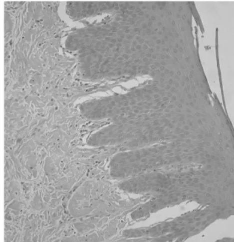
Cells and tissue were stained with hematoxylin and eosin, and this staining technique revealed the structural integr- ity of the epithelia and the absence or presence of inflammatory infiltrate in the gingiva.
Figure 1. Gingival inflammation classified as mild, ac- cording to inflammatory cell infilitration.
They were assessed with severity of inflammation under electron microscopy, and compared them with GI scores to find out whether this clinical index is reliable. The severity of gingival inflammation was classified(B) as normal, mild, moderate, and severe, according to the degree of inflammatory cell infiltra- tion, tissue destruction, vascular changes and scar- ring(Figure 1, 2, 3).
4. Gingival crevicular fluid (GCF) samp - ling and processing
GCF samples were collected using sterile paper strip(PROFLOWTM, New York), and the area was carefully isolated and gently dried by an air syringe to prevent samples from being con- taminated by saliva. The paper strips were inserted into the crevice until mild resistance was felt or in any event not more than 1mm, and left in place for 30 sec- onds. And they were immediately placed into a coded sterile microtube and stored at -70℃ until analysis.
GCF samples were collected from control sites(C), pre-surgical experi- mental sites (Pre) and post-surgical experimental sites(Post), and paper strips were used for four times res- pectively in every site to obtain suf-
ficient amount of MMPs or TIMP from GCF.
For the quantification of MMPs(-1, -8, -9, -13) and TIMP-1, Human Biotrak ELISA kit(amersham phar- macia biotech, UK) were used.
5. Statistical analysis
Wilcoxon signed rank test was employed to evalu- ate if there were any statistical differences in levels of MMPs (-1, -8, -9, -13) or TIMP-1 between control, pre- surgical, and post-surgical groups, as the sample size was not enough to do ANOVA. The criterion for sta- tistical significance was defined as a level of P<0.05.
The correlations among clinical para- meters(PD, GI) were analyzed using Spearman's correlation test.
For the statistical analysis between clinical parame- ters and MMPs or TIMP-1 levels, Spearman's correla- tion coef- ficient(r) was also employed, and a P value <0.05 was considered statistically significant.
III. Results
1. Level changes of MMPs and TIMP-1 after surgery
The results showed that all the MMPs(MMP-1, 8, Figure 2. moderate inflammation Figure 3. severe inflammation
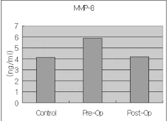
9, 13) levels in GCF were markedly decreased after periodontal flap surgery. But we found statistically significant difference in MMP-1(p=0.025), MMP- 9(p=0.016) and MMP-13(p=0.009) levels between pre-surgical(Pre) and post- surgical(Post) evalua- tion(Table 2, 3, Figure 4-8). Absolutely elevated level of MMPs was found in diseased experimental sites compared to healthy control sites, but we could find statistically significant difference only in MMP- 9(p=0.011) and MMP-13(p=0.026). Level of MMP-9
showed greatest amount among all other MMPs and TIMP levels, and MMP-9 level was even 500 to 1500 fold greater than MMP-1 level.
TIMP-1 levels were decreased after periodontal flap surgery, and showed highest levels in healthy control sites even though not statistically significant.
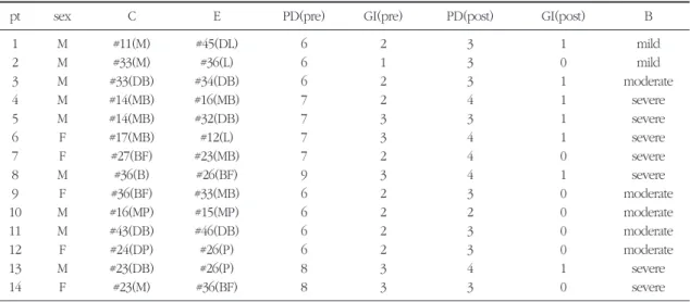
2. Relationships between clinical parameters After surgical treatment, there was absolute Table 1. Patients’whole data except MMPs and TIMP levels
pt sex C E PD(pre) GI(pre) PD(post) GI(post) B
1 M #11(M) #45(DL) 6 2 3 1 mild
2 M #33(M) #36(L) 6 1 3 0 mild
3 M #33(DB) #34(DB) 6 2 3 1 moderate
4 M #14(MB) #16(MB) 7 2 4 1 severe
5 M #14(MB) #32(DB) 7 3 3 1 severe
6 F #17(MB) #12(L) 7 3 4 1 severe
7 F #27(BF) #23(MB) 7 2 4 0 severe
8 M #36(B) #26(BF) 9 3 4 1 severe
9 F #36(BF) #33(MB) 6 2 3 0 moderate
10 M #16(MP) #15(MP) 6 2 2 0 moderate
11 M #43(DB) #46(DB) 6 2 3 0 moderate
12 F #24(DP) #26(P) 6 2 3 0 moderate
13 M #23(DB) #26(P) 8 3 4 1 severe
14 F #23(M) #36(BF) 8 3 3 0 severe
(C: Control site, E: Experimental site, pre: pre-operative data, post: post-operative data, B: severity of inflammation from biopsy)
Table 2. mean levels of MMPs and TIMP in each group (ng/ml)
MMP-1 MMP-8 MMP-9 MMP-13 TIMP-1
group cont pre post cont pre post cont pre post cont pre post cont pre post mean 0.04 0.08 0.02 4.13 5.85 4.20 20.20 54.17 27.30 0.05 0.19 0.04 3.77 3.64 3.30 SD 0.05 0.13 0.03 2.25 3.50 2.34 15.15 46.14 43.06 0.05 0.17 0.06 3.53 5.40 4.77 (cont: control site, pre: pre-operative data, post: post-operative data)
Table 3. p-vales by Wilcoxon Signed Rank Test between control (C), pre-operative (Pre) and post-operative (Post) in MMPs and TIMP-1 (p<0.05)
MMP-1 MMP-8 MMP-9 MMP-13 TIMP-1
C vs. Pre 0.114 0.177 0.011 0.026 0.875
C vs. Post 0.212 0.875 0.638 0.861 0.363
Pre vs. Post 0.025 0.158 0.016 0.009 0.551
decrease in pocket depth(PD) and gingival index(GI). But when we took them into considera- tions with stati- stical view, only pocket depths
showed significant difference(p=0.005). Maybe it can be easily expected, there was strong relation- ship between pocket depth and gingival index at baseline, and they were also statistically significant(p=0.000, r=0.808).
With the biopsy procedure, the severity of gingi- val inflammation was classified as normal, mild, moderate, and severe, according to the degree of inflammatory cell infiltration, and compared the results with gingival index(GI) whether the index is clinically reliable. Fortunately, we could find that there were strong positive relationships between them and surely they were statistically significant (p=0.001, r=0.773) even though study pool was small.
Figure 4. mean levels of MMP-1 Figure 5. mean levels of MMP
Figure 6 . mean levels of MMP-9
Figure 8. mean levels of TIMP-1
Figure 7. mean levels of MMP-13
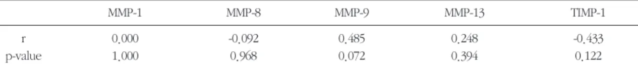
When we compared the MMPs and TIMP-1 levels with pre-operative pocket depth and gingival index, all the data showed no statistically significant dif- fer- ence, but only MMP-9 showed some positive rela- tionship with both baseline pocket depth and base- line gingival index(Table 4, 5).
IV. Discussion
As mentioned above, it is well known that extra- cellular matrix degradation in periodontal disease is mainly due to the increased level of host and micro- bial- derived proteinases such as MMPs, which are secreted during the interaction of plaque bacteria with resident gingival cells(fibroblast, epithelial cells) or inflam- matory cells such as PMN or monocyte/
macrophage(Birkedal-Hansen et al 1993, Sorsa et al 1992). They usually secreted in latent proforms and have a pro- peptide containing a conserved cystein in a region of homology. This cysteine is bonded to the Zn2+ of the active site in the latent form, and it is thought that dissociation of the cysteine thiol leads to exposure of the active site. Then the specific attack on matrix macromolecules take place, and this initial attack on the integrity of extracellular macromolecules facilitates further extracel- lular degradation.
There were many evidences that withstand the
fact that MMPs have intimate relationship with peri- odontal disease progression.
It was reported that elevated intersti- tial collage- nase(MMP-1, MMP-8) activities have been detected in GCF of adult periodontitis(AP) and localized juve- nile periodontitis(LJP), and these activities have been shown to decrease following periodontal treat- ment in both groups (Hakkarainen et al 1988).
Ingman(1996) also reported that MMP-8 and MMP-9 are the main collagenases in gingival tissue and GCF of AP patients, whereas the predominant collage- nase in GCF of LJP seems to be of the MMP-1 type.
He emphasized the importance of doxycycline, a known inhibitor of MMP-8 and MMP-9, as a possible adjunctive drug in the treatment of AP. However, the presence of MMP-1 in LJP GCF, whose MMP- type has been shown to be rather resistant to doxy- cycline inhibition at therapeutic levels, may suggest that doxycycline may be a less useful drug in the control of periodontal tissue destruc- tion in LJP than in AP.
On the other hand, Anne-Laure et al.(2003) revealed that MMP-9, MMP-13 and TIMP-1 increased significantly in the culture media using blot tech- nique with severe inflammation. Furthermore, their results seemed to be correlated with those reported by Makela et al.(1994), who demonstrated that MMP-9 was the major gelatinase present in the gin- Table 4. Spearman’s coefficient (r) of MMPs and TIMP-1 with PD (p<0.05)
MMP-1 MMP-8 MMP-9 MMP-13 TIMP-1
r 0.000 -0.092 0.485 0.248 -0.433
p-value 1.000 0.968 0.072 0.394 0.122
Table 5. Spearman’s coefficient (r) of MMPs and TIMP-1 with GI (p<0.05)
MMP-1 MMP-8 MMP-9 MMP-13 TIMP-1
r -0.074 -0.243 0.498 -0.440 -0.268
p-value 0.801 0.403 0.070 0.115 0.355
gival crevicular fluid of periodontal patients.
In this study, we got results that have something in common with former reportsin that all the MMPs from the inflamed experimental sites showed much elevated level compared to those from the healthy control sites, and PMN-type gelatinase(MMP-9) showed much higher level among other MMPs in GCF, from which we can conclude that MMP-9 is predominant matrix metalloproteinase in periodonti- tis patients.
After the flap surgery, all the MMPs (MMP-1,-8,-9,- 13) levels in GCF were markedly decreased. But we found statistically significant difference only in MMP- 1, MMP-9 and MMP-13 levels between pre-surgi- cal(Pre) and post- surgical (Post) evaluation. There were several reports which help us to understand this result. After establish- ment of a supragingivally clean oral environment, a rapid decrease of the col- lagenase activity took place following scaling and root planing of the root surfaces within the peri- odontal pockets. Also, occlusal adjustment of the hypermobile teeth with deep pathological pockets reduced the protein content and collagenase activity in sulcular fluid (Hakkarainen et al 1988). And Glay Tüter(2002) reported that levels of MMP-1 in GCF decreased and total levels of TIMP-1 in GCF increased after phase-I periodontal therapy. Those findings strongly suggests that such pathogen- induced MMPs can be well regulated by our con- ventional periodon- tal treatment.
For the regulation of destructive cascade by MMPs, there exists glyco- protein which called tissue inhibitor of matrix metalloproteinase (TIMP) that is synthesized and secreted by most connective tissue cells as well as by macrophages. TIMPs share a common two-domain structure in which only the inhibitory N-domain is capable of inhibiting MMPs.
The amino group of the N-terminal cysteine of TIMP co-ordinates with the active site Zn2+ of MMPs with
the adjacent residues of TIMP occupying the active site cleft of MMPs and contacting the surrounding surface of the catalytic domain of MMPs. They inac- tivate MMPs in that manner. But even though we clearly know the exact mechanism of how they work, whether TIMP level is higher or not in inflamed site than in healthy site is still controversial.
Nomura(1993) re- ported that TIMP was higher in inflamed sites than in healthy sites. He explained this result that when there is stimulation by bacterial colonization, MMP expression by host cell increases, and subsequent self tissue destruction occurs. So the host cells recognize the ongoing tissue destruction and try to defend the host tissue by producing TIMPs. Similarly, with elevated level of PMN elastase in inflamed sites, con- comitant elevated level of 1- proteinase inhibitor which regulates PMN elastase by forming an irreversible enzyme- inhibitor com- plex was detected in the GCF of periodontitis patients compared to controls(Ingman 1994).
On the contrary, Larivee et al.(1986) said that the activity of TIMP is relatively higher rather in healthy sites than in diseased sites. Moreover, Sorsa(1994) found that GCF of adult periodontitis patients con- tain PMN-derived MMP-8 and -9 but not recogniz- able amounts of TIMP-1. Thus it seems that PMNs do not contain and release TIMP-1 in amounts com- parable to MMP-8 or -9. Glay Tüter(2002) found lev- els of TIMP-1 increase significantly after phase-I therapy compared to baseline, and explained this phenomenon as a reduction of MMP-1, which bind to free TIMP (however, the regulation of TIMP-1 may not be solely dependent on the MMP-1).
Moreover, the decreased levels of TIMP-1 in peri- odontally diseased subjects may be due to the selec- tive degradation of TIMP-1 by neutrophil elastase of the inactivation of TIMP-1 by neutrophils themselves following oxidant release. Phase I therapy may have reduced the number of neutrophils and the elastase
released due to resolved gingival inflammation and tissue healing. Haerian et al.(1996) also reported that the GCF levels of stromelysin and TIMP were reduced by periodontal treatment. Our results are consistent with these previous studies in some extent that TIMP-1 levels were decreased after peri- odontal flap surgery, and showed highest levels in healthy control sites even though not statistically sig- nificant.
If we take a look at clinical parameters, though only pocket depths(PD) showed significant differ- ence(p=0.005), there was absolute decrease in PD and GI after surgical treatment as expected. And strong correlations between PD and GI (p=0.000, r=0.808) wasalso found. More- over, we could con- cludethat gingival index (GI) is pretty much reliable, when we compared them with the gingival biopsy specimens using electron microscopy. But there were no special correlations between MMPs or TIMP-1 levels and clinical parameters in the present study (only MMP-9 showed weak correlations).
Haerian(1996) found a moderate positive and signif- icant correlation between biochemical(GCF levels of stromelysin and TIMP) and clinical parameters when data from healthy, gingivitis and periodontitis sites were pooled. But Viella et al.(1987) reported a posi- tive but rather weak correlation between PD and GI and collagenase activity while Gangbar et al.(1990) and Teng et al.(1992) failed to find any correlations between them. It seems that different GCF sampling methods and laboratory techniques as well as varia- tions in reporting the results including different sta- tistical analysis methods may influence the presence and extent of correlation between clinical and bio- chemical para- meters.
In conclusion, when we think of the level changes of MMPs and TIMP-1 after periodontal flap surgery as well as the other results like correlation between clinical parameters or the actual high
amount we got, it is strongly suggested that MMP-9 and MMP-13 can be a possible marker in periodon- tal disease and controlled by conventional periodon- tal flap surgery. Other MMPs or TIMP-1 need to be examined for more information by further longitudi- nal study with more sample sizes.
V. References
1. Uitto VJ, Airola K, Vaalamo M, et al.
Collaganase-3(MMP-13) expression is induced in oral mucosal epithelium during chronic inflam- mation. Am J Pathol 1998;152:1489-1499.
2. Seguier S, Godeau G, Brousse N. Collagen fibers and inflammatory cells in healthy and diseased human gingival tissues: A comparative and quantitative study by immunohistoche- mistry and automated image analysis. J Periodontol 2000;71:1079-1085.
3. Makela M, Salo T, Uitto VJ, Larjava H. Matrix metalloproteinases(MMP-2 and MMP-9) of the oral cavity: Cellular origin and relationship to periodontal status. J Dent Res 1994;73:1397- 1406.
4. Golub LM, Sorsa T, Lee H-M, et al. Doxycycline inhibits neutrophil (PMN)- type matrix metallo- proteinases in human adult periodontitis gingi- va. J Clin Periodontol 1995;22:100-109.
5. Anne-Laure Ejeil, Sylvie Igondjo-Tchen, Sabah Ghomrasseni, Bernard Pellat, Gaston Godeau, and Bruno Gogly. Expression of MMPs and tis- sue inhibitors of matrix metalloproteinases (TIMPs) in healthy and diseased human gingiva.
J Peiodontol 2003;73:188-195.
6. Birkedal-Hansen, H. Role of matrix metallopro- teinases in human periodon- tal diseases. J Periodontol 1993;64: 474-484.
7. Villela B, Cogen RB, Bartolucci AA, Birkedal- Hansen, H. Collagenolytic activity in crevicular
fluid from patients with chronic adult periodonti- tis, localiz- ed juvenile periodontitis and gingivi- tis, and from healthy control subjects. J Periodont Res 1987;22:381-389.
8. Glay Tüter, Blent Kurtis, Muhittin Serdar. Effects of Phase I Periodontal treatment on gingival crevicular fluid levels of matrix metallopro- teinase-1 and tissue inhibitor of metallopro- teinase-1. J Periodontol 2002;73:487-493.
9. Hakkarainen K, Uitto V-J, Ainamo J. Collagenase activity and protein content of sulcular fluid after scaling and occlusal adjustment of teeth with deep periodontal pockets. J Periodont Res 1988;23:204-210.
10. Denis E Kinane. Regulators of tissue destruction and homeostasis as diag- nostic aids in peri- odontology. Perio- dontology 2000, vol24, 2000215-225.
11. John J. Reynolds, Murray C. Meikle.
Mechanisms of connective tissue matrix destruc- tion in periodontitis. Periodontology 2000, vol 14, 1997; 144-157.
12. Ingman T, Sorsa T, Suomalainen K et al.
Tetracycline inhibition and the cellular source of collagenase in gingival crevicular fluid in differ- ent periodontal diseases. A review article. J Periodontol 1993;64:82-88
13. Ingman T, Sorsa T, Kangaspunta P et al. Elastase and 1-proteinase inhibitor in gingival crevicular fluid and gingival tissue in adult and juvenile periodontitis. J Periodontol 1994;65:702-709.
14. Ingman T, Tervahartiala T, Ding Y et al. Matrix metalloproteinases and their inhibitors in gingi- val crevicular fluid and saliva of periodontitis patients. J Clin Periodontol 1996;23: 1127-1132.
15. Uitto VJ, Overall CM, McCulloch C. Proteolytic host cell enzymes in gingival crevice fluid.
Periodontology 2000, vol 31, 2003;77-104.
16. Sorsa T, Uitto VJ, Suomalainen K et al.
Comparison of interstitial col- lagenases from human gingiva, sulcular fluid and polymor- phonuclear leukocytes. J Periodont Res 1988;
23:386-393.
17. Meikle MC, Hembry RM, Holley J et al.
Immunolocalization of matrix metalloproteinases and TIMP-1(tissue inhibitors of metalloproteinas- es) in human gingival tissues from perio- dontitis patients. J Periodon Res 1994;29:118-126.
18. Teng YT, Sodek J, McCulloch C. Gingival crevic- ular fluid gelatinase and its relationship to peri- odontal disease in human subjects. J Perio- dont Res 1992;27:544-552.
19. Nomura T, Takahashi T, Hara K. Expression of TIMP-1, TIMP-2 and collagenase MRNA in peri- odontitis affected human gingival tissue. J Periodont Res 1993;28:354-360.
20. Haerian A, Adonogianaki E, Mooney J et al.
Gingival crevicular strom- elysin, collagenase and tissue inhibitor of metalloproteinases level in healthy and diseased sites. J Clin Periodontol 1995;22:505-509.
21. Larvee et al. Collagenase and col- lagenase inhibitor activities in crevicular fluid of patients receiving treatment for localized juvenile peri- dontitis. J Periodont Res 1986; 21:701-715.
22. Haerian A, Adonogianaki E, Mooney J, Manos A, Kinane DF. Effects of treatment on gingival crevicular col- lagenase, stromelysin and tissue inhibitor of metalloproteinases and their ability to predict response to treatment. J Clin Periodontol 1996; 23:83-91.
23. Gangbar S, Ovverall CM, McCulloch CAG, Sodek J. Identification of poly- morphonuclear leuko- cyte collagenase and gelatinase activities in mouthrinse samples: Correlation with periodon- tal disease activity in adult and juvenile peri- odontitis. J Peiodont Res 1990; 25:257-267.
- 국문초록 -
치주 수술이 치주염 환자의 치은 열구액 내의 MMPs와 TIMP-1에 미치는 영향
김지현1, 고재승2, 김현만2, 김태일1, 설양조1, 이용무1, 구 영1, 정종평1, 한수부1, 류인철1
1서울대학교 치과대학 치주과학교실
2서울대학교 치과대학 구강조직학교실
중등도 이상의 치주염 환자에서 치은 열구액내의 MMPs 및 TIMP-1과 치주염과의 연관성을 규명하고, 치주 수술이 MMPs 및 TIMP-1의 정량에 미치는 영향을 연구하고자 하였다.
총 14명의 치주낭 깊이 6mm 이상의 중등도 이상의 치주 질환 이환자에서 치아를 선정하여, 치주낭 심도, 치 은지수(gingival index)를 측정하고, 치은의 조직학적 염증의 정도를 측정하기 위해, 해당 치아의 치주낭 연조직 을 절취하여 H-E염색을 하고, 치은 절편에서 염증세포 침윤의 정도 및 분포를 비교하였다. Perio-paper를 이용 하여 치은열구액을 얻고, pyrogen-free water에서 추출하였다. 채취한 치은 열구액에서 ELISA-kit를 이용하여 MMP-1, 8, 9, 13과 TIMP-1을 측정하여 수술 전과, 수술 후, 그리고 건강한 조직인 대조군을 비교하였으며, 통계 처리는 Wilcoxon 검정을 사용하였다. 또한 MMPs 혹은 TIMP-1이 치주낭 심도나 치은지수등의 임상적 지표와 가지는 연관성을 Spearman’s correlation coefficient를 이용하여 알아보았다.
TIMP-1을 제외한 MMP-1, 8 ,9, 13 에서 수술 전보다 수술 후에 치은 열구액 내의 양이 현저하게 줄어든 것을 관찰할 수 있었으나, MMP-1(p=0.025), MMP-9(p=0.016) 와 MMP-13(p=0.009) 에서만 통계적으로 유의성있는 차이를 보였다. 한편 MMP-9 (p=0.011) 나 MMP-13(p=0.026) 은 건강한 대조군과 수술 전 사이에도 유의성있는 차이를 보였다. 연조직의 조직학적 관찰을 통하여 치은지수의 임상적 신뢰도를 평가한 결과 통계학적으로 유 의한 결과를 얻을 수 있었으며, 치주 치료 전의 치주낭 심도와 치은지수와의 관계나, 수술 전과 수술 후의 치주 낭 심도등의 변화도 통계적으로 유의성있는 결과를 보였다. 하지만 치주낭 심도나 치은지수등의 임상적 지표 는 MMPs나 TIMP의 정량과는 별다른 연관성을 보이지 않았다.
이 실험의 결과로 보아 MMP-1, MMP-9 나 MMP-13을 치주 수술 전과 수술 후의 치주염의 심도 변화를 반영할 수 있는 지표로 생각할 수 있으며, 특히 MMP-9와 MMP-13가 치주염과 가지는 연관성은 크다고 할 수 있겠다.
주요어: matrix metalloproteinase(MMP), tissue inhibitor of matrix metalloproteinase (TIMP), 치주낭 심도, 치은 지수, 치주 수술, 치은 열구액