서론
완전 무치악 환자의 수복 치료에 주로 사용되었던 치과용 임플란트가 최근에는 부분 무치악 환자뿐만 아니라 단일치 수복에서도 장기적으로 높은 성공률과 안정성을 보임이 확 인되었다1-4). 최근 여러 연구를 통해 고정성 부분 의치에 비 해 단일치 임플란트의 예후가 더 우수함이 보고되고 있고5), 경제적인 관점에서도 일반적인 고정성 보철물에 의한 수복 보다 단일치 임플란트의 수복이 경제성 대비 효과가 좋으며 특히 수복된 치아가 없거나 적으며, 골이 충분할 경우 임플 란트 치료가 더욱 추천된다고 하였다6).
그러나 구치부 단일치 임플란트의 경우에는 교합력으로 인한 나사 풀림, 변연골 소실, 고정체 파절 등의 문제점이 발생할 가능성이 높다고 알려져 있다7-10). 이러한 합병증의 원인으로는 첫째, 상악동과 하악관의 위치에 따른 가용골 높이의 부족과 불량한 골질 등의 해부학적 한계 요인과 둘 째, 구치부에 집중되는 큰 저작응력과 굴곡 모멘트(bending moment) 등의 생역학적 한계 요인 등을 들 수 있다11-13). 단일치 임플란트는 굴곡 모멘트에 취약하므로 교합력이 강 하게 작용하는 구치부에서 추천되지 않았으며13), 여러 문헌 에서 구치부에서 단일치 임플란트 수복은 전치부에 비해 성 공률이 낮고 많은 합병증을 유발할 수 있음이 보고되었다
14-16). 최근에는 하중 분산을 최적화시킨 임플란트 디자인과
표면 처리 개선, 내구성이 강한 지대주 나사 개발 등을 통해 구치부에서 단일치 임플란트의 성공률을 높이고 있다17-20).
그러나 최후방 구치부에 식립된 단일치 임플란트의 경우, 큰 교합력을 단일치 임플란트가 담당해야 한다. 한편 교합 면이 근원심으로 커져 원심측으로 캔틸레버가 작용하게 되 어 골 유착이 파괴되거나 임플란트의 파절을 야기할 수도
최후방 단일치 임플란트의 생존율에 대한 후향적 연구
정성우, 이재관, 엄흥식, 장범석
*강릉대학교 치과대학 치주과학교실
A retrospective study on survival rate of the most posterior single tooth implant Sung-Woo Jung, Jae-Kwan Lee, Heung-Sik Um, Beom-Seok Chang
*Department of Periodontology, College of Dentistry, Kangnung National University
ABSTRACT
Purpose: The purpose of this study was to assess the long term survival rates of the most posterior single tooth implant and to evaluate the influence of implant characteristics on implant survival.
Material and Methods: This retrospective report presents findings on 37 patients with 43 implants replacing single molars.
The inclusion criteria were having implants replacing a molar of the most posterior region and follow-up data over at least 6 months. Data were recorded regarding the incidence of complications and survival rates of these implants.
Results: The range of follow-up was from 9 to 66 months(mean: 40.2 months). The cumulative survival rate of total implants was 93.0% which reflects the loss of three implants: one had broken neck, one implant failed because of infection, one implant showed failed osseointegration. Abutment- screws loosening occurred in five implants(11.6%).
Conclusion: Within the limits of this study, a single tooth-implant can serve as a good long-term and predictable treatment modality to replace the most posterior teeth with low complication and failure rates.
(J Korean Acad Periodontol 2008;38:611-620)
KEY WORDS: Retrospective study; Implant survival rate; The most posterior single tooth implant
Correspondence: Dr. Beom-Seok Chang
Department of Periodontology, College of Dentistry, Kangnung National University, 123 Jibyun-dong, Gangneung, Gangwon-do, 210-702, Korea.
E-mail: [email protected], Tel: 82-33-640-3188, Fax: 82-33-640-3103
* 이 연구는 2008년 강릉대학교 치과병원의 지원에 의해 이루어졌음.
Received: Spe. 4, 2008; Accepted: Sep. 22, 2008
있다. 하악의 경우 장폭경의 임플란트를 식립하더라도 1개 의 임플란트로는 자연치와 같은 치근면적을 얻기 힘들어 2 개의 임플란트를 이용한 수복이 추천되기도 하였으며 캔틸 레버에 의한 굴곡 모멘트가 크게 작용하므로 생역학적으로 불리한 환경에 놓이게 된다21).
지금까지 구치부 단일치 임플란트의 단기간의 관찰 연구 에서 성공적인 결과를 보고하는 문헌들은 많이 있으나7,22), 기능적인 측면이 중요한 최후방 구치부 단일치 임플란트와 관련된 장기간의 보고는 거의 없다. 따라서 이 연구의 목적 은 최후방 구치부에 식립된 단일치 임플란트의 5년간 누적 생존율을 후향적 분석을 통하여 평가하는 것이다.
재료 및 방법
1. 연구대상 및 재료
2000년 1월부터 2005년 12월까지 강릉대학교 치과병원 치주과에 내원하여 최후방 구치부에 단일치 임플란트를 수 복한 환자를 대상으로 하였다. 43명에서 총 50개의 임플란 트가 식립되었다. 이 중에서 이전에 식립된 인접한 임플란 트와 연결되어 수복이 되거나 1차 수술 후 보철치료를 개인 의원으로 의뢰한 6명을 제외한 37명에서 43개의 임플란트 를 대상으로 하였다.
환자의 평균 나이는 52세(최소 36세에서 최대 75세)였다.
그 중 남자는 22명(59.5%), 여자는 15명(40.5%)이었다.
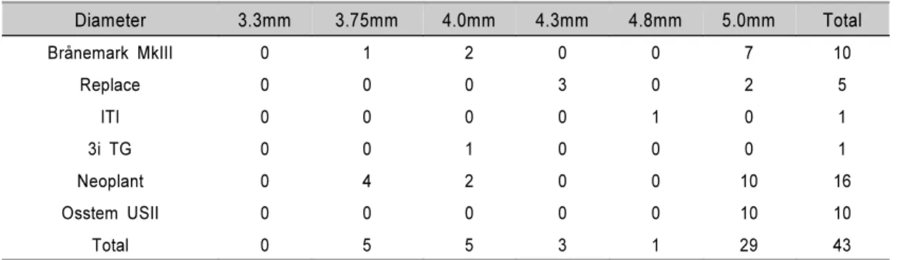
임플란트 시스템은 16개의 NeoplantⓇ(Neobiotech, Korea), 10개의 Brånemark MkIIIⓇ(Nobel Biocare, Göteborg, Sweden), 10개의 Osstem USIIⓇ(Osstem, Korea), 5개의 ReplaceⓇ(Nobel Biocare, Göteborg, Sweden), 1개 의 3i TGⓇ(Implant Innovations, Palm Beach Gardens, FL, USA), 1개의 ITIⓇ(Strauman Dental Implants;
Institut Straumann AG, Waldenburg, Switzerland)가 식
립되었다(Table 1). 표면처리에 따르면 기계 절삭형 임플란 트는 16개가 식립되었으며, 표면 처리된 임플란트는 27개가 식립되었다(Table 2).
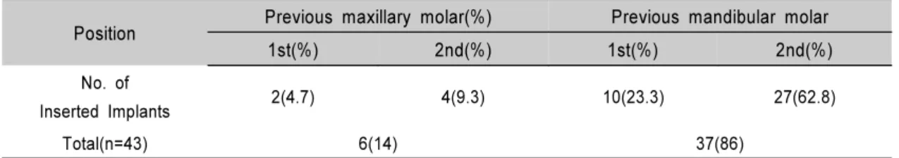
식립 부위는 상악 제 1 대구치 위치에 2개(4.7%), 제 2 대구치 위치에 4개(9.3%), 하악 제 1 대구치 위치에 10개 (23.3%), 제 2 대구치 위치에 27개(62.8%)의 임플란트가 식 립되었다(Table 3).
식립된 임플란트의 폭경은 5.0 mm가 29개(67.4%)로 가 장 많았고, 4.0mm, 3.75mm, 4.3 mm, 4.8mm 순이었다 (Table 4). 길이는 10 mm가 16개(37.2%)로 가장 많았으며 11.5 mm, 13mm, 12mm, 8mm 순이었다(Table 5).
Table 1. Distribution of Inserted fixtures according to system
Implant System No. of Inserted Implants
Neoplant 16
Brånemark MkIII 10
Osstem USII 10
Replace 5
3i TG 1
ITI 1
Total 43
Table 2. Distribution of inserted fixtures according to Implant surface
Implant surface No. of Inserted Implants Machined-surface implant 16
Treated-surface implant 27
Total 43
Table 3. Distribution of inserted fixtures according to position
Position Previous maxillary molar(%) Previous mandibular molar
1st(%) 2nd(%) 1st(%) 2nd(%)
No. of
Inserted Implants 2(4.7) 4(9.3) 10(23.3) 27(62.8)
Total(n=43) 6(14) 37(86)
2. 연구 방법
연구대상자의 의무기록지에 표기된 것을 기준으로 임플 란트 합병증 여부를 확인하였으며, 수술 부위, 임플란트 고 정체 길이와 폭경, 시스템, 가용골의 제한에 따른 복합 술식 여부 등에 따른 임플란트 합병증 여부를 검사하였다.
3. 평가 방법
임플란트의 생존율을 분석하기 위해서 임플란트를 보철 전 실패(식립 및 2 차 수술)와 보철 후 실패로 나누었고, 생 명표 분석을 사용하여 기능 후 5년까지의 누적 생존율을 조 사하였다.
결과
1. 임플란트의 누적 생존율
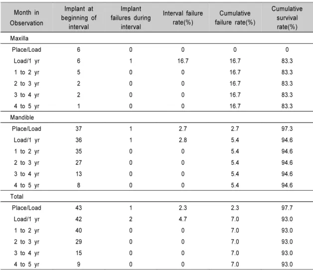
5년간 누적 생존율은 상악 83.3%, 하악 94.6%, 전체 93.0%이었다. 임플란트 식립부터 임시수복물을 장착하여
부하를 가하기까지의 기간은 상악은 평균 8.5개월, 하악은 평균 5.9개월, 전체는 평균 6.0개월이었다. 평균 관찰 기간 은 40.2개월로 최소 9개월에서 최대 66개월까지 관찰하였 다(Table 6, Fig. 1).
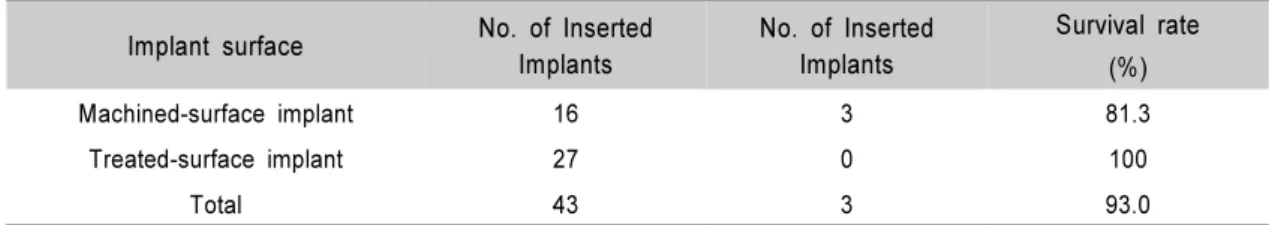
2. 표면처리에 따른 생존율
임플란트의 표면처리에 따른 생존율은 기계 절삭형 임플 란트가 16개 식립된 가운데 3개가 실패하였으며 81.3% 생존 율을 보였으며 표면처리된 임플란트는 27개가 식립되었으며 100% 생존율을 보였다.
3. 임플란트의 폭경과 길이에 따른 생존율
식립된 임플란트 중 단폭경은 없었으며 3.75~4.5 mm의 표준폭경에서는 13개 중 3개가 실패하여 76.9%의 생존율을 나타내었으며, 4.5mm 이상의 장폭경에서 30개의 임플란트 중 실패한 것은 없었다(Table 8). 10 mm, 11.5 mm, 15 mm 임플란트 각 1개씩이 실패하였다(Table 9).
Table 4. Distribution of inserted fixtures according to implant diameter
Diameter 3.3mm 3.75mm 4.0mm 4.3mm 4.8mm 5.0mm Total
Brånemark MkIII 0 1 2 0 0 7 10
Replace 0 0 0 3 0 2 5
ITI 0 0 0 0 1 0 1
3i TG 0 0 1 0 0 0 1
Neoplant 0 4 2 0 0 10 16
Osstem USII 0 0 0 0 0 10 10
Total 0 5 5 3 1 29 43
Table 5. Distribution of inserted fixtures according to implant length
Diameter 8mm 10mm 11.5mm 12mm 13mm 15mm Total
Brånemark MkIII 0 4 4 0 2 0 10
Replace 0 3 0 0 2 0 5
ITI 0 0 0 1 0 0 1
3i TG 0 0 0 0 1 0 1
Neoplant 1 5 4 0 5 1 16
Osstem USII 0 4 5 0 1 0 10
Total 1 16 13 1 11 1 43
Figure 1. Cumulative survival rate for total implants CSR = cumulative survival rate
Place = Placement of implant to time of loading Load/ 1 yr = time of loading to 1 year
Table 6. Cumulative survival rate for total implants
Month in Observation
Implant at beginning of
interval
Implant failures during
interval
Interval failure rate(%)
Cumulative failure rate(%)
Cumulative survival rate(%) Maxilla
Place/Load 6 0 0 0 0
Load/1 yr 6 1 16.7 16.7 83.3
1 to 2 yr 5 0 0 16.7 83.3
2 to 3 yr 2 0 0 16.7 83.3
3 to 4 yr 2 0 0 16.7 83.3
4 to 5 yr 1 0 0 16.7 83.3
Mandible
Place/Load 37 1 2.7 2.7 97.3
Load/1 yr 36 1 2.8 5.4 94.6
1 to 2 yr 35 0 0 5.4 94.6
2 to 3 yr 27 0 0 5.4 94.6
3 to 4 yr 13 0 0 5.4 94.6
4 to 5 yr 8 0 0 5.4 94.6
Total
Place/Load 43 1 2.3 2.3 97.7
Load/1 yr 42 2 4.7 7.0 93.0
1 to 2 yr 40 0 0 7.0 93.0
2 to 3 yr 29 0 0 7.0 93.0
3 to 4 yr 15 0 0 7.0 93.0
4 to 5 yr 9 0 0 7.0 93.0
Table 7. Survival rate of implants according to implant surface Implant surface No. of Inserted
Implants
No. of Inserted Implants
Survival rate (%)
Machined-surface implant 16 3 81.3
Treated-surface implant 27 0 100
Total 43 3 93.0
Table 8. Survival rate of implants according to diameter
Diameter No. of
Inserted Implants
No. of Failed Implants
Survival rate (%)
3.75mm 5 3 40
4.0mm 5 0 100
4.3mm 3 0 100
4.8mm 1 0 100
5.0mm 29 0 100
Total 43 3 93.0
Table 9. Survival rate of implants according to length
Length No. of
Inserted Implants
No. of Failed Implants
Survival rate (%)
8mm 1 0 100
10mm 16 1 93.8
11.5mm 13 1 92.3
12mm 1 0 100
13mm 11 0 100
15mm 1 1 0
Total 43 3 93.0
Table 10. Distribution of inserted fixtures according to implant system
Implant System No. of
Inserted Implants
No. of Failed Implants
Survival rate (%)
ITI 1 0 100
3i TG 1 0 100
Brånemark MkIII 10 0 100
Replace 5 0 100
Neoplant 16 3 81.3
Osstem USII 10 0 100
Total 43 3 93.0
4. 시스템에 따른 생존율
16개의 NeoplantⓇ 중 3개의 임플란트가 제거되었으며, 다른 시스템에서는 임플란트의 실패가 발생하지 않았다 (Table 10).
5. 수술 방법에 따른 생존율
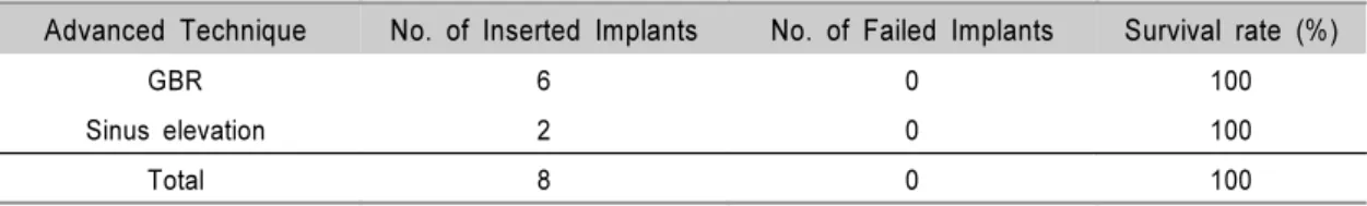
이회법으로 시술된 경우가 40 증례였고, 3 증례는 일회 법으로 시술되었다. 상악의 경우는 평균 5.0개월 후에 2 차 수술이 시행되었으며, 하악의 경우에는 평균 3.0개월 후에 2차 수술이 시행되었다. 골이식을 시행한 경우는 총 8 증례 이었고 이 중 6 증례는 임플란트 식립과 함께 골유도재생술 을 시행하였으며, 2 증례는 상악동거상술과 함께 이루어졌 다. 주로 이종골(BioOssⓇ Geistlich, Swiss)이 이식에 사용 되었으며, 차폐막을 사용한 경우에는 흡수성 콜라겐 차폐막 (BioGideⓇ, Geistlich, Swiss)이 주로 사용되었다. 이들 8 증례 모두 100% 생존율을 보였다(Table 11).
6. 합병증
치유기간 중 나사 풀림이 11.6%로 가장 높게 나타났고 그 다음으로 접촉점의 헐거워짐, 치유 지대주 풀림, 임플란트
주위염과 고정체 파절 등이 나타났다(Table 12). 합병증에 따른 임플란트는 중복표집하였다.
고찰
단일치 임플란트를 이용한 구치부의 수복은 단기간의 연구 들을 통해 효과적인 치료 방법으로 제시되고 있다24-25). 그러 나 교합력이 구치부에 집중되면 임플란트 주위골 뿐만 아니 라 임플란트의 각 구성 성분에도 영향을 끼치기 때문에 생역 학적인 부분에 대한 충분한 고려가 필요하다26). Parel27)은 구 치를 단일치 임플란트로 수복하는 경우 주의가 필요하며 교 합력이나 응력 분배 등의 문제가 발생할 수 있다고 하였다.
이번 연구에서 총 37명의 환자에게 식립된 43개의 임플 란트 중 실패로 간주된 것은 2명의 환자에서 3개 임플란트 였다. 모두 전신병력이나 특이한 병력은 없었으며, 실패한 부위의 재식립 후 골유착이 실패한 것을 제외하면 각 남, 여 1명에서 한 개의 임플란트가 부하 1년 이내에 실패하였으며, 전체 5년 누적 생존율은 93.0%로 다른 연구들과 유사한 결 과를 보였다7,22,24-25).
구치부에서 단일치 임플란트에 관한 문헌들을 살펴보면 그 대상이 상, 하악 또는 제 1 소구치부터 제 2 대구치까지 구분 없이 조사되었다. 그러나 각 부위는 해부학적 특징이 다르며 가해지는 교합력도 다르기 때문에 부위에 따라 성공 Table 11. Survival rate of implants according to the type of advanced technique
Advanced Technique No. of Inserted Implants No. of Failed Implants Survival rate (%)
GBR 6 0 100
Sinus elevation 2 0 100
Total 8 0 100
Table 12. Reported complication occurring during the 5-year period
Complication No. of Inserted Implants No. of Failed Implants
Peri-Implantitis 2 1
Screw loosening 5 2
Contact loosening 2 0
HA* loosening 2 0
Fixture fracture 1 1
Total 12 4
* HA - healing abutment
률은 차이가 있을 수 있다. 이 등28)은 하악 제 1, 2 대구치 위치에 따른 누적 성공률에서 제 1 대구치가 100%인 반면, 제 2 대구치에서는 70.37%로 낮음을 보고하였다. 그 원인 으로 제 2 대구치 부위는 전방 유도 교합시 측방 균형 간섭 이 잘 발생하며 제 1 대구치보다 10% 이상 교합력이 더 발 생하기 때문이라고 하였다. 이번 연구에서 상악의 경우 식 립된 임플란트 수가 적어 생존율을 비교할 수 없지만 하악 제 1 대구치와 2 대구치의 누적 생존율은 각각 100%와 92.6%로 다소 높은 수치가 나왔다. 재식립 후 골유착에 실 패한 것을 제외하면 거의 실패가 없는 것으로 생각할 수 있 다. 이 결과는 이 등3)이 보고한 하악 제 1 대구치와 제 2 대 구치의 누적 생존율 99%, 100%이었던 것과 유사한 결과이 다. 이것은 충분한 폭경과 길이의 임플란트가 식립되어 적 절한 하중 분산이 일어난 결과라고 생각할 수 있다.
단일치 임플란트 수복은 많은 장점과 함께 생역학적 문제 도 가지고 있다. 교합력이 구치부에 집중되어 하중 분산과 정에서 나사 풀림이나 파절 등이 보고되고 있으며 심각한 주위골 흡수 후에 고정체 파절이 일어날 수도 있다고 한다
13). 또한 근원심간 거리에 따라 근심 혹은 원심측으로 캔틸 레버 현상이 나타나 임플란트 고정체 파절의 원인이 될 수 도 있다21). 이번 연구에서 보철 수복 이후에 합병증으로 나 사 파절은 관찰되지 않았으나, 나사 풀림이나 임플란트 주 위염, 고정체 파절과 같은 다수의 합병증이 발견되었다. 실 패된 2개의 임플란트 모두에서 부하 후 나사 풀림이 관찰되 었으며 그 중 한 개의 임플란트는 정기적 내원 때마다 나사 풀림이 관찰되다 결국 부하 1년 후 고정체 파절로 제거하였 다. 나머지 한 개의 임플란트는 부하 초기에만 나사 풀림이 관찰되었으나 부하 1년 후 임플란트 주위염으로 제거하였 다. 2차 수술 때 골유착 실패로 제거한 임플란트의 경우 특 별한 합병증을 보이지는 않았다. 특히 실패한 임플란트에서 3회 이상의 반복적인 나사 풀림이 관찰되었다. 나사 풀림을 해결하기 위한 방법으로 한 개의 구치가 상실된 경우 두 개 의 임플란트를 이용하여 수복하는 것을 추천하였으며21,29), 철저한 교합 조정으로 임플란트에 가해지는 측방력을 작게 하여 굴곡 모멘트를 줄이고 원추형의 구조 등의 임플란트 사용을 추천하기도 하였다30). 또한 임플란트와 지대주를 연 결할 때 임플란트 및 지대주와 다른 재료로 된 나사를 이용 하여 조절된 회전력을 가한 후, 과도한 교합력을 제거할 경 우 나사 풀림을 줄일 수 있을 것이라고 보고하였다31).
임플란트의 성공률을 높이기 위하여 여러 가지 시스템이
소개되고 있으며 임플란트 디자인 및 표면처리 개선을 위한 많은 연구들이 이루어지고 있다. 임플란트 디자인은 하중 분산을 최적화시키기 위한 방향으로 연구되고 있으며, 임플 란트 표면처리는 임플란트의 초기 안정성을 높이고 빠른 부 하를 가능하게 하여 치료기간 단축을 통해 환자의 불편을 최소화하고 임플란트의 성공률을 높인다고 알려져 있다8,32). 초기의 산부식이나 블라스트(blast) 방법에서부터 발전하여 최근에는 산화막의 두께를 증가시킨 Ti-unite 표면 등의 표 면처리 기술들이 소개되고 있으며 제품마다 다양한 형태와 장점을 내세우며 높은 성공률을 보고하고 있다19-20,24,33-34). 구치부에 단일치 임플란트로 SLA 표면처리를 한 ITI TEⓇ 임플란트를 식립한 후 평균 21.2개월간 관찰한 결과 100%
생존율을 보였다32). 이번 연구에서 기계 절삭형 임플란트의 경우 81.3% 생존율을 보인 반면, 표면처리된 임플란트의 경 우 100% 생존율을 보였으며 표면처리된 경우 생존율이 높 음을 알 수 있었다. 또한 이 등28)의 보고에 따르면 표면처리 가 개선된 임플란트의 경우 구치부에서 생존율이 증가한다 고 하였다. 따라서 최후방 구치부에 기계 절삭형 임플란트 보다는 개선된 표면처리를 한 임플란트를 사용하는 것이 나 을 것이라고 생각된다.
장폭경 임플란트는 표준폭경에 비해 표면적이 늘어나 임 플란트-골 접촉이 증가되어 안정성이 증대된다고 하였으나
36), 그 유용성에 대해서는 상반된 의견들이 있다37-38). 이번 연구에서는 충분한 폭경의 잔존 치조골에 식립된 장폭경 임 플란트(68.2%)에서는 100% 생존율을 보인 반면, 표준폭경 임플란트에서는 76.9%의 생존율을 보였다.
이번 연구에서 최후방 구치부에 단일치 임플란트 식립 시 식립 부위, 저작 역학 그리고 임플란트 시스템이 생존율에 영향을 미칠 것으로 생각하였으나 그 대상 임플란트 수가 적었으며 하나의 시스템 임플란트에서만 실패가 관찰되어 다른 요인과 생존율과의 연관성을 보이는데 한계를 가진다.
비록 위에서 언급했듯이 각 요소에 따른 생존율과 합병증 등은 과거 연구에서 보고된 수치와 유사하나, 대상 임플란 트의 수가 적어 그 연구 결과와 동일하게 간주하기에는 한 계가 있다. 이와 같은 한계 속에서도 표면처리된 장폭경의 임플란트를 사용한 최후방 단일치 임플란트 수복은 예지성 있는 치료라고 생각한다. 그러나 이번 연구에서 다루어지지 않은 변연골 소실이나 보철물의 생존율 역시 임플란트 생존 율 못지않게 고려되어져야 하며 보다 많은 자료에 대한 조 사와 장기간의 연구가 뒤따라야 할 것으로 사료된다.
결론
이번 연구에서는 37명의 환자에 식립된 43개의 임플란트 를 대상으로 5년간의 누적 생존율과 합병증을 조사한 결과, 누적 생존율은 93.0%이었다. 구치부의 최후방 단일치 임플 란트 치료는 해부학적, 생역학적 요인들을 충분히 고려하여 시행된다면 예지성 있는 치료라고 생각한다.
참고문헌
1. Jivraj S, Chee W. Rationale for dental implants. Br Dent J 2006;200:661-665.
2. Priest G. Single-tooth implants and their role in preserving remaining teeth: a 10-year survival study. Int J Oral Maxillofac Implants 1999;14:181-188.
3. Lee SM, Chae GY, Jung UW et al. Retrospective study on ITI SLA(sand-blasted, large-grit, acid-etched) implant for mandibular posterior single tooth replacement. J Korean Acad Periodontol 2006;36:661-670.
4. Levin L, Laviv A, Schwartz-Arad D. Long term success of implants replacing a single molar. J Periodontol 2006;77:
1528-1532.
5. Berglundh T, Persson L, Klinge B. A systematic review of the incidence of biological and technical complications in implant dentistry reported in prospective longitudinal stud- ies of at least 5 years. J Clin Periodontol 2002;29(Suppl.
3):197-212.
6. Bragger U, Krenander P, Lang NP. Economic aspects of single-tooth replacement. Clin Oral Impl Res 2005;16:
335-341.
7. Becker W, Becker BE. Replacement of maxillary and man- dibular molars with single endosseous implant restorations:
a retrospective study. J Prosthet Dent 1995;74:51-55.
8. Jemt T, Lekholm U, Gröndalhl K. A 3 year follow-up study of early single implant restoration and modum Brånemark. Int J Periodontics Restorative Dent 1990;10:
341-349.
9. Jemt T, Laney WR, Harris D et al. Osseointegrated im- plants for single tooth replacement: a 1-year report from a multicenter prospective study. Int J Oral Maxillofac Implants 1991;6:29-36.
10. Jemt T, Petterson P. A 3-year follow-up study on single
implant treatment. J Dent 1993;21:203-208.
11. Howell AH, Brudevold F. Vertical forces used during chewing of food. J Dent Res 1950;29:133-136.
12. Okeson JP. Management of temporomandibular disorders:
Functional Neuroanatomy and Physiology of the Masticatory system. 4th ed. St. Louis, Mo:Mosby-Year Book, Inc 1998:29-65.
13. Rangert B, Krogh PH, Langer B et al. Bending overload and fixture fracture, a retrospective clinical analysis, Int J Oral Maxillofac Impl 1995;10:326-334.
14. Carvalho W, Casado PL, Caula AL, Barboza EP. Implants for single first molar replacement: Important treatment concerns. Implant Dent 2004;13:328-335.
15. Haas R, Mensdroff-Pouilly N, Mailath G et al. Brånemark single tooth implants: a preliminary report of 76 implants.
J Prosthet Dent 1995;73:274-279.
16. Parein AM, Eckert SE, Wollan PC et al. Implants re- construction in the posterior mandible: a long term retro- spective study. J Prosthet Dent 1997;78:34-42.
17. Jprmeis L, Jemt T, Carlsson L. Loads and designs of screw joints for single crowns supported by osseointegrated implants. Int J Oral Maxillofac Implants 1992;7:353-359.
18. Palacci P, Ericsson I, Engstrand P. Optimal implant posi- tioning & soft tissue management for the Brånemark sys- tem Chicago: Quintessence; 1995;35-39.
19. Chang M, Odman PA, Wennstrom JL, Andersson B.
Esthetic outcome of implant-supported single-tooth replace- ments assessed by the patient and by prosthodontists. Int J Prosthodont 1999;12:335-341.
20. Chang M, Wennstrom JL, Odman PA, Andersson B, Implants supported single-tooth replacements compared to contralateral natural teeth. Crown and soft tissue dimensions. Clin Oral Implants Res 1999;10:185-194.
21. Petropoulos VC, Wolfinger GJ, Balshi TJ. Complication of mandibular molar replacement with a single implant: a case report. J Can Dent Assoc 2004;70:238-242.
22. Jo SJ, Lee KW, Cho KS, Moon IS. A Retrospective study on upper single tooth implants. J Korean Acad Periodontol 2003;33:383-393.
23. Albreksson T, Sennerby L.: ʻʻState of the art in oral im- plantsʼʼ. J Clin Periodontol 1991;18:471-481.
24. Schmitt A, Zarb GA. The longitudinal clinical effectiveness of osseointegrated dental implants for single-tooth replacement. Int J Prosthodont 1993;6:197-202.
25. Laney WR, Jemt T, Harris D, et al. Osseointegrated im- plants for single-tooth replacement: progress report from a multicenter prospective study after 3 years. Int J Oral Maxillofac Implants 1994;9:49-54.
26. Haraldson T, Carlsson GE, Ingervall B. Functional state, bite force and postural activity in patients with osseointe- grated oral implant bridges. Acta Odontol Scand 1979;37:
195-206.
27. Parel S. Personal commucication, Nobelpharma Team Day.
Sidney Australia, 1993.
28. Lee HB, Paik JW, Kim CS et al. The Comparison between the success rates of single implants replacing the man- dibular first and second molar. J Korean Acad Periodontol 2004;34:101-111.
29. Balshi TJ, Hernandez RE, Pryszlak MC, Rangert B. A comparative study of one implant versus two replacing a single molar. Int J Oral Maxillofac Implants 1996;11:372-378.
30. Norton MR. The Astra Tech Single-tooth implant system: a report on 27 consecutively placed and restored implants. Int J Periodontics Restorative Dent 1997;17:574-583.
31. Chang MT. A literature review on the survival rate of sin- gle implant-supported restorations. J Korean Acad Periodontol 2002;32:69-87.
32. Suh HK, Chae GJ, Jung UW et al. A retrospective clinical study of survival rate of the ITI TEⓇ implant. J Korean Acad Periodontol 2006;36:673-682.
33. Albrektsson T, Branemark PI, Hansson HA, Lindstrom J.
Osseointegrated titanium implants. Requirements for ensur- ing a long-lasting, direct bone-to-implant anchorage in man.
Acta Orthop Scand 1981;52:155-170.
34. Park J, Kim T, Jung U et al. Long-term evaluation of IMZ implants: Success and survival rates. J Korean Acad Periodontol 2005;35:1039-1052.
35. Nam KY, Chang BS, Um HS. A Two-year Retrospective Study on the Clinical Success of the Korean Implant Systems. J Korean Acad Periodontol 2003;33:37-47.
36. Langer B, Langer L, Herrman I et al. The wide fixture: a solution of special bone situations and a rescue for the compromised implant, Int J Oral Maxillofac Impl 1993;8:
400-448.
37. Friberg B, Ekestubbe A, Sennerby L. Clinical outcome of Brånemark System Implants of various diameters: a retro- spective study. Int J Oral Maxillofac Implants 2002;17:
671-677.
38. Ivanoff CJ, Grondahl K, Semmerby L, Bergastrom C, Lekholm U. Influence of variations in implant diameters: a 3- to 5- year retrospective clinical report. Int J Oral Maxillofac Implants 1999;14:173-180.
39. Sullivan DY. Wide implants for wide teeth, Dent Econ 1994;84:82-83.
40. Rangert B, Forsmalm B. Nobelpharma News 1994;8:7.