73
Received:October 22, 2019, Revised:December 2, 2019, Accepted:December 3, 2019 Corresponding to:Jinseok Kim http://orcid.org/0000-0001-7518-3284
Division of Rheumatology, Department of Internal Medicine, Jeju National University School of Medicine, 15 Aran 13-gil, Jeju 63241, Korea. E-mail:[email protected]
Copyright ⓒ 2020 by The Korean College of Rheumatology. All rights reserved.
This is an Open Access article, which permits unrestricted non-commerical use, distribution, and reproduction in any medium, provided the original work is properly cited.
Clinical Image
pISSN: 2093-940X, eISSN: 2233-4718
Journal of Rheumatic Diseases Vol. 27, No. 1, January, 2020 https://doi.org/10.4078/jrd.2020.27.1.73
Figure 1. Magnetic resonance imaging demonstrated focal cortical disruption at anterior mid aspect of right distal clavi- cle and decreased bone mar- row signal intensity on T1 im- age, and right sternoclavicular joint arthritis, which suggested septic arthritis with osteomyeli- tis or malignancy (A∼C, arrows).
A Case of Acute Lymphoblastic Leukemia Presenting as Unilateral Sternoclavicular Joint Arthritis and Mass
Eun-Jung Park, M.D.1, Chang Lim Hyun, M.D., Ph.D.2, Jinseok Kim, M.D., Ph.D.3
1Division of Rheumatology, Department of Internal Medicine, National Medical Center, Seoul, 2Department of Pathology and 3Division of Rheumatology, Department of Internal Medicine, Jeju National University School of Medicine, Jeju, Korea
We report a patient with acute lymphoblastic leukemia of bone and joint, which is very rare, presenting as mono- arthritis [1-4]. A 57-year-old woman presented with the complaints of right sternoclavicular (SC) joint area pain with swelling, malaise, and intermittent fever lasting for two weeks. She had no past medical history and denied trauma or injection of affected joint. The patient had epi- sodes of fever up to 39.2oC, and mild tachycardia. On physical examination, localized heat in the right SC joint and a 2×3 cm soft and tender mass around the joint were detected. Laboratory results showed hemoglobin 10.6 g/dL, hematocrit 30.3%, platelets 242,000/mm3, and white blood cell count 5,400/mm3 including absolute neutrophil count 870/μL, segment neutrophil 39.6%, and monocyte 18.9%. Erythrocyte sedimentation rate was 28 mm/hour and C-reactive protein was 6.34 mg/dL.
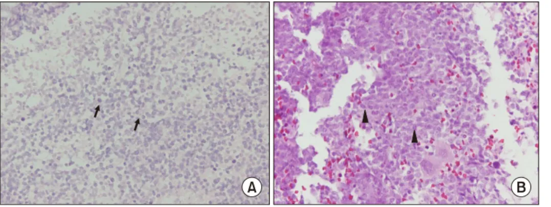
Otherwise, blood laboratory findings and urinalysis were unremarkable. Plain radiography of clavicle and sternum showed no abnormal finding. Magnetic resonance imag- ing demonstrated focal cortical disruption at ante- romedial aspect of right distal clavicle, decreased bone marrow signal intensity on T1 image, and right SC joint arthritis, which suggested septic arthritis with osteomye- litis or malignancy (Figure 1A∼C, arrows). Bone biopsy of right distal clavicle revealed increased atypical cell in- filtration with necrosis, which was consistent with leuke- mic infiltration (Figure 2A; H&E stain, ×400, arrows).
The bone marrow biopsy showed nearly packed marrow with myelofibrosis (Figure 2B; H&E stain, ×400, arrow- heads). On aspirate smears, blasts were present in up to 87.8% of all marrow nucleated cells. They were small to large-sized with round nuclei, dispersed chromatin and
Eun-Jung Park et al.
74 J Rheum Dis Vol. 27, No. 1, January, 2020
Figure 2. Bone biopsy of right distal clavicle revealed increased atypical cell infiltration with ne- crosis, which was consistent with leukemic infiltration (A, H&E stain, ×400, arrows). The bone marrow biopsy showed nearly packed marrow with myelofibrosis (B, H&E stain,
×400, arrowheads).
inconspicuous nucleoli. Normal erythroid and myeloid elements were markedly suppressed. Cytochemical stain on aspiration and biopsy demonstrated negative MPO and PAS and positive CD 34 and CD 20. All findings sug- gested B-lymphoblastic leukemia with chloroma of right distal clavicle and SC joint. This case indicates that malig- nancy should also be considered in evaluation of mono- arthritis, and that chloroma of bone can precede the onset of acute lymphoblastic leukemia.
ACKNOWLEDGMENTS
This work has supported by the 2019 education, re- search and student guidance grant funded by Jeju National University.
CONFLICT OF INTEREST
No potential conflict of interest relevant to this article was reported.
AUTHOR CONTRIBUTIONS
Among the authors, E.J.P. appointed topic, wrote and re- vised the manuscript. C.L.H. was responsible for the anal- ysis and the interpretation of the pathology. J.S.K. de- termined and adjusted the topic, concept and drafting of the manuscript.
REFERENCES
1. Kubota H, Saida S, Kouzuki K, Hamabata T, Daifu T, Kato I, et al. [Pediatric acute lymphoblastic leukemia presenting with bone and joint pain]. Rinsho Ketsueki 2018;59:
167-73. Japanese.
2. Pécheux L, Forget P, Geurten C, Rausin L, Nicolescu R, Hoyoux C. [Bone disorders and complications of pediatric acute lymphoblastic leukemia : monocentric study and re- view of the literature]. Rev Med Liege 2018;73:575-82.
French.
3. Rehman A, Abbas N, Saba T, Rahman SIU, Mehmood Z, Kolivand H. Classification of acute lymphoblastic leukemia using deep learning. Microsc Res Tech 2018;81:1310-17.
4. Tragiannidis A, Vasileiou E, Papageorgiou M, Damianidou L, Hatzipantelis E, Gombakis N, et al. Bone involvement at diagnosis as a predictive factor in children with acute lym- phoblastic leukemia. Hippokratia 2016;20:227-30.